
Approach to fever with thrombocytopenia.
fever thrombocytopenia diagnosis approach
fever with thrombocytopenia differential diagnosis approach algorithm
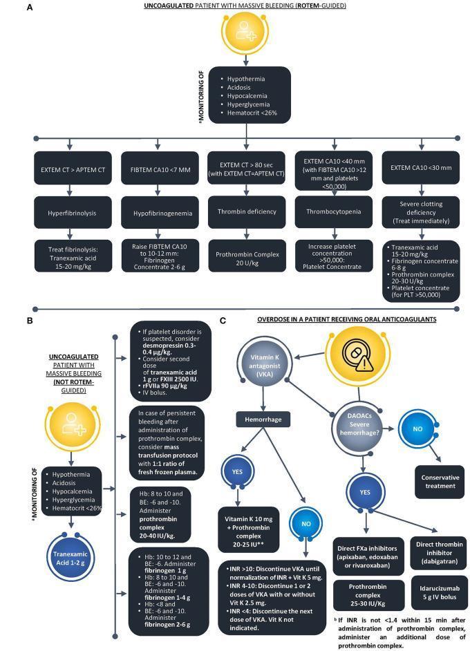
This medical infographic presents three clinical algorithms (A, B, and C) for managing massive bleeding in acute settings. Algorithm A details a ROTEM-guided approach for uncoagulated patients, using viscoelastic parameters (EXTEM, FIBTEM, APTEM) to differentiate and treat hyperfibrinolysis (tranexamic acid), hypofibrinogenemia (fibrinogen concentrate), thrombin deficiency (prothrombin complex), and thrombocytopenia. Algorithm B outlines management for uncoagulated patients when ROTEM is unavailable, focusing on monitoring for the 'lethal triad' (acidosis, hypothermia, coagulopathy), lab values like hemoglobin and base excess, and the administration of blood products or PCC. Algorithm C addresses oral anticoagulant overdose, branching by agent type: Vitamin K Antagonists (VKA) or Direct Oral Anticoagulants (DOACs). It provides decision nodes based on INR levels and hemorrhage severity, recommending reversal agents such as Vitamin K, prothrombin complex (PCC), or Idarucizumab for dabigatran. The diagrams use standardized medical terminology and dosage guidelines suitable for emergency medicine, hematology, and trauma surgery education.
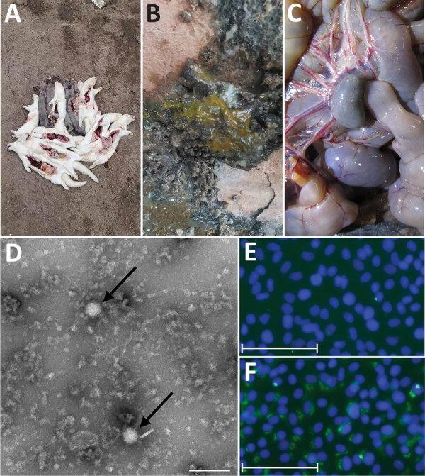
This composite educational image illustrates the clinical, gross pathological, and microscopic features of Severe Fever with Thrombocytopenia Syndrome Virus (SFTSV) in a zoonotic context. Panels A-C provide clinical photographs: (A) shows morbidity/mortality in an animal cohort; (B) displays mucoid, yellowish-green loose stools indicative of viral enteritis; (C) presents a gross pathological view of internal viscera showing mesenteric lymph node enlargement (lymphadenopathy) and hyperemia. Panel D is a diagnostic Transmission Electron Microscopy (TEM) micrograph showing typical Bunyaviridae morphology, characterized by enveloped, spherical virions (indicated by arrows) approximately 100 nm in diameter. Panels E and F represent an Indirect Immunofluorescence Assay (IFA) in Vero cells. Panel E serves as a negative control with blue DAPI staining of nuclei, while Panel F demonstrates positive SFTSV infection, visualized by granular green fluorescence indicating the presence of viral nucleoprotein. This figure serves as a diagnostic reference for identifying SFTSV through clinical presentation, ultrastructural morphology, and molecular confirmation.
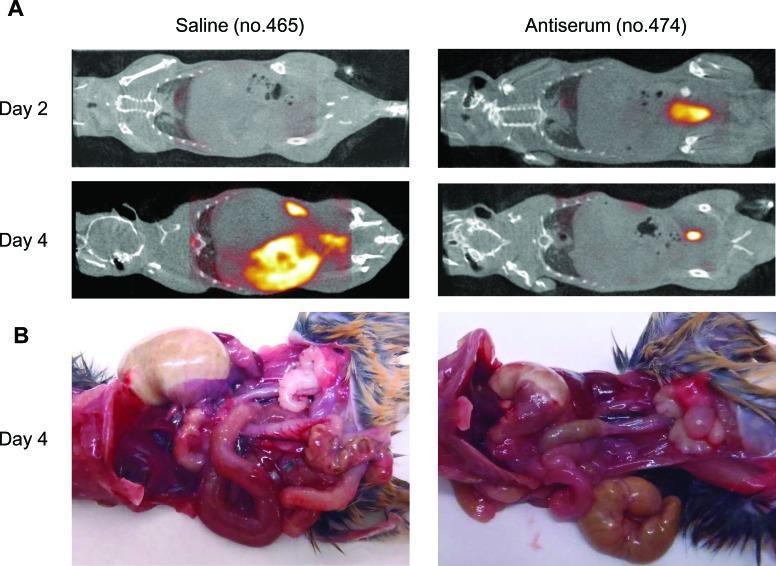
This figure presents a comparative study of disease progression and therapeutic efficacy in a mouse model of Severe Fever with Thrombocytopenia Syndrome Virus (SFTSV) infection. Panel A displays whole-body PET/CT imaging at days 2 and 4 post-infection (pi) using 18F-FDG as a metabolic tracer. The saline-treated control shows progressively increasing, high-intensity FDG accumulation in the abdominal region by day 4, indicative of severe inflammatory or viral metabolic activity. In contrast, the antiserum-treated mouse exhibits significantly reduced tracer uptake, demonstrating therapeutic mitigation. Panel B shows gross pathology specimens at day 4 pi. The saline-treated specimen reveals marked gastric and intestinal distension (bloating) and visceral congestion. Conversely, the antiserum-treated specimen shows relatively normal organ morphology with minimal distension. These images highlight the use of PET/CT as a non-invasive diagnostic tool to monitor gastrointestinal pathology and therapeutic response in viral infections.
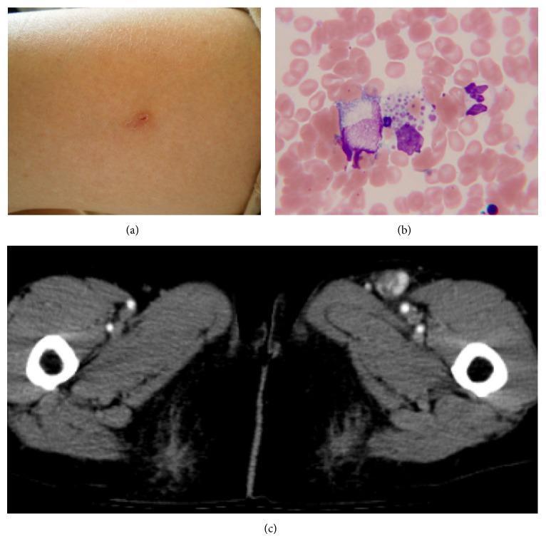
A composite of three images documenting a case of Severe Fever with Thrombocytopenia Syndrome Virus (SFTSV) infection. (a) Clinical photograph of the lateral left thigh showing a small, focal tick bite site, characterized by a minor cutaneous puncture with localized mild erythema and no surrounding rash or wheal. (b) Light microscopy of a Giemsa-stained bone marrow aspirate demonstrating hemophagocytosis; specifically, a large macrophage containing engulfed platelet-specific material, characteristic of the hematologic complications of SFTSV. (c) Axial contrast-enhanced computed tomography (CT) scan of the pelvic/inguinal region revealing significant unilateral left inguinal lymphadenopathy. The enlarged lymph node shows contrast enhancement compared to surrounding soft tissue. These panels collectively illustrate the primary inoculation site, systemic hematologic response, and localized lymphatic involvement in a viral zoonotic infection.
fever with thrombocytopenia clinical approach differential diagnosis 2024
Approach to Fever with Thrombocytopenia
Pathophysiology of Thrombocytopenia in Febrile States
| Mechanism | Example |
|---|---|
| Decreased production | Bone marrow suppression (viruses, alcohol, B12 deficiency, drugs) |
| Increased destruction/consumption | Sepsis, DIC, TTP, immune-mediated (ITP, SLE, drugs) |
| Splenic sequestration | Splenomegaly from portal hypertension, malaria, leishmaniasis (platelet count usually stays >40,000/µL) |
Differential Diagnosis: Etiology-Based Framework
1. Tropical / Vector-Borne Infections (Most Common in Endemic Areas)
- Mild-to-moderate thrombocytopenia is almost universal; a normal platelet count should actively question the diagnosis. Severe DIC occurs in <5% of cases.
- Classic periodicity (tertian/quartan), splenomegaly, anemia, hemolytic jaundice.
- Key lab: peripheral smear, malaria antigen RDT, PCR. - Harrison's Principles of Internal Medicine 22E, Laboratory Findings in Acute Malaria
- Fever, severe headache, retro-orbital pain, myalgia, rash (maculopapular/petechial), leukopenia, and thrombocytopenia.
- Dengue Hemorrhagic Fever (DHF): hemorrhagic phenomena + plasma leakage (rising hematocrit, pleural effusion, ascites, hypoalbuminemia) + platelet <100,000/µL.
- Dengue Shock Syndrome (DSS): DHF + circulatory failure. - Brenner & Rector's The Kidney and Rosen's Emergency Medicine
- Orientia tsutsugamushi (scrub typhus): eschar at bite site, high fever, rash, lymphadenopathy, thrombocytopenia, elevated LFTs.
- Rocky Mountain Spotted Fever (RMSF): Rickettsia rickettsii; petechial rash starting at wrists/ankles then spreading centripetally to involve palms and soles; leukopenia, thrombocytopenia. Diagnosis: immunofluorescent staining of skin biopsy.
- Ehrlichiosis/Anaplasmosis: Ehrlichia chaffeensis / Anaplasma phagocytophilum; fever, headache, confusion, nausea; 20% have maculopapular/petechial rash; leukopenia + thrombocytopenia are classic; elevated LFTs. - Harrison's Principles of Internal Medicine 22E, Differential Diagnosis
- Freshwater exposure, fever, headache, myalgia, conjunctival suffusion, acute kidney injury, moderate hepatocellular injury, hyponatremia, thrombocytopenia.
- Weil's disease: jaundice + AKI + bleeding (very high bilirubin + thrombocytopenia is characteristic). - Frameworks for Internal Medicine and Brenner & Rector's The Kidney
2. Viral Hemorrhagic Fevers
- Common features: fever, thrombocytopenia, elevated aminotransferases, hemorrhagic manifestations.
- Must be distinguished from: rickettsial infections, leptospirosis, meningococcemia, typhoid fever, falciparum malaria (Goldman-Cecil Medicine).
- Caused by Dabie bandavirus (phenuivirus); tick-transmitted; East Asia.
- Fever, gastrointestinal symptoms (nausea, vomiting, diarrhea), leukopenia + thrombocytopenia, elevated LFTs, lymphadenopathy.
- Bone marrow biopsy may show hemophagocytosis.
- Diagnosis: RT-PCR in early phase; serology (IFA/ELISA) less sensitive in first 2 weeks. - Jawetz Melnick Medical Microbiology 28E
3. Bacterial Sepsis and DIC
- Any severe bacterial infection can produce thrombocytopenia via platelet consumption + DIC.
- Meningococcemia: petechial/purpuric rash, septic shock, DIC.
- Typhoid: relative bradycardia, stepladder fever, rash (rose spots), leukopenia, mild thrombocytopenia.
4. Non-Infectious Causes (Do Not Miss)
| Condition | Clues |
|---|---|
| ITP | Isolated thrombocytopenia, no anemia, no organomegaly; can present with "fever" if concurrent infection |
| TTP | Microangiopathic hemolytic anemia (MAHA) + thrombocytopenia ± fever, neurological signs, renal failure (pentad) |
| HUS | MAHA + thrombocytopenia + AKI (usually post-diarrheal, E. coli O157:H7) |
| SLE | Multi-system features, ANA +ve, thrombocytopenia from autoimmunity |
| Lymphoma/Leukemia | Lymphadenopathy, hepatosplenomegaly, bone marrow infiltration |
| Drug-induced | Heparin (HIT), quinine, sulfonamides, phenytoin |
| Visceral Leishmaniasis | Prolonged fever, massive splenomegaly, pancytopenia, weight loss |
| Hemophagocytic Lymphohistiocytosis (HLH) | Ferritin >500 (often >10,000), splenomegaly, cytopenias ≥2, hemophagocytosis on marrow biopsy |
Clinical Approach: Step-by-Step
Step 1: History - "THREADS" Framework
- Travel history (tropics, endemic areas, freshwater exposure)
- Habitat / occupation (farmers, soldiers, outdoor workers)
- Rash (when, where, spreading pattern)
- Exposure (ticks, mosquitoes, animal contact, unpasteurized food)
- Animal contact (ungulates → Q fever; rodents → hantavirus, leptospirosis)
- Drug history (heparin, quinine, sulfonamides)
- Sexual history, IV drug use (HIV, hepatitis C → secondary thrombocytopenia)
Step 2: Physical Examination - Key Findings
| Finding | Points Toward |
|---|---|
| Eschar | Scrub typhus, RMSF, CCHF |
| Petechiae/purpura (palms, soles) | RMSF, meningococcemia |
| Conjunctival suffusion | Leptospirosis |
| Maculopapular rash | Dengue, ehrlichiosis, typhoid (rose spots) |
| Splenomegaly | Malaria, visceral leishmaniasis, EBV, typhoid |
| Jaundice | Malaria, leptospirosis (Weil's), yellow fever, viral hepatitis |
| Lymphadenopathy | EBV, scrub typhus, SFTS, lymphoma |
| Hepatomegaly + splenomegaly | Visceral leishmaniasis, malaria, EBV |
| Mucosal bleeding | DHF, TTP, DIC |
Step 3: Investigations - Tiered
- CBC with differential (leukopenia + thrombocytopenia vs. leukocytosis)
- Peripheral blood smear (malaria parasites, fragmented RBCs for MAHA, leukemic blasts, morulae in ehrlichiosis)
- Blood culture x2 (aerobic + anaerobic)
- LFTs, RFTs, LDH, uric acid
- Coagulation profile: PT, aPTT, fibrinogen, D-dimer (for DIC)
- Urine routine/microscopy
- Malaria: RDT + thick/thin peripheral smear (repeat 3 times if initially negative) + PCR
- Dengue: NS1 antigen (days 1-5), IgM/IgG ELISA (from day 5); dengue PCR
- Scrub typhus/RMSF: Weil-Felix (screening), Scrub typhus IgM ELISA; skin biopsy immunofluorescence for RMSF
- Ehrlichia/Anaplasma: PCR (most sensitive in acute phase); morulae on smear
- Leptospirosis: Microscopic agglutination test (MAT) - gold standard; IgM ELISA for rapid screening; urine/blood culture
- SFTS: RT-PCR (early phase); serology (late phase)
- HIV: 4th generation Ag/Ab combo
- EBV/CMV: monospot, EBV VCA IgM, CMV IgM
- Bone marrow aspiration + trephine biopsy (leishmaniasis amastigotes, hemophagocytosis in HLH, marrow infiltration in lymphoma/leukemia)
- ANA, anti-dsDNA, complement levels (SLE)
- Ferritin (>500 raises HLH concern; >10,000 strongly suggestive)
- ADAMTS13 activity (TTP if severely reduced <10%)
- Hantavirus serology (IgM), viral hemorrhagic fever panel (if exposure history)
- CT chest/abdomen/pelvis (lymphadenopathy, splenomegaly, effusions)
Red Flags Requiring Urgent Action
- Platelet count <20,000/µL with active bleeding → platelet transfusion, consider TTP/DIC/HLH
- Signs of shock (hypotension, cold extremities, altered sensorium) → DHF/DSS, septic shock, meningococcemia
- Purpuric non-blanching rash → meningococcemia or RMSF: do NOT wait; start empirical IV ceftriaxone / doxycycline immediately
- Pulmonary edema or respiratory distress with fever + low platelets → dengue pulmonary manifestation, hantavirus pulmonary syndrome
- Pentad of TTP (MAHA + thrombocytopenia + AKI + fever + neuro signs) → emergency plasma exchange
Empirical Treatment While Awaiting Results
| Suspected etiology | First-line treatment |
|---|---|
| Malaria | Artesunate-based combination (severe: IV artesunate) |
| Dengue | Supportive (fluids, paracetamol; avoid NSAIDs and aspirin) |
| Rickettsial / Ehrlichia / Anaplasma | Doxycycline 100 mg BD (drug of choice; covers all) |
| Scrub typhus | Doxycycline 100 mg BD (or azithromycin in pregnancy) |
| Leptospirosis | Mild: doxycycline 100 mg BD; Severe (Weil's): IV penicillin G or ceftriaxone |
| Meningococcemia | IV ceftriaxone 2 g BD |
| Sepsis + DIC | Source control + broad-spectrum antibiotics; FFP/cryoprecipitate for active bleeding |
| TTP | Urgent plasma exchange (PE) + steroids; avoid platelet transfusion |
A Practical Diagnostic Algorithm
Fever + Thrombocytopenia
         |
         ├── Travel to tropics / endemic area?
         |       ├── YES → Smear for malaria (first priority)
         |       |           + Dengue NS1/PCR
         |       |           + Scrub typhus / Rickettsial serology
         |       |           + Leptospirosis serology
         |       |
         |       └── NO → Consider:
         |                   Sepsis + DIC (blood cultures, coagulation)
         |                   Viral infections (EBV, CMV, HIV)
         |                   Drug-induced (HIT, quinine)
         |                   TTP/HUS (MAHA on smear, ADAMTS13)
         |                   HLH (ferritin, bone marrow)
         |
         ├── Eschar present? → Scrub typhus / RMSF (start doxycycline)
         ├── Purpuric rash? → Meningococcemia / RMSF (start antibiotics NOW)
         ├── Jaundice + AKI? → Leptospirosis (Weil's disease)
         ├── MAHA on smear? → TTP / HUS / DIC
         ├── Massive splenomegaly + long fever? → Visceral leishmaniasis
         └── Ferritin >10,000 + pancytopenia? → HLH (bone marrow biopsy)
Key Mnemonics
- M - Malaria
- I - ITP / Immune causes
- L - Leptospirosis
- D - Dengue
- R - Rickettsial (RMSF, scrub typhus, ehrlichia)
- A - Arboviral (Chikungunya, Zika, Yellow fever, Hantavirus)
- S - Sepsis + DIC
- H - HLH / HUS
- T - TTP / Typhoid
- I - Infections: HIV, EBV, CMV
- P - Para-infectious / Drug-induced