
Amelogenesis kuhs university bds first year essay
Reading File
Now let me search the medical library for amelogenesis content.
Finding Sources
Finding Sources
The Histology textbook has the most relevant content. Let me read those sections.
Reading File
Reading File
Reading File
Reading File
Finding Sources
I now have comprehensive information. Let me also fetch the key diagram images to include in the answer.
Loading Image
Loading Image
Excellent! I now have comprehensive textbook content and diagrams. Let me compose the full BDS first-year essay on amelogenesis.
Amelogenesis
A BDS First Year Essay - Oral Histology & Embryology
Definition
Amelogenesis is the process of enamel formation by ameloblasts. It is a matrix-mediated biomineralization process that produces dental enamel - the hardest tissue in the human body, composed predominantly of calcium phosphate hydroxyapatite crystals.
- Histology: A Text and Atlas - Ross & Pawlina, p. 1445
Prerequisite: Development of the Enamel Organ
Before amelogenesis begins, the enamel organ passes through three developmental stages:
1. Bud Stage
The oral epithelium proliferates downward into the underlying ectomesenchyme, forming a tooth bud. The mesenchyme condenses beneath it.
2. Cap Stage
The tooth bud invaginates to form a cap-like structure. Cells in the concavity differentiate into tall columnar inner enamel epithelium (IEE). The condensed mesenchyme forms the dental papilla (future dentin and pulp). The enamel organ at this stage has four layers:
- Outer enamel epithelium (OEE) - convex surface
- Inner enamel epithelium (IEE) - concave surface; becomes ameloblasts
- Stratum intermedium - cell layer internal to IEE; contains alkaline phosphatase, important for calcification
- Stellate reticulum - star-shaped cells occupying the inner portion
3. Bell Stage
Connection with oral epithelium is nearly severed. The enamel organ is fully differentiated. The dental lamina degenerates, leaving the tooth primordium detached from its origin - this occurs just before dentinogenesis and amelogenesis begin.
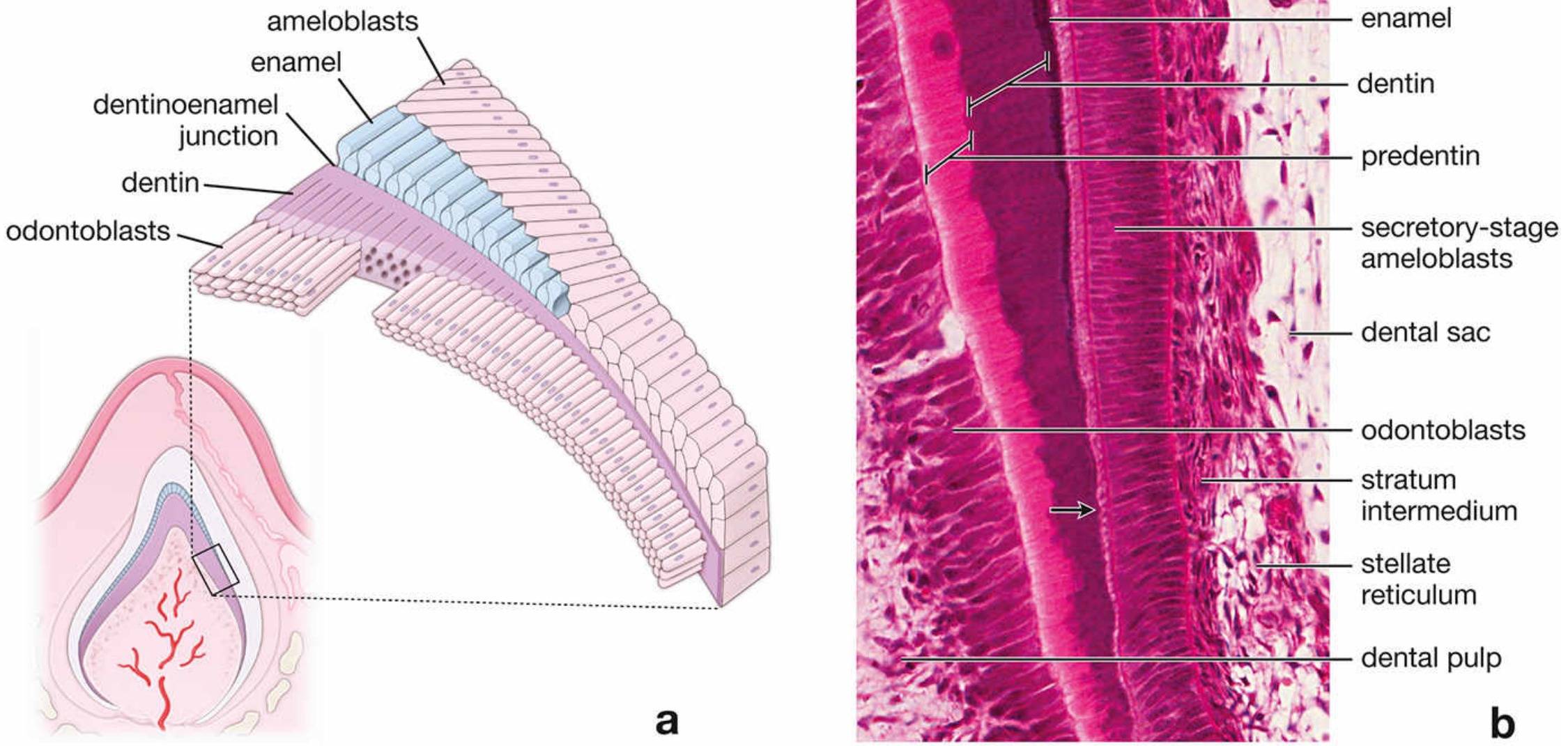
Figure: Cellular relationships during enamel formation. (a) Schematic of ameloblasts overlying dentin at the dentinoenamel junction. (b) H&E photomicrograph showing secretory-stage ameloblasts, predentin, dentin, stratum intermedium, stellate reticulum, and dental pulp.
Stages of Amelogenesis
Stage 1 - Secretory (Matrix Production) Stage
Key points:
- Dentin is always formed first (dentinogenesis precedes amelogenesis)
- Partially mineralized enamel matrix is then deposited directly on the surface of the previously formed dentin
- Cells at this stage are called secretory-stage ameloblasts
- These cells are tall, polarized columnar cells
- They produce organic proteinaceous enamel matrix using the rER, Golgi apparatus, and secretory granules - a mechanism similar to osteoblast bone production
- Secretion continues until the full thickness of the future enamel is achieved
Tomes Process:
- At the apical pole of each secretory-stage ameloblast is a cytoplasmic extension called the Tomes process
- The Tomes process is surrounded by developing enamel
- It contains matrix-laden secretory vesicles
- The direction of the enamel rod in mature enamel is a direct record of the path taken by the secretory-stage ameloblast
- A distal terminal web is found between the Tomes process and the main cell body, and a proximal terminal web is at the base - both appear as pink lines on H&E staining due to actin filament accumulation
Enamel Rod (Prism) Formation:
- The enamel rod is formed in the "wake" of the moving ameloblast
- Enamel rods extend from the dentinoenamel junction (DEJ) to the outer surface of the enamel
- Hydroxyapatite crystals are deposited within the organic matrix
Role of stratum intermedium:
- The base of the secretory-stage ameloblasts is adjacent to the stratum intermedium
- The plasma membrane of stratum intermedium cells contains alkaline phosphatase, an enzyme active in calcification
- External to the stratum intermedium lies the stellate reticulum, separated from adjacent blood vessels by a basal lamina
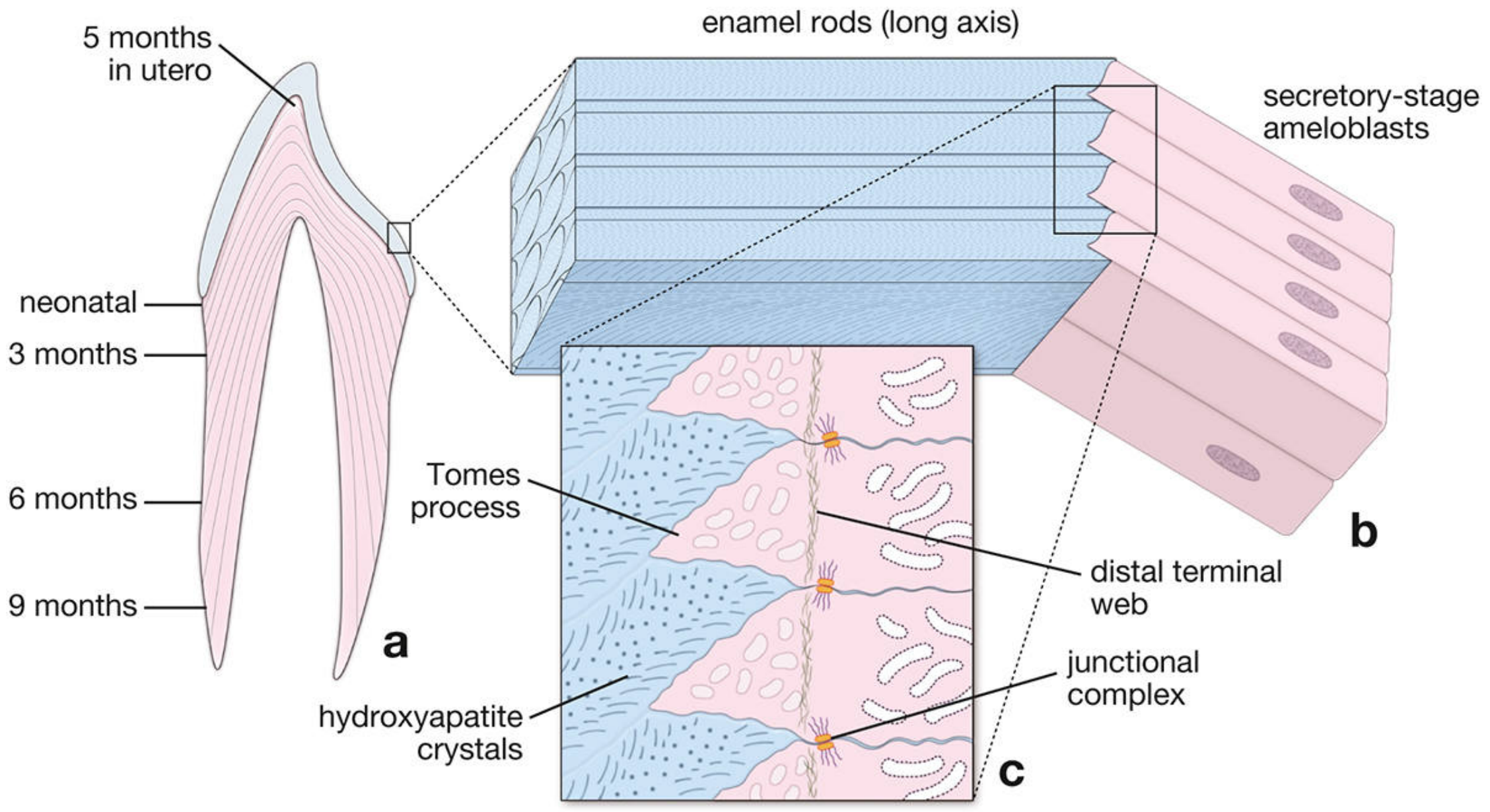
Figure: (a) Cross-section of a partially formed incisor showing enamel rod deposition at successive time points. (b) Enamel rods running from the DEJ to the enamel surface, produced by secretory-stage ameloblasts. (c) High-magnification view of Tomes processes, distal terminal web, junctional complexes, and hydroxyapatite crystal deposition.
Stage 2 - Maturation Stage
Key points:
- Cells in this stage are called maturation-stage ameloblasts
- They differentiate from secretory-stage ameloblasts
- Their primary function is as a transporting epithelium - moving substances into and out of the maturing enamel
Process of maturation:
- Involves removal of organic material (the original proteinaceous matrix)
- Continued influx of calcium (Ca²⁺) and phosphate ions into the mineralizing enamel
- Maturation-stage ameloblasts undergo cyclical modulation - they alternate between displaying a striated (ruffled) border and a smooth border on their apical surface:
- Striated border (70% of cells): responsible for secreting bicarbonate ions (HCO₃⁻); contains plasma membrane Ca²⁺-ATPases (PMCA) that actively transport Ca²⁺ into the maturing enamel
- Smooth border (30% of cells): produce and secrete enzymes to degrade and reabsorb the extracellular matrix no longer needed
Changes in enamel organ at this stage:
- The stratum intermedium is no longer present
- Stellate reticulum, stratum intermedium, and outer dental epithelium collapse and reorganize
- Blood vessels invaginate into this layer, forming the papillary layer containing stellate papillary cells adjacent to maturation-stage ameloblasts
- Maturation-stage ameloblasts and papillary cells contain numerous mitochondria - reflecting high metabolic activity
Fate of Ameloblasts
After enamel formation is complete, ameloblasts become part of the reduced enamel epithelium, which fuses with the oral epithelium at the time of tooth eruption. Once the tooth erupts, ameloblasts are shed and lost permanently.
This is why:
Enamel cannot be repaired or regenerated once formed - there are no ameloblasts remaining in the erupted tooth.
Enamel Matrix Proteins
During the secretory stage, ameloblasts produce specific proteins:
| Protein | Function |
|---|---|
| Amelogenin | Major matrix protein (~90%); controls crystal shape and spacing |
| Enamelin | Minor protein; involved in crystal elongation |
| Ameloblastin (amelin) | Cell adhesion and signaling |
| MMP-20 (Enamelysin) | Protease that degrades amelogenin during maturation |
| KLK4 (Kallikrein-4) | Protease responsible for final matrix removal in maturation stage |
Clinical Correlation: Amelogenesis Imperfecta
Genetic mutations in enamel matrix proteins or ameloblast function result in amelogenesis imperfecta (AI), a group of hereditary enamel disorders.
-
Incidence: ranges from approximately 1 in 700 to 1 in 14,000 depending on population
-
The enamel may be hard or soft, pitted or smooth, thin or normal in thickness - depending on the type
-
Types include hypoplastic (defective secretory stage), hypomaturation (defective removal of matrix), and hypocalcification (defective mineralization) forms
-
The Developing Human - Moore & Persaud, p. clinical box
Summary Table
| Feature | Secretory Stage | Maturation Stage |
|---|---|---|
| Cell type | Secretory-stage ameloblast | Maturation-stage ameloblast |
| Key structure | Tomes process | Ruffled/striated border |
| Activity | Matrix protein secretion | Matrix removal + Ca²⁺/phosphate influx |
| Adjacent layer | Stratum intermedium | Papillary layer |
| End result | Full thickness enamel (incompletely mineralized) | Fully mineralized enamel (96% mineral) |
Key Points to Remember for KUHS Exams
- Amelogenesis = enamel formation by ameloblasts (derived from IEE of the enamel organ)
- Dentin always forms before enamel - the DEJ is the starting point
- Two main stages: Secretory (matrix production) and Maturation (mineralization)
- Secretory-stage hallmark = Tomes process at the apical pole
- Maturation-stage hallmark = striated/ruffled border with cyclic modulation
- Ca²⁺ transport into enamel = PMCA (Ca²⁺-ATPases) in striated-border ameloblasts
- After eruption, ameloblasts are lost permanently - enamel cannot self-repair
- Genetic defects in this process = Amelogenesis Imperfecta
Sources: Histology: A Text and Atlas with Correlated Cell and Molecular Biology (Ross & Pawlina, 8th edition), pp. 1443-1450; The Developing Human: Clinically Oriented Embryology (Moore & Persaud)
This is a shared conversation. Sign in to Orris to start your own chat.