
Volume of distribution and its importance pharmacology Assinments with index and refrance
Reading File
Now let me search the medical library for volume of distribution content, in parallel with a web search for supplementary info.
Finding Sources
Finding Sources
Searching the Web
volume of distribution pharmacology definition formula clinical importance
Now let me read the core textbook sections in parallel for deep content:
Reading File
Reading File
Reading File
Finding Sources
Reading File
Finding Sources
Reading File
Loading Image
Finding Sources
Finding Sources
I now have comprehensive content to write the full pharmacology assignment. Let me compose it:
Volume of Distribution and Its Importance in Pharmacology
A Pharmacology Assignment
Index
- Introduction
- Definition of Volume of Distribution (Vd)
- Formula and Calculation
- Units and Types of Vd
- Physical Body Compartments and Drug Distribution
- Factors Affecting Volume of Distribution
- Clinical Importance of Vd
- 7.1 Loading Dose Calculation
- 7.2 Relationship to Half-Life
- 7.3 Therapeutic Drug Monitoring (TDM)
- 7.4 Extracorporeal Drug Removal (Dialysis)
- 7.5 Drug Overdose Estimation
- Vd in Special Populations
- Worked Clinical Examples
- Summary Table of Drug Vd Values
- References
1. Introduction
Pharmacokinetics describes what the body does to a drug after administration, encompassing four processes: absorption, distribution, metabolism, and excretion (ADME). Among the parameters used to quantify drug behavior in the body, volume of distribution (Vd) is one of the most fundamental. It is a "primary" pharmacokinetic parameter - meaning it depends directly on the physiological properties of the body and the physicochemical properties of the drug, and is not derived from other parameters. Understanding Vd allows clinicians to calculate appropriate loading doses, predict drug half-life, interpret plasma drug concentrations, and make dosing adjustments in altered physiological states.
2. Definition of Volume of Distribution
Volume of distribution (Vd) is defined as the apparent volume into which a drug disperses in order to produce the observed plasma concentration. It is a proportionality constant that relates the total amount of drug in the body to the plasma (or blood) concentration of the drug at a given time.
"The volume of distribution relates the amount of drug in the body to the concentration of drug in the plasma." - Goldman-Cecil Medicine, 26th Edition
"Volume of distribution (V) relates the amount of drug in the body to the concentration of drug (C) in blood or plasma." - Katzung's Basic and Clinical Pharmacology, 16th Edition
The key qualifier is "apparent" - Vd is not a real physiological volume. It is a mathematical construct (a hypothetical volume) that explains the plasma concentration observed given the total amount of drug administered. As stated in Barash's Clinical Anesthesia:
"The apparent volume of distribution is a numeric index of the extent of drug distribution that does not have any relationship to the actual volume of any tissue or group of tissues."
Vd can be smaller than plasma volume (for large protein molecules like antibodies), or it can vastly exceed total body volume (for drugs like chloroquine ~250 L/kg), which is physically impossible - illustrating that "apparent" is a critical modifier.
3. Formula and Calculation
The standard formula is:
$$V_d = \frac{\text{Amount of drug in the body}}{\text{Plasma concentration of the drug}}$$
Or equivalently, calculated after IV bolus administration:
$$V_d = \frac{\text{Dose}}{C_{p_0}}$$
Where Cp₀ is the plasma concentration extrapolated back to time zero (derived by back-extrapolating the linear elimination phase on a log-concentration vs. time graph).
Example (Barash Clinical Anesthesia):
- If 10 mg of drug is administered and the plasma concentration is 2 mg/L:
- Vd = 10 mg ÷ 2 mg/L = 5 L
- If the concentration was 4 mg/L instead:
- Vd = 10 mg ÷ 4 mg/L = 2.5 L
The graph below (from Goldman-Cecil Medicine) illustrates the two phases of plasma drug concentration over time - the distribution phase and elimination phase - from which Cp₀ and half-life can be derived:
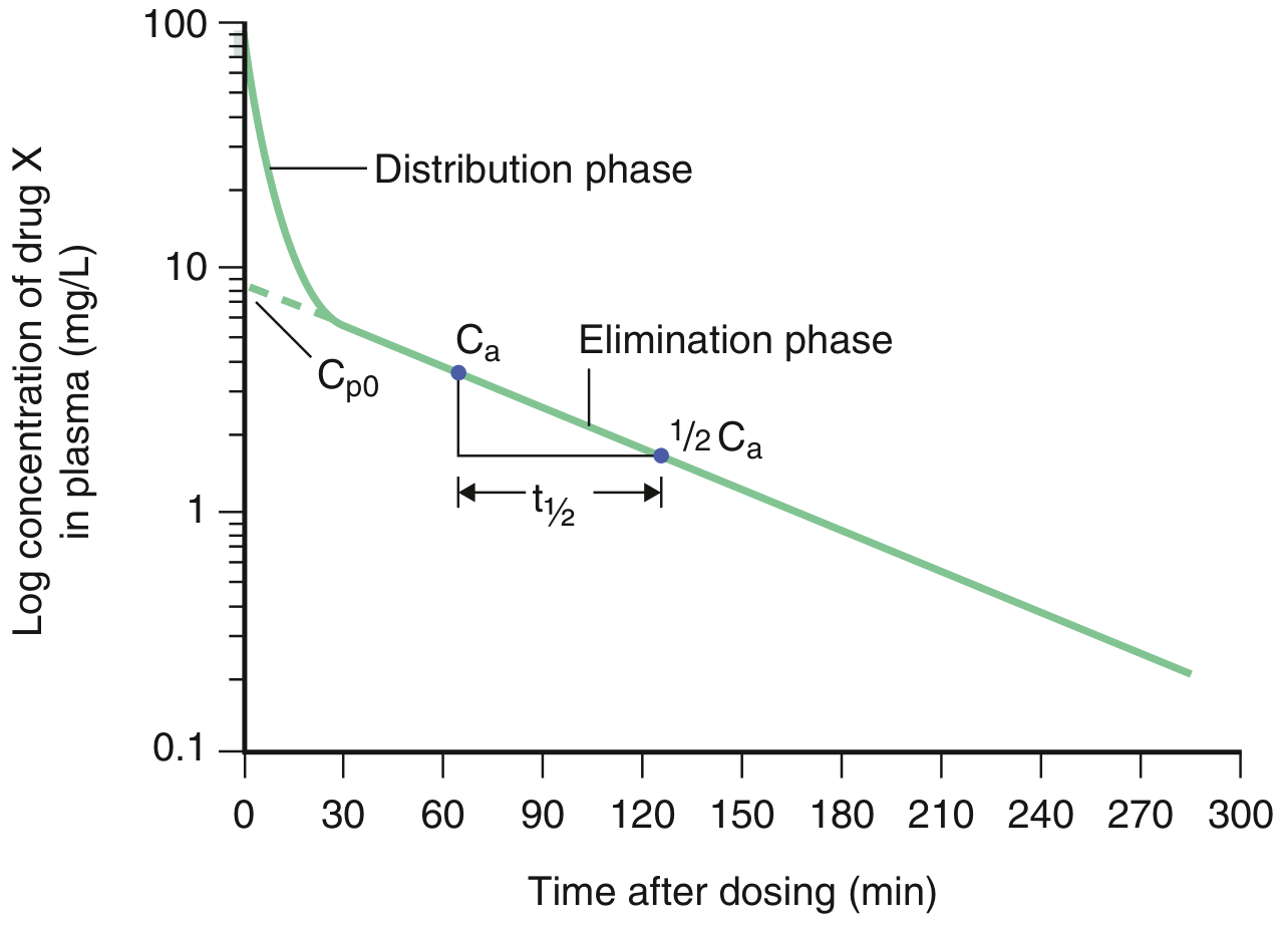
Figure: Log-linear plot of plasma drug concentration versus time after IV bolus. Cp₀ is obtained by back-extrapolation of the elimination phase. The half-life (t½) is read directly from the elimination phase. - Goldman-Cecil Medicine
4. Units and Types of Vd
Vd is expressed in:
- Absolute volume (litres, L) - useful for individual patients
- Weight-indexed (L/kg) - useful for comparing across patients and populations
There are several distinct Vd values used in multi-compartment pharmacokinetic models (from Miller's Anesthesia, 10e):
| Parameter | Description |
|---|---|
| V_initial (Vdα) | Volume of distribution of the central compartment (from the rapid distribution phase) |
| V_extrap (Vdz or Vβ) | Volume of distribution of the tissue compartment (from the terminal elimination phase, extrapolated) |
| V_ss | Volume of distribution at steady state - the most physiologically relevant |
5. Physical Body Compartments and Drug Distribution
To understand what Vd means clinically, comparing it to known body fluid compartments is helpful. From Katzung's Basic and Clinical Pharmacology (16th Edition), Table 3-2:
| Compartment | Volume (L/kg) | Examples of Drugs |
|---|---|---|
| Plasma | 0.04 L/kg | Large protein molecules (e.g., antibodies, heparin) |
| Extracellular water | 0.2 L/kg | Larger water-soluble molecules (e.g., gentamicin) |
| Total body water | 0.6 L/kg | Small water-soluble molecules (e.g., ethanol) |
| Fat | 0.2-0.35 L/kg | Highly lipid-soluble molecules (e.g., diazepam) |
| Total body | ~0.6 L/kg (42 L in 70-kg person) | - |
Interpretation of Vd ranges (StatPearls/NCBI, 2026):
- Low Vd (<0.6 L/kg or ~42 L total): Drug is largely confined to the plasma or extracellular fluid. Examples: warfarin (~8 L), heparin (~4 L).
- High Vd (>5 L/kg or >350 L total): Drug distributes extensively into body tissues, fat, or muscle. Examples: chloroquine (~250 L/kg), digoxin (~7 L/kg), amiodarone (~70 L/kg).
"Drugs with very high volumes of distribution have much higher concentrations in extravascular tissue than in the vascular compartment." - Katzung's Basic and Clinical Pharmacology, 16th Edition
6. Factors Affecting Volume of Distribution
Several physicochemical and physiological factors determine the Vd of a drug:
6.1 Physicochemical Properties of the Drug
| Factor | Effect on Vd |
|---|---|
| Lipophilicity | Lipophilic drugs partition into fat and tissues → higher Vd |
| Protein binding (plasma) | High plasma protein binding keeps drug in plasma → lower Vd |
| Tissue binding | Binding to tissue proteins/receptors draws drug out of plasma → higher Vd |
| Molecular size | Large molecules (>1 kDa) stay in vascular compartment → lower Vd |
| Ionization (pKa) | Ionized drugs at physiologic pH have limited membrane permeation → lower Vd |
"The apparent volume of distribution reflects a balance between binding to tissues, which decreases plasma concentration and makes the apparent volume larger, and binding to plasma proteins, which increases plasma concentration and makes the apparent volume smaller." - Katzung's Basic and Clinical Pharmacology, 16th Edition
6.2 Physiological and Pathological Factors
- Obesity: Increases Vd of lipophilic drugs (e.g., benzodiazepines) due to greater adipose tissue mass
- Aging: Lean body mass decreases and body fat increases with age, affecting Vd for both lipophilic and hydrophilic drugs. The half-life of many drugs is increased with aging as a consequence of larger apparent Vd and decreased hepatic or renal clearance (Goldman-Cecil Medicine)
- Hepatic disease (cirrhosis): Often increases Vd of water-soluble drugs and decreases plasma protein levels (reduced albumin → decreased plasma protein binding → altered distribution)
- Renal failure (nephrotic syndrome): Reduced plasma protein levels raise free drug fraction, increasing effective Vd
- Edema/ascites: Increases total body water → increases Vd of water-soluble drugs
- Cardiac failure: Decreased cardiac output reduces tissue perfusion → may reduce Vd
- Gender: Women have higher body fat proportion → larger Vd for lipophilic drugs (e.g., diazepam) (Kaplan & Sadock's Comprehensive Textbook of Psychiatry)
- Neonates/pediatrics: Have a larger total body water proportion, decreased protein binding, decreased body fat, and immature organ function → altered Vd compared to adults (Barash Clinical Anesthesia)
7. Clinical Importance of Volume of Distribution
7.1 Loading Dose Calculation
The most direct clinical application of Vd is calculating the loading dose - the initial large dose given to rapidly achieve a target therapeutic plasma concentration. From Barash's Clinical Anesthesia:
$$\text{Loading Dose} = V_d \times \text{Target Concentration}$$
From Goldman-Cecil Medicine:
$$\text{Loading Dose} = \text{Desired Concentration} \times V_d$$
Clinical Example - Phenytoin (Goldman-Cecil Medicine):
- Target concentration: 10-20 mg/L (minimum therapeutic)
- Vd of phenytoin: ~0.6 L/kg
- Loading dose for a 70-kg adult: 0.6 L/kg × 70 kg × 10 mg/L = 420 mg
- However, IV bolus of 420 mg phenytoin risks cardiac arrest - so oral administration (taking advantage of lower bioavailability F=0.8 and slower absorption) allows safe loading at 500 mg orally.
Key insight: Any drug with a large Vd requires a larger loading dose to "fill the box" and achieve the same plasma concentration. Conversely, a drug with a small Vd needs a smaller loading dose.
From Katzung's Basic and Clinical Pharmacology, for theophylline:
- Loading dose = 35 L × 10 mg/L = 350 mg for a 70-kg person
"An increase in the volume of distribution means that a larger loading dose will be required to 'fill up the box' and achieve the same concentration." - Barash Clinical Anesthesia, 9th Edition
7.2 Relationship to Drug Half-Life
Vd is directly related to the elimination half-life (t½) through the equation (Goldman-Cecil Medicine):
$$t_{1/2} = \frac{0.693 \times V_d}{Clearance (CL)}$$
This means:
- Increase in Vd → longer t½ (drug persists longer in the body)
- Decrease in Vd → shorter t½
- A disease may affect Vd and clearance differently, so t½ may increase, decrease, or remain unchanged
This relationship is critical for predicting how long a drug remains active, when to redose, and when steady-state will be reached (approximately 4-5 half-lives).
7.3 Therapeutic Drug Monitoring (TDM)
Vd is essential for interpreting plasma drug concentration measurements in TDM. It enables clinicians to:
- Calculate whether a measured plasma level is expected for a given dose
- "Boost" a subtherapeutic drug level to the desired range: $$\text{Boost Dose} = (C_{target} - C_{current}) \times V_d$$
Example (Goldman-Cecil Medicine): If a patient's phenytoin is 5 mg/L (target: 15 mg/L), the boost dose for a 70-kg patient:
- (15 - 5) mg/L × 0.6 L/kg × 70 kg = 420 mg
7.4 Extracorporeal Elimination - Dialysis Decisions
Vd is a key determinant of whether hemodialysis or other extracorporeal techniques will effectively remove a drug. From Goodman & Gilman's Pharmacological Basis of Therapeutics:
"A drug amenable to removal by hemodialysis has a low molecular weight, a low volume of distribution, high solubility in water, and minimal protein binding."
Rationale: A high Vd means most of the drug is sequestered in tissues, not in the plasma. Since hemodialysis only clears what is in the plasma compartment, high-Vd drugs (e.g., beta-blockers, tricyclic antidepressants, digoxin) are not significantly removed by dialysis despite being renally cleared under normal circumstances.
From Tintinalli's Emergency Medicine: Beta-blockers with high protein binding, lipid solubility, and large Vd render extracorporeal drug removal useless for most drugs in this class (except acebutolol, atenolol, nadolol).
7.5 Drug Overdose Assessment
Vd allows retrospective estimation of the magnitude of a drug overdose when plasma concentrations are measured:
$$\text{Amount in body} = V_d \times C_{plasma}$$
This is useful in emergency and toxicology settings to gauge severity of poisoning and choose appropriate management.
8. Vd in Special Populations
8.1 Elderly Patients
From Goldman-Cecil Medicine: "The elimination half-life of many drugs is increased with aging as a consequence of a larger apparent volume of distribution and a decreased hepatic or renal clearance."
From Goodman & Gilman's: Advanced age is associated with reduced lean body mass and increased body fat, both of which affect Vd. Hepatic blood flow decreases by 40%, further reducing clearance.
8.2 Neonates and Pediatrics
From Barash Clinical Anesthesia: Factors affecting drug metabolism in neonates include:
- Larger total body water → larger Vd for water-soluble drugs
- Decreased protein binding → more free drug available
- Decreased body fat → smaller Vd for lipophilic drugs
- Immature renal and hepatic function → reduced clearance
8.3 Pregnancy
- Plasma volume expands by ~50% in pregnancy
- Increased body fat stores
- Decreased plasma albumin
- All of these increase Vd for most drugs
8.4 Obesity
Lipophilic drugs (e.g., benzodiazepines, propofol) have a significantly larger Vd in obese patients due to expanded adipose tissue compartment. This prolongs their effect and t½.
8.5 Hepatic and Renal Disease
From Morgan and Mikhail's Clinical Anesthesiology: "Cirrhotic liver disease and chronic kidney failure often result in an increased volume of distribution and a lower plasma concentration for a given dose of water-soluble drugs, such as muscle relaxants."
9. Summary Table of Selected Drug Vd Values
From Katzung's Basic and Clinical Pharmacology, 16th Edition (Table 3-1) and clinical references:
| Drug | Vd (L/kg) | Interpretation |
|---|---|---|
| Heparin | ~0.06 L/kg | Confined to plasma (large molecule) |
| Warfarin | ~0.14 L/kg | Highly plasma protein bound |
| Gentamicin | ~0.25 L/kg | Extracellular fluid distribution |
| Theophylline | ~0.5 L/kg | Total body water distribution |
| Phenytoin | ~0.6 L/kg | Total body water + moderate tissue binding |
| Ethanol | ~0.6 L/kg | Total body water (small, water-soluble) |
| Diazepam | ~1-2 L/kg | Distributes into fat |
| Digoxin | ~7 L/kg | Extensive tissue binding (Na/K-ATPase) |
| Amiodarone | ~60 L/kg | Massive tissue/fat sequestration |
| Chloroquine | ~250 L/kg | Extreme tissue accumulation |
10. Worked Calculations Summary
| Clinical Task | Formula |
|---|---|
| Calculate Vd | Vd = Dose / Cp₀ |
| Calculate loading dose | Loading Dose = Vd × Target Concentration |
| Calculate t½ | t½ = 0.693 × Vd / CL |
| Calculate CL from t½ | CL = 0.693 × Vd / t½ |
| "Boost" dose (TDM) | Boost Dose = (C_target - C_current) × Vd |
| Estimate amount in body | Amount = Vd × C_plasma |
References
-
Katzung BG, Vanderah TW. Basic and Clinical Pharmacology, 16th Edition. McGraw-Hill; 2021. Chapter 3: Pharmacokinetics & Pharmacodynamics.
-
Goldman L, Cooney KA (eds). Goldman-Cecil Medicine, International Edition. Elsevier; 2024. Chapter 25: Principles of Pharmacokinetics.
-
Barash PG, et al. Barash, Cullen, and Stoelting's Clinical Anesthesia, 9th Edition. Wolters Kluwer; 2022. Chapter 11: Pharmacokinetics and Pharmacodynamics. pp. 746-749.
-
Brunton LL, Knollmann BC (eds). Goodman & Gilman's The Pharmacological Basis of Therapeutics, 14th Edition. McGraw-Hill; 2023. Chapters 2-3: Pharmacokinetics.
-
Miller RD, et al. Miller's Anesthesia, 10th Edition (2-Volume Set). Elsevier; 2025. Chapter 22: Pharmacokinetics.
-
Sadock BJ, Sadock VA, Ruiz P (eds). Kaplan & Sadock's Comprehensive Textbook of Psychiatry, 11th Edition. Wolters Kluwer; 2022. Pharmacologic Interventions.
-
Tintinalli JE, et al. Tintinalli's Emergency Medicine: A Comprehensive Study Guide, 9th Edition. McGraw-Hill; 2020. Toxicology Chapters.
-
Bisht S, Chauhan V, Patel P. Clinical Significance of Volume of Distribution in Pharmacotherapy. StatPearls. NCBI Bookshelf. Updated March 22, 2026.
-
Berezhkovskiy LM. Volume of distribution at steady state for a linear pharmacokinetic system with peripheral elimination. J Pharm Sci. 2004.
-
Holford NHG. Volume of Distribution. Translational and Clinical Pharmacology. 2016;24(2):74-79. tcpharm.org.
This assignment was prepared using Katzung's Basic and Clinical Pharmacology (16th Ed.), Goldman-Cecil Medicine, Barash Clinical Anesthesia (9th Ed.), Goodman & Gilman's (14th Ed.), Miller's Anesthesia (10th Ed.), and StatPearls as primary authoritative sources.
This is a shared conversation. Sign in to Orris to start your own chat.