
Vitiligo
vitiligo depigmented skin patches clinical photo
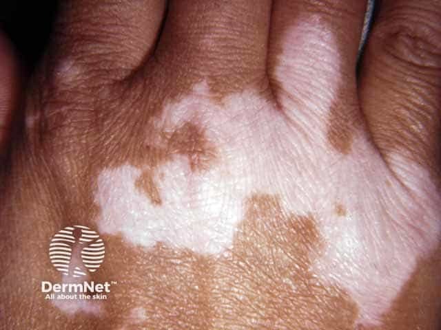
Clinical photography of a dorsal hand demonstrating vitiligo with Koebnerization on knuckle skin. The image displays several well-defined, milky white depigmented macules and patches over the dorsal metacarpophalangeal and interphalangeal regions, with sharp, irregular margins and no scale or erythema. Peripheral skin is variably hyperpigmented, creating a contrasted patchwork appearance. The lesions coalesce in places, forming larger depigmented areas that highlight focal loss of pigmentation rather than true hypopigmentation. The clinical context suggests Koebner phenomenon, where traumatic microinjury to the skin (e.g., friction on knuckles) triggers new depigmented plaques in susceptible individuals. This pattern is characteristic of vitiligo, an autoimmune pigmentary disorder driven by melanocyte destruction with loss of epidermal melanocytes and melanin pigment. The image is not of histology or radiology; it is a gross dermatologic photograph; no staining or contrast used. Relevance: aids diagnosis of vitiligo and Koebnerization, guides patient counseling, photoprotection, and potential systemic evaluation for associated autoimmune conditions (thyroid disease, type 1 diabetes). In differential diagnosis, consider tinea versicolor, postinflammatory hypopigmentation, and less likely leprosy or pityriasis alba. The photo supports educational demonstrations of lesion morphology, distribution, and the Koebner response in pigmentary disorders. High-contrast images enhance recognition of margins and sparing of surrounding pigmented skin.
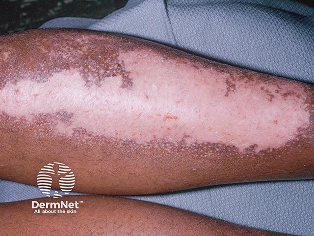
Clinical photograph of forearm skin showing vitiligo camouflage patches with concurrent allergic contact dermatitis to cosmetic camouflage pigments. Imaging modality: Clinical photography; technique: Standardized dermatologic photography. Forearm skin demonstrates large, well-demarcated hypopigmented macules consistent with vitiligo camouflage; pattern borders the depigmented zones, creating a patchy mosaic with variable pigmentation and a paler central area. Surrounding erythema, scaling, and crusting indicate an inflammatory response consistent with allergic contact dermatitis to camouflage constituents. The dermatitis is most pronounced at the periphery of the camouflage application, indicating sensitization to a cosmetic dye, binder resin, fragrance, or preservative component. Textural change and surface desquamation contribute to skin irritation and potential secondary infection risk if scratching occurs. Anatomical localization is cutaneous, in the superficial to mid-dermis, on the volar aspect of the forearm. Clinically, this finding raises suspicion for a cosmetic-related ACD in vitiligo patients and highlights the need for allergen identification via patch testing. Management implications include avoidance of suspected agents, topical corticosteroids or calcineurin inhibitors for inflammation, barrier repair emollients, and potential referral to dermatology for patch and photo-patch testing. Documenting this image supports differential diagnoses, clinical correlation, and patient education on cosmetic safety and vitiligo camouflage.
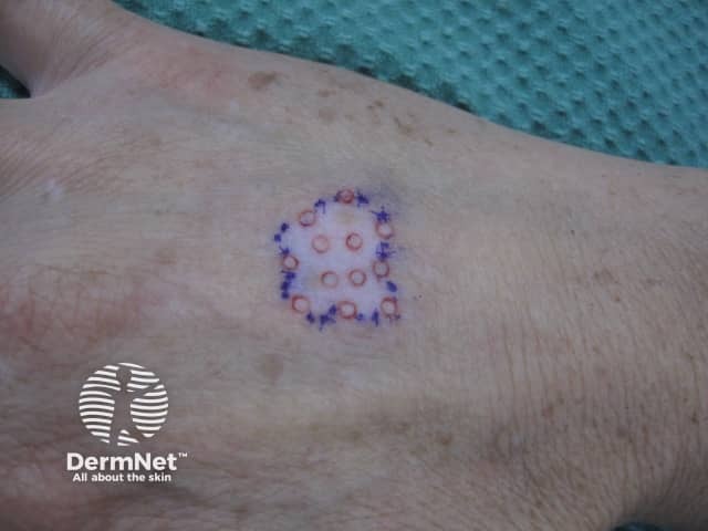
Clinical photography of vitiligo surgical planning on the dorsal hand. The area is demarcated with a round recipient-site grid drawn in blue ink; within the circle, small circular recipient holes are distributed in a uniform lattice. Red circular marks inside indicate planned grafts or donor sites. The surrounding skin shows depigmented patches characteristic of vitiligo with sharp borders and even texture in adjacent normal skin. The markings provide a standardized template for punch grafting or epidermal cell transplantation to achieve repigmentation by repopulating epidermis with melanocytes. The technique is used for stable, localized vitiligo and aims to establish contiguous pigment across the affected patch via transplanted melanocytes from pigmented donor skin. The image documents preoperative planning or intraoperative marking. It highlights key steps in vitiligo surgery: selecting a favorable recipient area, ensuring adequate coverage density, maintaining graft orientation, and minimizing graft rejection or failure. Clinically, this photo is suitable for dermatology, dermatologic surgery, and plastic surgery education, as well as research on graft take, repigmentation rates, and end-point outcomes. It also illustrates the importance of patient counseling regarding procedure duration, downtime, and expected cosmetic results. Documentation of instrument handling and markings can aid reproducibility across centers.
vitiligo treatment
Vitiligo
Epidemiology
- Affects roughly 0.5-1% of the population worldwide, with higher rates (>8%) reported in parts of India - Andrews' Diseases of the Skin
- Onset most often in childhood to young adulthood (peak age 10-30); average onset around age 30 in other series - Dermatology 2-Volume Set, 5th ed.
- No clear sex predilection in true prevalence, though women present for care more often
- Can be induced via bone marrow transplant/lymphocyte infusion from a vitiligo-affected donor, supporting an immune-mediated mechanism
Pathogenesis
- Multifactorial: genetic susceptibility (7% of first-degree relatives affected; ~23% monozygotic twin concordance) plus environmental/immune triggers
- Melanocytes have intrinsic vulnerability to oxidative stress; this impairs WNT signaling (needed for melanocyte precursor differentiation and repigmentation)
- Autoimmune destruction of melanocytes is central - associated genes overlap with other autoimmune/autoinflammatory conditions
- Koebner phenomenon: new lesions can arise at sites of physical trauma/friction (classic example: knuckles)
Clinical Features
- Depigmented macules/patches with a normal or hyperpigmented border; sometimes an intermediate tan zone ("trichrome vitiligo") in early lesions
- Hair within patches (leukotrichia) often turns white due to follicular melanocyte loss
- Six clinical subtypes based on distribution:
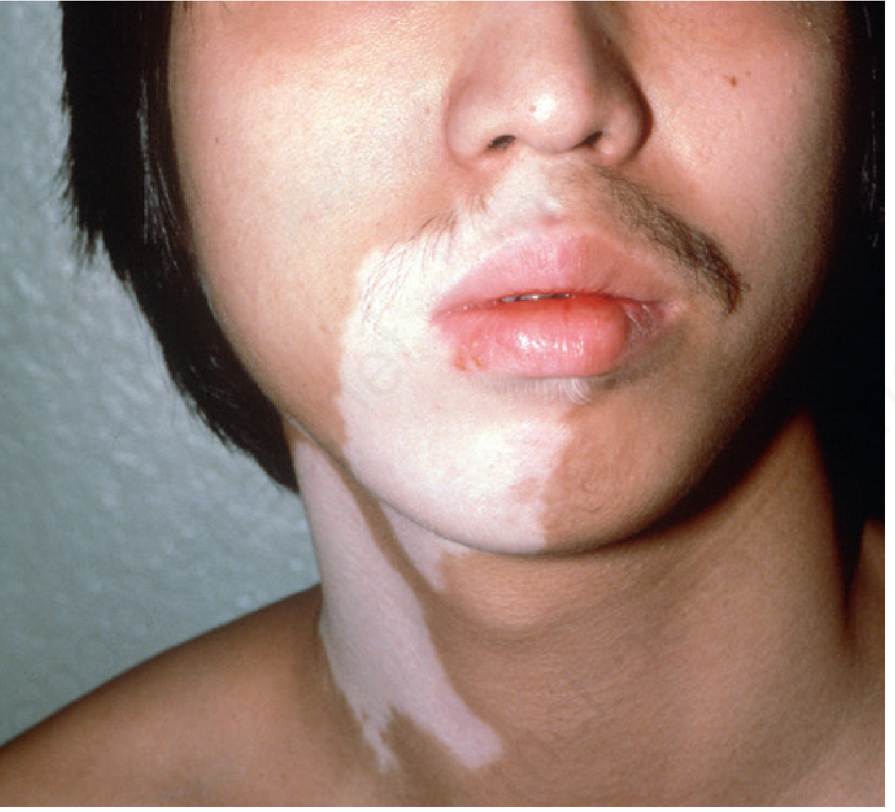
- Localized/focal - one or few macules in one area (common in children, often trigeminal area)
- Segmental - follows a unilateral, non-dermatomal band
- Generalized (most common) - symmetric, favoring face, chest, dorsal hands, axillae, groin, and periorificial skin (eyes, mouth, nose, nipples, umbilicus, genitals)
- Universal - near-total body depigmentation
- Acrofacial - distal fingers and facial orifices
- Mucosal
- Favors trauma-prone areas (elbows, knees) due to Koebnerization
Diagnosis
- Largely clinical: history, physical exam, and Wood's lamp (~365nm) examination, which sharpens contrast between depigmented/hypopigmented skin and normal skin - especially useful in light-skinned patients (phototype I/II) and neonates
- Wood's lamp also helps distinguish true melanocyte loss from hemoglobin-related leukodermas (e.g., nevus anemicus disappears under Wood's lamp)
- Biopsy is rarely needed but can help exclude inflammatory hypopigmenting conditions (sarcoidosis, lichen sclerosus, mycosis fungoides) when the picture is atypical
- Associated with other autoimmune conditions (thyroid disease, type 1 diabetes) - screening is often warranted
Treatment (from textbook + confirmed by recent systematic reviews)
- Topical corticosteroids and topical calcineurin inhibitors (tacrolimus, pimecrolimus) - first-line for localized disease
- Phototherapy: narrowband UVB is the mainstay for widespread disease; PUVA also used
- Systemic therapy: for progressive/widespread disease - oral corticosteroids (mini-pulse), and other agents such as methotrexate, azathioprine, cyclosporine used in select cases
- JAK inhibitors (e.g., ruxolitinib topical) have emerged as an important newer option
- Surgical repigmentation (punch grafting, melanocyte-keratinocyte transplantation) for stable, localized/segmental vitiligo unresponsive to medical therapy
- Camouflage cosmetics can help but carry a risk of contact dermatitis to dyes/preservatives in sensitized patients
Recent Evidence Check (last 2 years, systematic reviews/meta-analyses)
- Regenerative medicine approaches (platelet-rich plasma, stem cell/keratinocyte-melanocyte transplantation) show promise for repigmentation - PMID 39509558
- A meta-analysis of RCTs found simvastatin has been studied as an adjunct for vitiligo with mixed safety/efficacy data - PMID 40252138
- Systemic oral/injectable therapies (methotrexate, azathioprine, mycophenolate, minocycline, alefacept, cyclosporine, corticosteroids) reviewed across case series and trials, reinforcing corticosteroids and immunosuppressants as options for progressive disease - PMID 38454597