
Neuromuscular junction image
Searching Images
neuromuscular junction diagram anatomy
Searching Images
neuromuscular junction motor end plate acetylcholine
Reading File
Finding Sources
Reading File
Reading File
I found excellent textbook images. Let me fetch them now.
Loading Image
Loading Image
Loading Image
Here are three high-quality neuromuscular junction images from Ganong's Review of Medical Physiology:
Figure A - Scanning Electron Micrograph
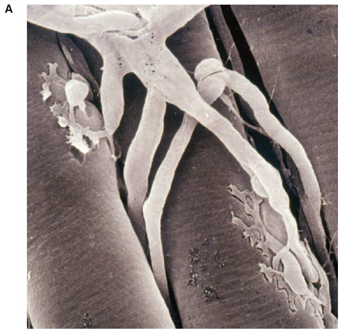
Scanning electron micrograph showing branching of motor axons with their terminals embedded in grooves on the muscle fiber surface.
Figure B - Structural Diagram of the NMJ
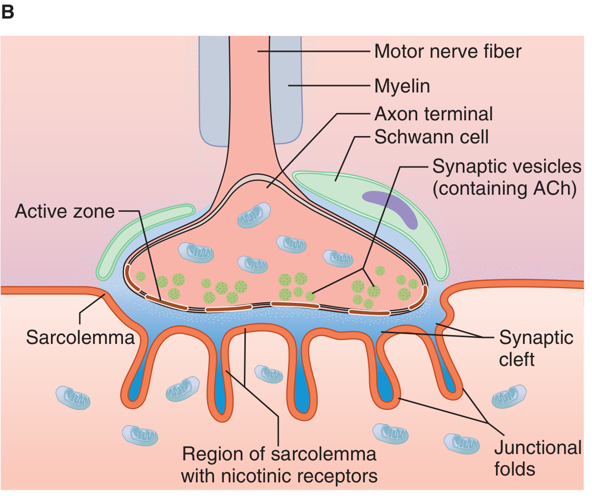
Structure of the neuromuscular junction showing the key anatomical components: motor nerve fiber, myelin sheath, axon terminal with synaptic vesicles (containing ACh), Schwann cell cap, active zones, synaptic cleft, junctional folds of the sarcolemma, and nicotinic receptor regions.
Figure C - Transmission Events at the NMJ
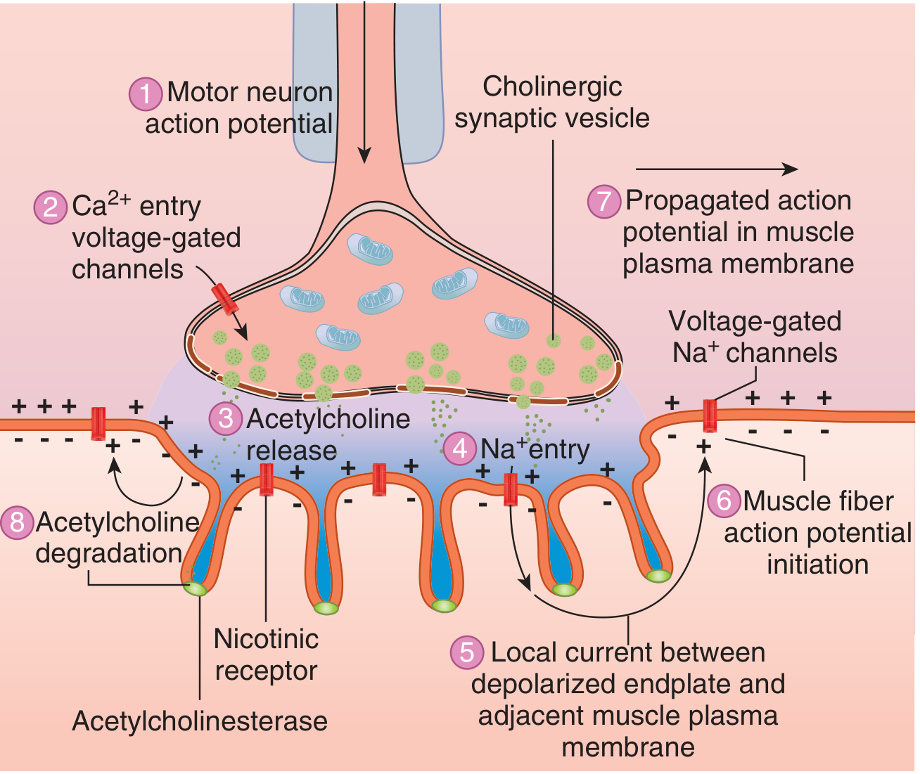
The sequence of events at the NMJ leading to muscle fiber activation:
- Motor neuron action potential arrives
- Ca²+ entry via voltage-gated channels
- ACh release by exocytosis
- Na+ entry through nicotinic receptors
- Local current between depolarized endplate and adjacent membrane
- Muscle fiber action potential initiation
- Propagated action potential along the muscle plasma membrane
- ACh degradation by acetylcholinesterase
Source: Ganong's Review of Medical Physiology, 26th Edition, Figure 6-11 and 6-12.
This is a shared conversation. Sign in to Orris to start your own chat.