Now I have all the material needed. Let me compile the comprehensive viva answer.
🧠 BRAINSTEM SYNDROMES — Complete Neurosurgery Viva Guide
Sources: Localization in Clinical Neurology 8e, Adams & Victor's Neurology 12e, Bradley & Daroff's Neurology, Harrison's 22e, Plum & Posner, Neuroanatomy through Clinical Cases 3e, Schwartz's Surgery 11e
PART 1: FOUNDATION — Anatomy & Vascular Supply
Brainstem Anatomy Overview
The brainstem = Midbrain + Pons + Medulla. Each level has a ventral base (motor tracts) and dorsal tegmentum (sensory tracts + CN nuclei). The principle of crossed syndromes (alternating hemiplegia) is the hallmark: ipsilateral CN deficit + contralateral long tract signs.
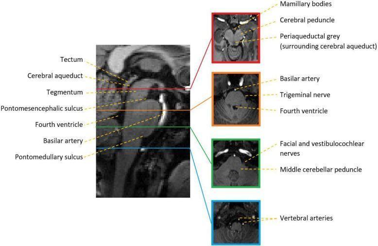
Sagittal MRI with corresponding axial sections showing midbrain (cerebral peduncle, periaqueductal grey), pons (basilar artery, CN V, IV), and medulla (vertebral arteries). — Localization in Clinical Neurology
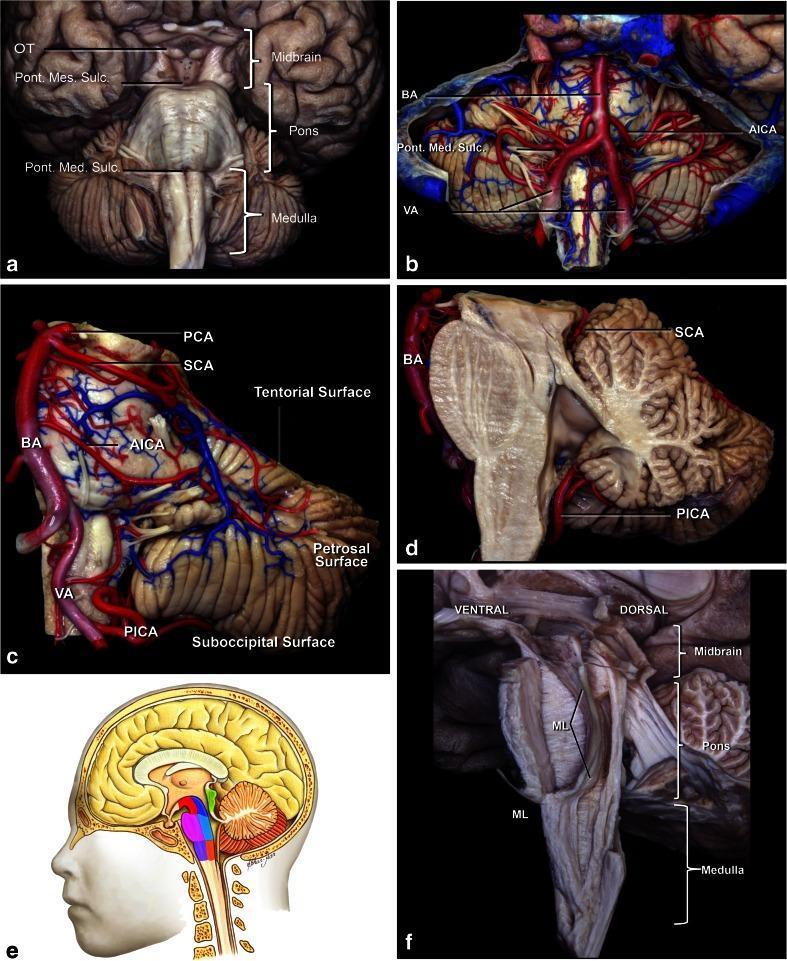
Cadaveric specimen showing brainstem vascular supply: BA = basilar artery, PICA = posterior inferior cerebellar artery, AICA = anterior inferior cerebellar artery, SCA = superior cerebellar artery, VA = vertebral artery
Vascular Supply — Key Points
| Level | Artery | Territory |
|---|
| Midbrain | Posterior cerebral artery (PCA) — paramedian perforators | Cerebral peduncle, CN III fascicle, red nucleus |
| Pons (medial) | Paramedian branches of basilar artery | Corticospinal tract, CN VI, VII nuclei, PPRF |
| Pons (lateral) | AICA | Lateral pons, CN VII, VIII territory |
| Medulla (lateral) | PICA / vertebral artery | Lateral medullary wedge (spinothalamic, CN V spinal, sympathetics, nucleus ambiguus, vestibular nuclei) |
| Medulla (medial) | Anterior spinal artery | Pyramid, medial lemniscus, CN XII |
PART 2: CLASSIFICATION OF BRAINSTEM SYNDROMES
I. By Level
MIDBRAIN PONS MEDULLA
───────── ──────── ──────────
Weber Millard-Gubler Wallenberg (Lateral)
Benedikt Raymond-Foville Dejerine (Medial)
Claude Locked-In
Nothnagel (bilateral pons)
Parinaud (dorsal)
II. By Mechanism
- Vascular (most common): ischaemic infarct > haemorrhage
- Demyelinating: MS
- Compressive: tumour, AVM, abscess
- Traumatic: severe TBI, pontine haematoma
PART 3: INDIVIDUAL SYNDROMES — Full Viva Detail
🔴 SYNDROME 1: WEBER SYNDROME (Ventral Midbrain)
Demography
- Age: 50–70 years (atherosclerotic) | Young adults: PCA dissection, embolism
- Sex: M > F
- Risk factors: HTN, DM, hyperlipidaemia, atrial fibrillation
Lesion Location
- Ventromedial midbrain — damages the cerebral peduncle (corticospinal + corticobulbar) AND the CN III fascicle
- Artery: Paramedian perforators of PCA or top of basilar
Pathology of Clinical Features
| Structure Damaged | Clinical Sign |
|---|
| CN III fascicle (ipsilateral) | Ipsilateral ptosis, eye "down and out," dilated unreactive pupil |
| Corticospinal tract (ipsilateral peduncle) | Contralateral hemiplegia (UMN) |
| Corticobulbar fibers | Contralateral lower facial weakness, tongue deviation |
Clinical Features
- Ipsilateral (same side as lesion):
- Complete CN III palsy: ptosis, eye deviated "down and out," mydriasis (dilated unreactive pupil), loss of accommodation
- Contralateral (opposite side):
- UMN hemiparesis/hemiplegia (arm, leg, lower face)
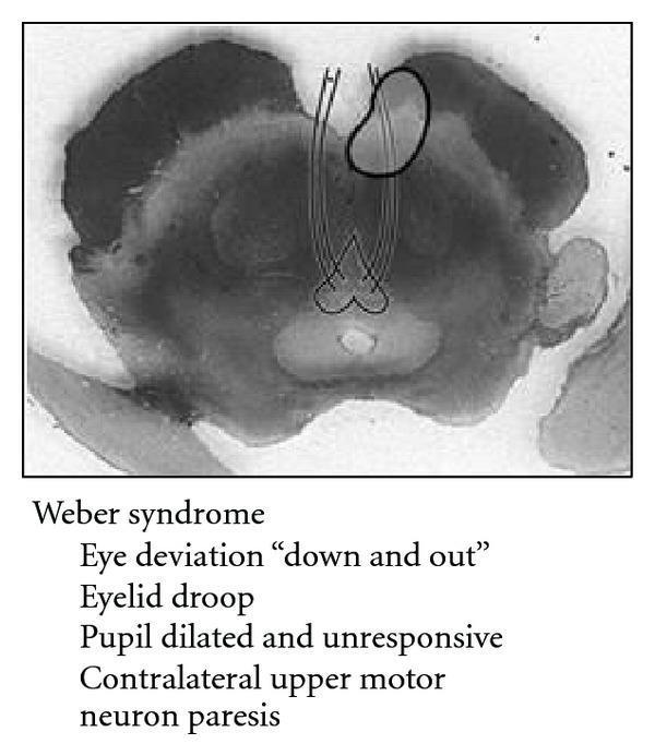
Weber syndrome: ventromedial midbrain infarct. Eye deviation "down and out," eyelid droop, dilated pupil + contralateral UMN paresis. — Localization in Clinical Neurology 8e
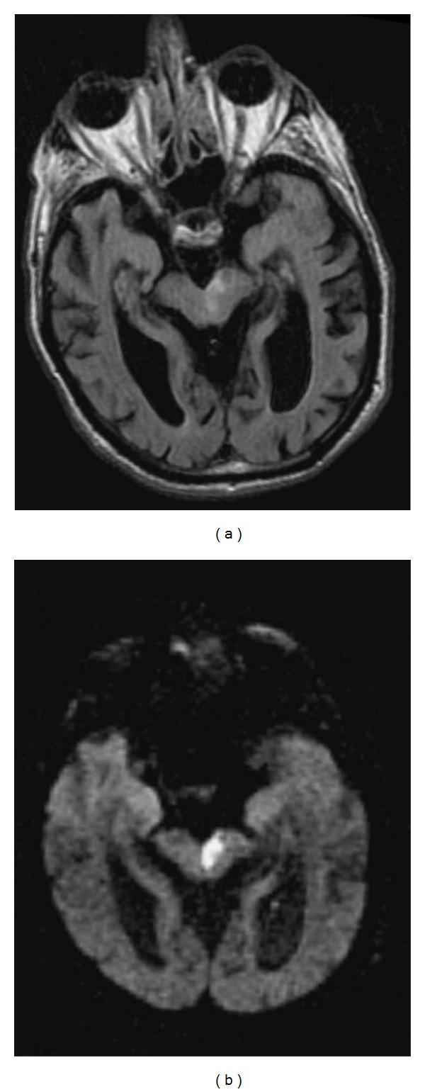
MRI of Weber syndrome: FLAIR (a) and DWI (b) showing focal acute infarct in the ventromedial midbrain, involving CN III fascicle and cerebral peduncle
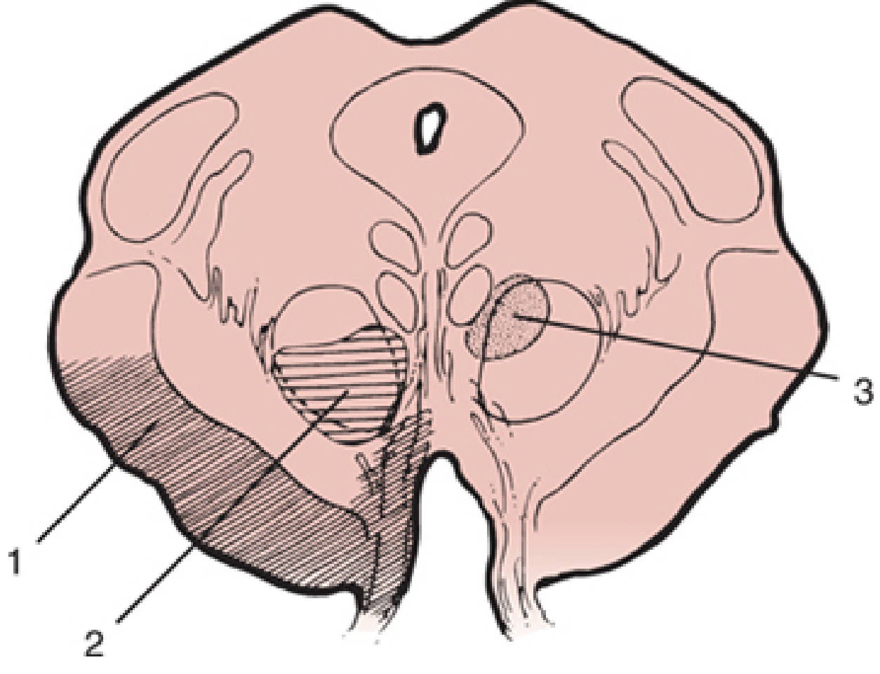
Midbrain section: (1) Weber syndrome zone — ventromedial; (2) Benedikt zone — tegmentum + red nucleus; (3) Claude zone — dorsal tegmentum. — Localization in Clinical Neurology 8e
🟠 SYNDROME 2: BENEDIKT SYNDROME (Midbrain Tegmentum)
Demography
- Same age/risk as Weber. Rarer — caused by PCA branch occlusion to tegmentum
Lesion Location
- Midbrain tegmentum — damages CN III fascicle + red nucleus + brachium conjunctivum (superior cerebellar peduncle)
Pathology of Clinical Features
| Structure Damaged | Clinical Sign |
|---|
| CN III fascicle | Ipsilateral CN III palsy |
| Red nucleus | Contralateral hemiataxia + intention tremor (rubral tremor) |
| Corticospinal tract (partial) | Contralateral hemiparesis + hyperreflexia |
Clinical Features — The Classic Triad
- Ipsilateral CN III palsy (ptosis, mydriasis, "down and out")
- Contralateral hemiataxia with intention tremor (rubral/Holmes tremor — coarse, 3–5 Hz)
- Contralateral hemiparesis (milder than Weber)
Memory aid: Benedikt = CN III + Red nucleus involvement (contralateral Big tremor)
🟡 SYNDROME 3: CLAUDE SYNDROME (Dorsal Midbrain)
Lesion Location
- Dorsal midbrain tegmentum — CN III fascicle + brachium conjunctivum (superior cerebellar peduncle)
Clinical Features
- Ipsilateral CN III palsy
- Contralateral cerebellar ataxia (prominent, no hemiplegia)
- Dysmetria, dysdiadochokinesia
Claude = "Cerebellar" signs dominant (no tremor, no hemiplegia)
🟢 SYNDROME 4: PARINAUD SYNDROME (Dorsal Midbrain / Tectum)
Also called: Sylvian Aqueduct Syndrome / Koerber–Salus–Elschnig Syndrome / Pretectal Syndrome
Demography
- Children/Young adults: Pineal tumour (germinoma, pineocytoma)
- All ages: Hydrocephalus (aqueductal stenosis), AVM, MS, haemorrhage
- Elderly: Midbrain infarct
Lesion Location
- Dorsal rostral mesencephalon — damages the posterior commissure and rostral interstitial nucleus of MLF (riMLF) — the vertical gaze centre
- NOT a crossed syndrome (bilateral structure compressed from above)
- Caused by compression from pineal region or hydrocephalus — NOT typically a stroke
Pathology of Clinical Features
| Structure Damaged | Clinical Sign |
|---|
| Posterior commissure / riMLF | Loss of upgaze (conjugate vertical gaze palsy) |
| Edinger-Westphal nucleus compression | Large pupils with light-near dissociation (react to near but not light) |
| Nucleus of Darkschewitsch | Convergence-retraction nystagmus on upward gaze |
| Superior colliculus | Collier's sign (eyelid retraction) |
| Aqueduct compression | Hydrocephalus → papilloedema, headache, vomiting |
Clinical Features (COONS mnemonic)
- Convergence-retraction nystagmus (pathognomonic — on attempted upgaze)
- Ophthalmoplegia — bilateral upgaze palsy (can look down, not up)
- Other: Eyelid retraction (Collier's sign = "sunset sign" or lid retraction on downgaze)
- Near-light dissociation of pupils (pupils react to convergence not to light)
- Signs of hydrocephalus (headache, vomiting, papilloedema)
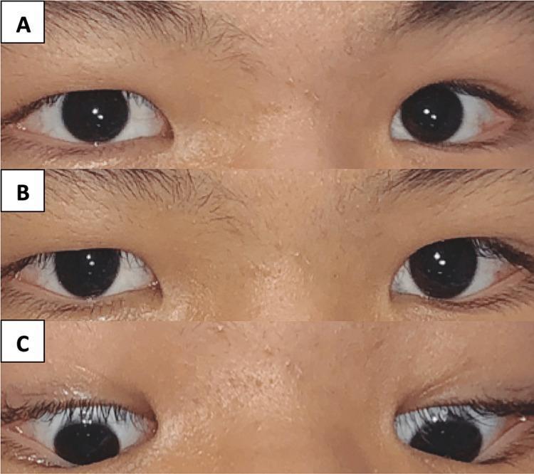
Clinical photographs of Parinaud syndrome. A: Absent upgaze bilaterally. B: Normal primary gaze. C: Preserved downgaze. Associated with pineal germinoma.
Investigations for Parinaud Syndrome
Indications: Young patient + upgaze palsy + hydrocephalus → rule out pineal tumour
| Investigation | Indication | Positive Findings |
|---|
| MRI brain (gadolinium) | First-line | Pineal mass, tectal compression, aqueductal obstruction, hydrocephalus |
| CT head | Emergency | Hydrocephalus, calcified pineal mass |
| Serum/CSF tumour markers | Germ cell tumour | AFP ↑ (teratoma), β-hCG ↑ (germinoma) |
| Visual field testing | All patients | Superior field cut from superior colliculus involvement |
| Lumbar puncture | After imaging (if no herniation) | Cytology for CSF seeding |
Management of Parinaud (Pineal Region)
Step 1: Call neurosurgery + neuro-oncology
Step 2: Treat hydrocephalus urgently — EVD or VP shunt
Step 3: Tissue diagnosis
Surgical Options
A. Endoscopic Third Ventriculostomy (ETV) + Biopsy
- Indications: Obstructive hydrocephalus from pineal/tectal mass; avoids CSF shunt
- Steps: Kocher's point burr hole → rigid endoscope into lateral ventricle → foramen of Monro → third ventricle floor fenestration → simultaneous biopsy via separate working channel
- Advantage: Diagnose AND treat in one session
B. VP Shunt (if ETV fails or not feasible)
C. Open Resection (Supracerebellar Infratentorial approach)
- Indications: Large benign tumour (pineocytoma, teratoma), failed biopsy, residual disease
- Patient position: Sitting or park-bench position
- Steps: Midline incision → infratentorial craniotomy → approach under tentorium above cerebellum → microsurgical resection
- Contraindications: Radiosensitive tumour (germinoma responds to RT), poor performance status
D. Stereotactic biopsy (if lesion in deep tectum, not accessible)
Non-surgical:
- Germinoma: Radiation ± chemotherapy (cisplatin, etoposide) — highly radiosensitive
- Dexamethasone 0.1 mg/kg/dose QDS: reduces perilesional oedema (MOA: inhibits phospholipase A2, reduces arachidonic acid → ↓ prostaglandins + vascular permeability)
🔵 SYNDROME 5: MILLARD-GUBLER SYNDROME (Ventral Pons)
Demography
- Age: 45–70 years
- Cause: Paramedian basilar perforator infarct, tumour, MS
Lesion Location
- Ventral pons — damages CN VII fascicle + CN VI nucleus/fascicle + corticospinal tract
Pathology of Clinical Features
| Structure Damaged | Clinical Sign |
|---|
| CN VII fascicle (LMN) | Ipsilateral LMN facial palsy (entire face — forehead + lower) |
| CN VI (abducens) nucleus | Ipsilateral lateral rectus palsy (can't abduct eye) → diplopia |
| Corticospinal tract | Contralateral hemiplegia |
Clinical Features
- Ipsilateral complete LMN CN VII palsy (brow, eye, mouth all involved)
- Ipsilateral CN VI palsy (esotropia, diplopia on lateral gaze)
- Contralateral UMN hemiparesis
Raymond Syndrome = CN VI palsy + contralateral hemiplegia (no VII involvement)
Foville Syndrome = CN VII palsy + conjugate horizontal gaze palsy (toward lesion) + contralateral hemiplegia (PPRF involvement)
🟣 SYNDROME 6: LOCKED-IN SYNDROME (Bilateral Ventral Pons)
Demography
- Age: Any age; typically 40–60 (stroke), younger (trauma or basilar thrombosis)
- Cause: Bilateral basilar perforator infarction / basilar artery thrombosis / pontine haemorrhage / severe TBI
- Mortality: Extremely high acutely; survivors may live years with preserved consciousness
Lesion Location
- Bilateral ventral pons — bilateral corticospinal + corticobulbar tract destruction
- Spares: Dorsal pons (ARAS intact → consciousness preserved), vertical gaze pathways (riMLF in midbrain intact)
Pathology of Clinical Features
| Structure Damaged | Clinical Sign |
|---|
| Bilateral corticospinal tracts | Quadriplegia |
| Bilateral corticobulbar tracts | Anarthria (no speech), aphonia |
| CN VI nuclei (bilateral) | Cannot move eyes horizontally |
| ARAS spared | Consciousness fully preserved |
| Vertical gaze (riMLF, midbrain) spared | Vertical eye movement + blinking = ONLY communication |
Clinical Features
- Awake and aware — fully conscious, cannot communicate verbally or move limbs
- Quadriplegia (all 4 limbs)
- Aphonia (no phonation)
- Preserved: Vertical gaze (upward + downward), blinking — used as YES/NO communication
- Preserved hearing, vision, reading ability
- Autonomic instability (HR, BP fluctuations)
Classic teaching: Patient appears comatose but is fully awake — ALWAYS test vertical eye movement in apparent coma!
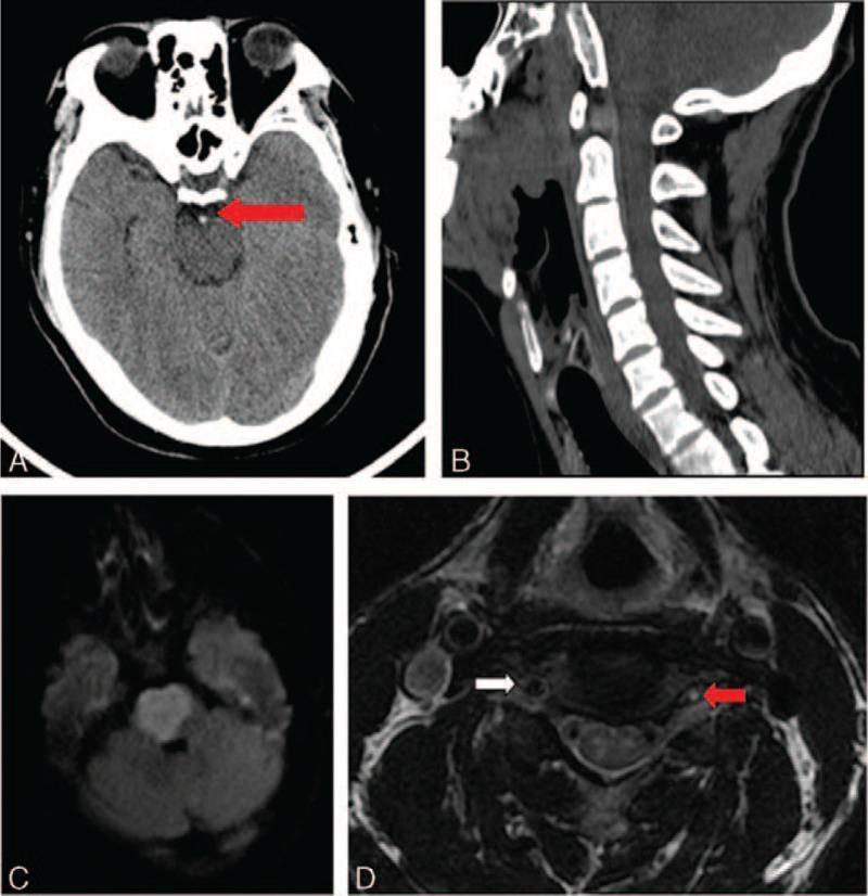
Locked-in syndrome: A = CT showing hyperdense basilar artery (thrombosis, red arrow). C = DWI showing bilateral pontine infarct. D = T2 MRI showing left vertebral artery thrombosis (red arrow) vs patent right VA (white arrow).
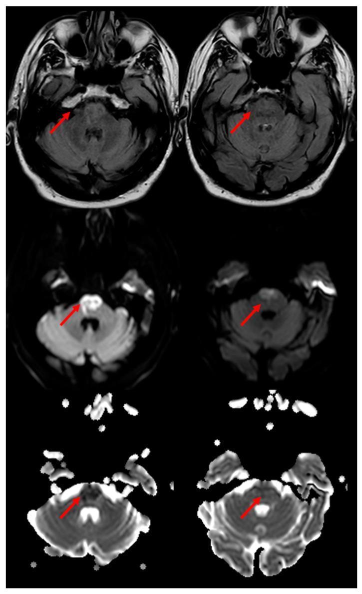
Bilateral pontine paramedian infarct: "heart appearance sign" on FLAIR (top), DWI (middle), ADC map (bottom) — characteristic of locked-in syndrome.
🟤 SYNDROME 7: WALLENBERG SYNDROME (Lateral Medullary)
The MOST COMMON and Clinically Important Brainstem Stroke Syndrome
Demography
- Age: 50–70 years (atherosclerotic); 30–50 years (vertebral artery dissection — after neck manipulation, trauma, chiropractic)
- Sex: M > F (2:1)
- Aetiology (from 130-patient series — Localization in Clinical Neurology):
- Large vessel atherosclerosis: 50%
- Arterial dissection: 15%
- Small vessel disease: 13%
- Cardioembolism: 5%
Lesion Location
- Lateral medulla — a wedge-shaped territory supplied by PICA and/or vertebral artery
- Structures damaged: spinothalamic tract, spinal nucleus/tract CN V, nucleus ambiguus, inferior cerebellar peduncle, descending sympathetic fibres, vestibular nuclei, dorsal motor nucleus of vagus
Pathology of Clinical Features
| Structure Damaged | Clinical Sign | Side |
|---|
| Spinal nucleus + tract of CN V | Facial pain/temperature loss | Ipsilateral |
| Spinothalamic tract | Body pain/temperature loss | Contralateral |
| Nucleus ambiguus (CN IX, X) | Dysphagia, dysarthria, palatal palsy, hoarseness | Ipsilateral |
| Descending sympathetic fibres | Horner syndrome (ptosis, miosis, anhidrosis) | Ipsilateral |
| Vestibular nuclei | Vertigo, nausea, vomiting, nystagmus | — |
| Inferior cerebellar peduncle | Cerebellar ataxia of ipsilateral limbs | Ipsilateral |
| No pyramidal tract involvement | Motor power PRESERVED (pyramids in medial medulla) | — |
The Classic TRIAD (diagnostic):
Ipsilateral Horner + Ipsilateral ataxia + Contralateral hypoalgesia
Full Clinical Features (9 features — WALLED mnemonic)
- Weakness — NOT a feature (pyramids spared!)
- Ataxia ipsilateral + vertigo/nystagmus
- Loss of pain/temp — crossed (face ipsilateral, body contralateral)
- Laryngeal/palatal palsy → dysphagia, dysarthria
- Eye signs: Horner (ipsilateral), skew deviation, ocular ipsipulsion
- Dysphagia (aspiration risk — major complication!)
Additional signs:
- Hiccups (singultus) — lateral dorsal medulla involvement
- Ocular ipsipulsion — saccades veer toward side of lesion (damage to climbing fibres from inferior olivary nucleus)
- Environmental tilt — room appears tilted or upside-down ("floor-on-ceiling" phenomenon)
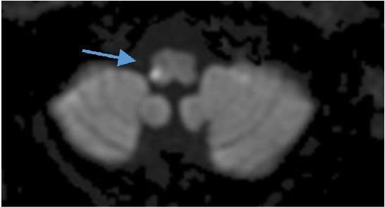
DWI MRI: Right lateral medullary hyperintensity (blue arrow) = acute Wallenberg syndrome infarct in PICA territory
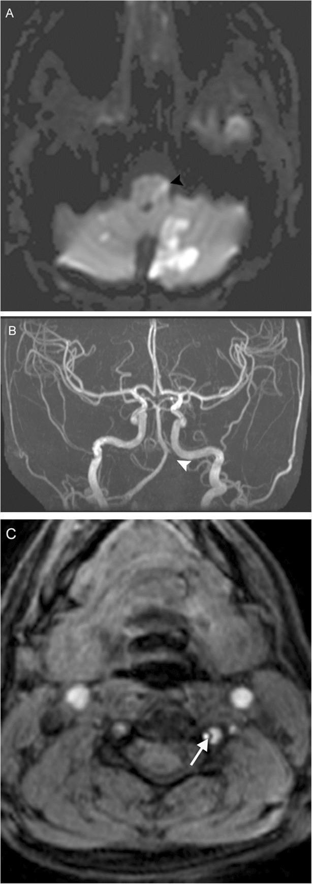
Wallenberg from left vertebral artery dissection: A = DWI showing lateral medullary + bilateral cerebellar infarcts (arrowhead). B = MRA showing absent left vertebral/PICA flow (arrowhead). C = Fat-saturated T1 showing intramural haematoma (crescent sign, white arrow).
🔵 SYNDROME 8: DEJERINE SYNDROME (Medial Medullary)
Demography
- Rarer than Wallenberg
- Cause: Anterior spinal artery or vertebral artery occlusion (medial branches)
Lesion Location
- Medial medulla — damages: pyramid (corticospinal), medial lemniscus, CN XII nucleus/fascicle
Pathology of Clinical Features
| Structure | Sign | Side |
|---|
| Pyramid (corticospinal) | Hemiplegia (UMN) | Contralateral |
| Medial lemniscus | Loss of proprioception + vibration | Contralateral |
| CN XII | Tongue deviation + atrophy (LMN) | Ipsilateral |
| No spinothalamic involvement | Pain/temp PRESERVED | — |
Classic Triad:
Ipsilateral CN XII LMN palsy + Contralateral UMN hemiplegia + Contralateral proprioception/vibration loss
PART 4: APPROACH TO THE PATIENT
Step-by-Step Clinical Approach
STEP 1: CALL FOR HELP
→ Neurosurgery + Neurology + ICU
STEP 2: AIRWAY + BREATHING (most critical)
→ Nucleus ambiguus lesion → dysphagia → aspiration
→ Locked-in → respiratory failure
→ GCS ≤8 → INTUBATE immediately
→ Position: 30° head elevation
STEP 3: RAPID NEUROLOGICAL ASSESSMENT
→ Level of consciousness (GCS)
→ Pupils (size, reactivity, Horner)
→ Eye movements (gaze palsy, nystagmus)
→ Cranial nerve function (IX, X, XII, V, VI, VII)
→ Motor power + reflexes (crossed?)
→ Sensory (crossed pattern?)
STEP 4: VITAL SIGNS
→ Cushing response (HTN + bradycardia + irregular breathing) =
impending brainstem compression!
STEP 5: INVESTIGATIONS (see below)
STEP 6: MANAGEMENT
PART 5: INVESTIGATIONS
Scoring Systems
Glasgow Coma Scale (GCS)
- E1–4 + V1–5 + M1–6 = 3–15
- GCS ≤8 = severe brain injury / coma
- Use: Initial assessment, monitor deterioration, intubation threshold (GCS ≤8)
NIH Stroke Scale (NIHSS)
- 0–42 score; brainstem strokes can score disproportionately high
- Used to guide thrombolysis eligibility
NIHSS > 4 = consider IV thrombolysis if within window
Investigations Table
| Investigation | Indication | Positive Findings |
|---|
| Non-contrast CT head | Emergency — first imaging in ALL patients | Hyperdense basilar artery (thrombosis), haemorrhage, mass effect, hydrocephalus |
| MRI brain (DWI + FLAIR + T2 + MRA) | Gold standard for brainstem ischaemia; CT misses 40-50% posterior fossa infarcts | DWI hyperintensity confirms acute infarct; FLAIR for subacute; MRA shows vessel stenosis/occlusion |
| MRA/CTA neck + brain | Suspected vertebral artery dissection or stenosis | Irregular vessel lumen, "string sign," absent flow |
| MRI fat-saturated T1 neck | Young patient + Wallenberg + neck pain | Crescent sign = intramural haematoma = dissection |
| ECG | All patients (cardioembolism source?) | AF, LVH |
| Echocardiogram (TTE/TOE) | Cardioembolic source | Thrombus, PFO, valvular disease |
| Holter monitor 24–72h | Paroxysmal AF | |
| Carotid/vertebral Doppler USS | Extracranial vessel disease | Stenosis >70%, dissection |
| FBC, U&E, glucose, lipids, INR/PT | Baseline + risk factors | |
| Thrombophilia screen | Young patient, no vascular risk factors | Factor V Leiden, protein C/S, antiphospholipid abs |
| Tumour markers (AFP, β-hCG) | Parinaud syndrome in young | Raised in germinoma, teratoma |
| Lumbar puncture | Suspected demyelination (MS), vasculitis | Oligoclonal bands, raised protein, cells |
PART 6: MANAGEMENT
A. MEDICAL MANAGEMENT
1. Acute Ischaemic Stroke (Brainstem)
Thrombolysis — IV Alteplase (tPA)
- MOA: Tissue plasminogen activator → converts plasminogen → plasmin → dissolves fibrin clot
- Indication: Ischaemic stroke ≤4.5 hours from onset, no contraindications; NIHSS ≥4
- Dose: 0.9 mg/kg IV (max 90 mg); 10% bolus over 1 min, rest over 60 min
- Special points: Contraindicated if: BP >185/110 (treat first), bleeding, recent surgery, INR >1.7, glucose <2.8 or >22.2 mmol/L
Mechanical Thrombectomy (EVT)
- Indication: Basilar artery occlusion (LOCKED-IN syndrome risk) — large vessel occlusion; within 24h (posterior circulation may have extended window)
- MOA: Catheter-based stent retriever or aspiration to retrieve clot
- Special points: Basilar artery thrombosis has mortality >90% without recanalization — KEY VIVA POINT
2. Antiplatelet Therapy
- Aspirin 300 mg loading → 75 mg OD maintenance
- MOA: Irreversibly inhibits COX-1 → ↓ TXA2 → ↓ platelet aggregation
- Start within 24h (if no thrombolysis)
3. Anticoagulation (Vertebral Artery Dissection)
- Heparin (IV infusion) or LMWH → then transition to warfarin or NOAC for 3–6 months
- MOA: Heparin — activates antithrombin III → inhibits thrombin + Xa; NOAC (e.g. rivaroxaban) — direct Xa inhibitor
4. Mannitol (ICP control)
- MOA: Osmotic diuretic → creates osmotic gradient across BBB → draws free water out of brain → ↓ ICP; also transient plasma expansion → ↓ viscosity → ↑ CBF
- Dose: 0.25–1 g/kg IV bolus over 15–20 minutes; repeat Q4–6h
- Serum osmolality target: 300–315 mOsm/L
- Special points: Avoid in hypotension; monitor renal function; avoid if intracranial haemorrhage before decompression (may expand haematoma)
5. Dexamethasone
- MOA: Reduces vasogenic oedema (tumour-related) by stabilising BBB; inhibits phospholipase A2 → ↓ arachidonic acid → ↓ prostaglandin-mediated vascular permeability
- Dose: 0.1 mg/kg/dose QDS (paediatric); Adults 4–8 mg IV QDS
- Special points: NOT effective for cytotoxic oedema (ischaemic stroke). Use only for tumour/abscess-related oedema and Parinaud syndrome from pineal tumour
6. Antihypertensives in Acute Stroke
- Target BP <180/105 during thrombolysis window; do NOT aggressively lower in first 24h (permissive hypertension)
- After 24h: target <130/80
B. SURGICAL MANAGEMENT
1. ICP Monitoring — External Ventricular Drain (EVD)
Indications:
- GCS 3–8 with abnormal CT scan
- Obstructive hydrocephalus from Parinaud/posterior fossa lesion
- Need for CSF drainage to control ICP
EVD Placement — Steps (Kocher's Point)
- Patient supine, head in neutral position, head elevated 30°
- Shave and prep scalp
- Kocher's point: 1 cm anterior to coronal suture, 3 cm lateral to midline (mid-pupillary line)
- Local anaesthetic ± GA if agitated
- Burr hole drilled at Kocher's point
- Catheter directed toward ipsilateral medial canthus in coronal plane, nasion in sagittal plane — depth ≈5.5–6.5 cm
- Stylet removed → CSF should flow; connect to closed drainage system
- Zero at level of external auditory meatus (EAM)
- Drain at ICP threshold >20–25 mmHg
Contraindications: Coagulopathy (relative), bilateral cortical injury, brain death
Target ICP: < 20 mmHg; Cerebral Perfusion Pressure (CPP) > 60 mmHg
2. Suboccipital Craniectomy (Posterior Fossa Decompression)
Indications:
- Large cerebellar infarct/haemorrhage causing brainstem compression
- Fourth ventricle obstruction → hydrocephalus
- Cerebellar herniation through foramen magnum
Steps:
- Patient: Park-bench or prone position
- Midline incision from inion to C2
- Expose posterior fossa bone
- Drill/remove posterior fossa bone (suboccipital craniectomy ≥3 × 4 cm)
- Open dura (Y-shaped incision) + duraplasty if needed
- Decompress cerebellum; if haematoma → evacuate
- If hydrocephalus → concurrent EVD or VP shunt
Contraindications: Brain death, herniation with no recoverable function, coagulopathy (correct first)
3. Decompressive Hemicraniectomy (Malignant MCA or Basilar territory)
- Not directly for brainstem syndromes but for their supratentorial correlates causing transtentorial herniation
C. REHABILITATION
- Swallowing assessment (SALT) within 24h — Wallenberg (nucleus ambiguus) = aspiration risk
- Nasogastric tube if unsafe swallow
- PEG if prolonged dysphagia (>3 weeks)
- Eye patch (diplopia from CN VI palsy)
- Physiotherapy, OT, speech therapy
PART 7: FOLLOW-UP
| Condition | Modality | Frequency |
|---|
| Brainstem stroke | MRI DWI → then MRI FLAIR + MRA | Repeat MRI at 3 months to assess penumbra/infarct evolution |
| Vertebral dissection | MRI fat-sat + MRA neck | 3 and 6 months — most recanalize by 6 months |
| Parinaud/Pineal tumour | MRI with gadolinium | Post-op at 3 months, then 6-monthly × 2y, then annually |
| VP shunt (Parinaud + hydrocephalus) | CT head | Any new symptom (shunt blockage) + annually |
| Secondary prevention | Clinic + bloods (lipids, HbA1c, INR if on warfarin) | 1 week, 1 month, 3 months, then 6-monthly |
📸 IMAGE GALLERY SUMMARY
All syndromes illustrated:
| Image | Syndrome |
|---|
| Midbrain zones (1,2,3) | Weber / Benedikt / Claude |
| Weber diagram + clinical signs | Weber syndrome |
| Weber MRI (FLAIR + DWI) | Weber — acute infarct |
| Parinaud eye movements | Parinaud syndrome |
| Wallenberg DWI | Lateral medullary infarct |
| Wallenberg with VA dissection | Vertebral artery dissection cause |
| Locked-in CT + DWI | Basilar thrombosis + bilateral pons |
| Bilateral pontine "heart sign" | Locked-in bilateral pons |
| Brainstem MRI anatomy | Orientation reference |
| Brainstem vascular anatomy | PICA/AICA/SCA/BA |
📋 SUMMARY TABLE — All Brainstem Syndromes at a Glance
| Syndrome | Level | Artery | Ipsilateral CN | Contralateral Signs | Key Feature |
|---|
| Weber | Ventral midbrain | PCA perforators | CN III palsy | Hemiplegia | "Down & out" eye + hemiplegia |
| Benedikt | Midbrain tegmentum | PCA | CN III palsy | Hemiataxia + tremor | Rubral (Holmes) tremor |
| Claude | Dorsal midbrain tegmentum | PCA | CN III palsy | Cerebellar ataxia | Ataxia dominant, no hemiplegia |
| Nothnagel | Dorsal midbrain | Pineal/tectal area | CN III (uni or bilateral) | Cerebellar ataxia | Rare, tumour-related |
| Parinaud | Dorsal midbrain/tectum | Compression/hydrocephalus | Upgaze palsy (bilateral) | Hydrocephalus | Convergence-retraction nystagmus, pineal tumour |
| Millard-Gubler | Ventral pons | Basilar perforators | CN VI + VII (LMN) | Hemiplegia | Facial + abducens + hemiplegia |
| Raymond | Ventral pons | Basilar perforators | CN VI only | Hemiplegia | Abducens + hemiplegia (no VII) |
| Foville | Pons (PPRF + CN VII) | Basilar | CN VII + gaze palsy toward lesion | Hemiplegia | Horizontal gaze palsy + VII |
| Locked-in | Bilateral ventral pons | Bilateral basilar | All except vert. gaze + blink | Quadriplegia + anarthria | Conscious, communicates by eyes only |
| Wallenberg | Lateral medulla | PICA / vertebral | CN V (face), Horner, IX/X, ataxia | Pain/temp loss (body) | NO HEMIPLEGIA; crossed sensory; dysphagia |
| Dejerine | Medial medulla | Anterior spinal artery | CN XII (tongue) | Hemiplegia + proprioception loss | Tongue deviation + crossed motor/sensory |
🔑 VIVA RAPID-FIRE POINTS
- Brainstem rule: Ipsilateral CN + Contralateral long tract = lesion IN brainstem
- Wallenberg: ONLY brainstem syndrome without hemiplegia (pyramids in medial medulla)
- Parinaud: NOT a stroke syndrome — think PINEAL TUMOUR; check AFP/hCG; treat hydrocephalus first
- Locked-in: Patient is CONSCIOUS — always test vertical gaze in apparent coma
- Basilar thrombosis: Hyperdense basilar on plain CT → emergency thrombectomy
- Vertebral dissection (Wallenberg in young): neck pain + Horner → MRI fat-sat T1 → crescent sign
- Parinaud pupils: Light-near dissociation = pupils react to convergence/accommodation NOT to light (opposite of Argyll Robertson pupil of syphilis which is the same phenomenon but different aetiology)
- Kocher's point for EVD: 1 cm anterior to coronal suture, 3 cm lateral to midline — aim at ipsilateral medial canthus
- Dysphagia in Wallenberg = aspiration risk = NG tube + SALT assessment within 24h
- Rubral tremor in Benedikt: Coarse, 3–5 Hz, worsens with intention — different from essential tremor
Sources: Localization in Clinical Neurology 8e (Brazis), Adams & Victor's Principles of Neurology 12e, Harrison's Principles of Internal Medicine 22e, Plum & Posner's Stupor and Coma, Schwartz's Principles of Surgery 11e, Neuroanatomy through Clinical Cases 3e (Blumenfeld), Bradley & Daroff's Neurology in Clinical PracticeHere is your complete viva guide. Let me highlight the key architecture:
What's Covered
8 Brainstem Syndromes with full detail:
| # | Syndrome | Level |
|---|
| 1 | Weber | Ventral midbrain |
| 2 | Benedikt | Midbrain tegmentum |
| 3 | Claude | Dorsal midbrain |
| 4 | Parinaud | Dorsal tectum/pineal region |
| 5 | Millard-Gubler (+ Raymond + Foville) | Ventral pons |
| 6 | Locked-in | Bilateral ventral pons |
| 7 | Wallenberg | Lateral medulla |
| 8 | Dejerine | Medial medulla |
For each syndrome you get:
- Demography + age + causative artery
- Pathology table linking structure → sign
- Full clinical features
- Investigations with indications + positive findings
- Drugs with MOA + dose + special points (Alteplase, Aspirin, Mannitol, Dexamethasone)
- Surgical options with steps/indications/contraindications (EVD at Kocher's point, Suboccipital craniectomy, ETV for Parinaud)
- Follow-up schedule + modality
- Images for every syndrome
Top 3 viva traps to remember:
- Wallenberg = NO hemiplegia (pyramids spared)
- Locked-in = conscious patient — test vertical gaze
- Parinaud = PINEAL TUMOUR not a stroke — treat hydrocephalus first, check AFP/β-hCG