
Neural pathways and pharmacological basis of epidural anesthesia 20 mark answer
Reading File
Now let me search the medical library in parallel across all relevant topics.
Finding Sources
Finding Sources
Finding Sources
Excellent. Now let me read the key sections in parallel - the main epidural anesthesia sections from Miller's, Morgan & Mikhail, and Goodman & Gilman.
Reading File
Reading File
Reading File
Excellent content retrieved. Let me now get more specific content on neural pathways and the mechanism sections.
Reading File
Finding Sources
Finding Sources
Reading File
Reading File
Searching Images
epidural anesthesia spinal cord anatomy neural pathways diagram
Reading File
Finding Sources
Loading Image
I have comprehensive content from the textbooks. Let me fetch one more key image (the midline approach/epidural space diagram) and get some more content on mechanisms of local anesthetic action, then compose the full answer.
Loading Image
Reading File
I now have all the content needed for a comprehensive 20-mark answer. Let me compose it.
Neural Pathways and Pharmacological Basis of Epidural Anesthesia
1. Introduction
Epidural anesthesia is a neuraxial technique in which local anesthetic (with or without adjuvants) is injected into the epidural space to produce reversible blockade of spinal nerve roots, the spinal cord, and paravertebral nerves. It can be performed at the cervical, thoracic, lumbar, or sacral (caudal) levels, and is used for surgical anesthesia, obstetric analgesia, postoperative pain control, and chronic pain management. A catheter placed in the epidural space allows continuous infusions or intermittent boluses, extending both the duration and flexibility of the block. - Morgan and Mikhail's Clinical Anesthesiology, 7e
2. Relevant Anatomy
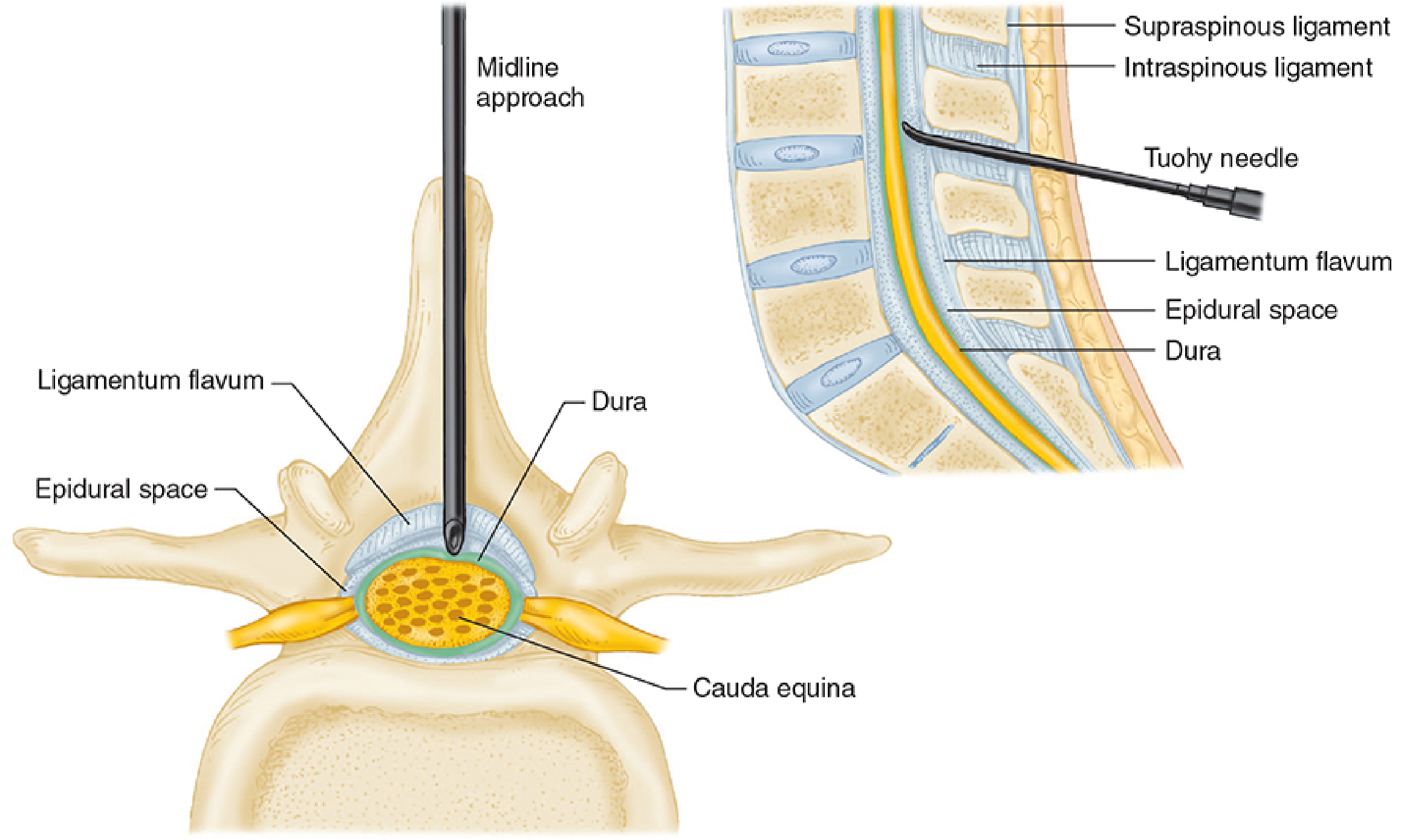
Epidural Space
The epidural space is a potential space within the spinal canal that lies outside the dura mater. It is bounded:
- Posteriorly by the ligamentum flavum
- Laterally by the spinal periosteum and pedicles
- Anteriorly by the dura mater
Contents include fatty connective tissue, lymphatics, and the Batson venous plexus (an extensive epidural venous network). Spinal nerve roots travel through this space as they exit laterally through the intervertebral foramina to form peripheral nerves. The presence of connective tissue septa occasionally explains unilateral epidural blocks.
Spinal Meninges
Three meningeal layers cover the spinal cord: the pia mater (innermost, adherent to cord), the arachnoid mater, and the dura mater (outermost). The subarachnoid space (between pia and arachnoid) contains CSF. The epidural space lies external to the dura.
Spinal Cord and Nerve Roots
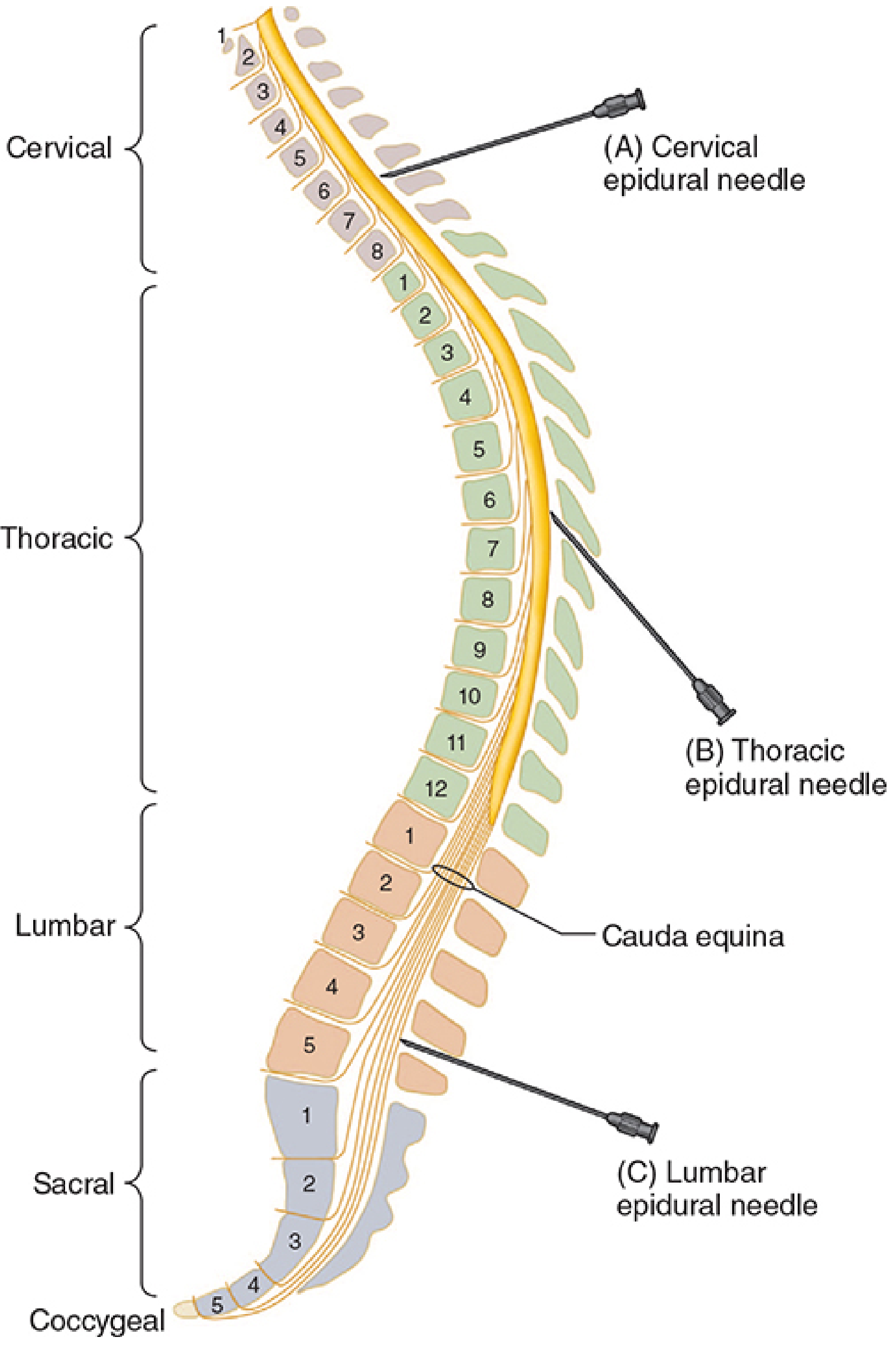
The spinal cord terminates at L1 in adults (L3 in children). Below this, the cauda equina (nerve roots of L2-S5) fills the spinal canal, making lumbar epidural placement safer. Nerve roots exit through the intervertebral foramina as:
- Dorsal (posterior) roots - sensory (afferent)
- Ventral (anterior) roots - motor (efferent)
3. Neural Pathways Blocked in Epidural Anesthesia
Primary Site of Action
The principal site of action is the spinal nerve root - at least during initial onset. Local anesthetics also act secondarily on the spinal cord itself and on paravertebral nerves. - Goodman & Gilman's Pharmacological Basis of Therapeutics
Fiber Type Classification (Order of Block)
| Fiber Type | Function | Diameter | Myelination | Sensitivity to LA |
|---|---|---|---|---|
| B fibers | Preganglionic sympathetic | < 3 µm | Lightly myelinated | Most sensitive |
| C fibers | Pain, temperature, postganglionic sympathetic | 0.2-1.5 µm | Unmyelinated | Very sensitive |
| Aδ fibers | Sharp pain, temperature | 1-4 µm | Thinly myelinated | Sensitive |
| Aβ fibers | Touch, pressure, proprioception | 5-12 µm | Myelinated | Intermediate |
| Aα fibers | Motor, proprioception (Ia) | 12-20 µm | Heavily myelinated | Least sensitive |
The order of blockade typically proceeds: sympathetic → sensory (temperature, then pain, then touch) → motor. This forms the basis of differential nerve block.
Differential Blockade
A hallmark of epidural anesthesia is differential blockade:
- Sympathetic block (measured by skin temperature increase) extends up to 6 spinal segments above the motor block
- Sensory block (pinprick/sharp sensation loss) extends 2 or more segments cephalad to motor block
- Motor block is the most restricted, as large Aα fibers are most resistant
This differential is exploited clinically - dilute local anesthetic with opioid can produce analgesia without motor block, as used in labor epidurals and postoperative pain management. - Barash, Cullen, and Stoelting's Clinical Anesthesia, 9e
Sympathetic Pathways
Blockade of efferent autonomic transmission at the spinal nerve roots produces sympathetic block, with profound physiological consequences:
- Cardiovascular: Vasodilatation → decreased SVR → hypotension; blockade of cardiac accelerator fibers (T1-T4) → bradycardia (Bezold-Jarisch reflex)
- GI: Vagal dominance → small, contracted gut, active peristalsis; postoperative epidural analgesia hastens return of bowel function
- Urinary: Blockade of lumbar and sacral sympathetic/parasympathetic → urinary retention
- Neuroendocrine: Blockade of somatic and visceral afferents attenuates the surgical stress response - reducing cortisol, catecholamines, ACTH, and vasopressin - Morgan and Mikhail's Clinical Anesthesiology, 7e
Segmental Block
Because the drug is deposited near its point of injection, it is possible to achieve a segmental block - a well-defined band of anesthesia at certain nerve roots while leaving roots above and below unaffected. A thoracic epidural, for example, can provide upper abdominal anesthesia while sparing cervical and lumbar roots.
4. Mechanism of Action of Local Anesthetics
Resting Membrane Potential
Neurons maintain a resting membrane potential of -60 to -70 mV, generated by the Na⁺-K⁺-ATPase pump (extruding 3 Na⁺ for every 2 K⁺) and selective K⁺ leak channels.
Voltage-Gated Na⁺ Channels and Action Potential
Excitable cells generate action potentials via voltage-gated Na⁺ channels. When a depolarizing stimulus activates these channels, Na⁺ floods inward, transiently raising the membrane potential to +35 mV. The channel then inactivates, restoring the resting potential. This wave of depolarization propagates as a nerve impulse.
Local Anesthetic Action
Local anesthetics are weak bases (pKa typically 7.6-8.9). Their mechanism involves:
- Penetration: In the ionized (protonated) form they cannot cross the lipid membrane; the unionized base form crosses into the axoplasm
- Re-ionization: Inside the axoplasm (lower pH), the drug becomes re-ionized (cationic form)
- Channel binding: The cationic form enters the inner mouth of the voltage-gated Na⁺ channel and blocks Na⁺ influx
- Prevention of depolarization: With Na⁺ entry blocked, an action potential cannot be generated or propagated
This is the sodium channel blockade theory of local anesthetic action. The block is use-dependent (frequency-dependent) - channels that are being repetitively activated (open/inactivated states) are more susceptible to block than channels in the resting closed state. - Morgan and Mikhail's Clinical Anesthesiology, 7e
Structure-Activity Relationships
Local anesthetics consist of:
- A lipophilic aromatic ring (benzene derivative)
- An intermediate chain (ester or amide linkage)
- A hydrophilic amine group
Ester-type: Cocaine, procaine, chloroprocaine, tetracaine - metabolized by plasma pseudocholinesterase; more allergenic (PABA metabolites)
Amide-type: Lidocaine, bupivacaine, ropivacaine, levobupivacaine - metabolized by hepatic CYP enzymes; less allergenic; preferred for epidural use
5. Pharmacology of Epidural Agents
Choice of Agent: Duration-Based
| Agent | Class | Onset | Duration | Concentration for Epidural | Notes |
|---|---|---|---|---|---|
| Chloroprocaine | Ester | Rapid | Very short | 2-3% | Rapid onset; neurotoxicity risk if intrathecal |
| Lidocaine | Amide | Moderate | Intermediate | 1.5-2% | Most commonly used intermediate agent |
| Bupivacaine | Amide | Slow | Long | 0.0625-0.75% | Gold standard for labor; 0.75% contraindicated in obstetrics |
| Ropivacaine | Amide | Moderate | Long | 0.2-0.75% | Less cardiotoxic than bupivacaine; preferred where cardiac safety is paramount |
| Levobupivacaine | Amide | Moderate | Long | 0.25-0.75% | S-enantiomer; reduced CNS/cardiac toxicity vs. racemic bupivacaine |
Concentration and Block Type
The concentration of local anesthetic determines which fiber types are blocked (from Goodman & Gilman):
- High concentration (e.g., bupivacaine 0.5%): blocks sympathetic + sensory + motor
- Intermediate (e.g., 0.25%): sensory anesthesia without significant motor block
- Low concentration (e.g., 0.0625%): only preganglionic sympathetic fibers blocked
Dose-Segment Relationship
Each 1-1.5 mL of 2% lidocaine produces approximately one additional dermatome segment of block in healthy adults aged 20-40 years. The volume required decreases with age, in pregnancy, and in children (due to reduced epidural space compliance and altered CSF volume). - Goodman & Gilman's Pharmacological Basis of Therapeutics
Systemic Absorption and Toxicity
Epidural anesthesia uses 10-20x greater drug volumes than spinal anesthesia. Peak blood concentrations of lidocaine after 400 mg epidural injection average 3-4 µg/mL; for bupivacaine 150 mg, approximately 1 µg/mL. The rate of systemic absorption follows: intercostal > paracervical > epidural > brachial plexus > sciatic > subcutaneous.
Cardiotoxicity of bupivacaine deserves special mention: accidental intravascular injection can cause severe left ventricular depression, AV block, ventricular tachycardia/fibrillation. CNS toxicity (seizures) occurs at lower blood concentrations than cardiac toxicity; cardiac toxicity requires ~3x the concentration needed for seizures. - Morgan and Mikhail's Clinical Anesthesiology, 7e
6. Pharmacological Adjuvants in Epidural Anesthesia
Epinephrine
- Acts via α₁-adrenoceptors to cause local vasoconstriction
- Reduces systemic absorption → lower peak blood concentrations, reduced toxicity
- Prolongs duration: extends lidocaine block by ≥50% (less effect on bupivacaine)
- Acts as a marker for inadvertent intravascular injection (tachycardia test dose)
- May contribute to analgesia via α₂-adrenoceptor activation
Opioids (Fentanyl, Morphine, Sufentanil)
- Bind to µ-opioid receptors in the dorsal horn of the spinal cord (Rexed laminae I, II - substantia gelatinosa)
- Inhibit release of substance P and glutamate from primary afferents, and hyperpolarize second-order neurons
- Synergize with local anesthetics, allowing dose reduction of both
- Fentanyl (lipophilic): rapid onset, segmental effect, minimal rostral spread
- Morphine (hydrophilic): delayed onset, wide spread, risk of delayed respiratory depression (up to 24 hours)
Clonidine / Dexmedetomidine
- α₂-adrenoceptor agonists acting at the dorsal horn and intermediolateral cell column
- Reduce norepinephrine release, hyperpolarize neurons
- Prolong sensory and motor block, reduce LA requirements
- Dexmedetomidine: 8x more selective than clonidine for α₂ receptors
Dexamethasone
- Mechanism not fully established; likely involves glucocorticoid receptor-mediated suppression of nociceptive pathways and inhibition of potassium channels in nerve fibers
- Prolongs block duration by up to 50%
7. Physiological Effects of Epidural Anesthesia
| System | Effect | Mechanism |
|---|---|---|
| Cardiovascular | Hypotension, bradycardia (T1-T4 block) | Sympathectomy → ↓ SVR, vagal predominance |
| Respiratory | Minimal change with low/mid blocks | Diaphragm (C3-C5) spared; ↓ expiratory muscle function with high blocks |
| GI | Contracted gut, enhanced peristalsis | Vagal dominance; hastens return of bowel function |
| Renal | Preserved with maintained BP | Autoregulation; urinary retention if sacral roots blocked |
| Neuroendocrine | Attenuation of stress response | Blocks afferent signaling via somatic and visceral fibers |
| Coagulation | Favorable (reduces DVT risk) | Sympathectomy improves venous blood flow |
8. Onset and Factors Affecting Block Level
Epidural anesthesia has an onset of 10-20 minutes (slower than spinal anesthesia) and may not be as dense. Factors determining the spread of epidural block:
- Volume injected - primary determinant of craniocaudal spread
- Concentration - determines depth of block (fiber type specificity)
- Level of injection - thoracic vs. lumbar
- Age - reduced epidural space compliance → wider spread in elderly
- Pregnancy - epidural venous engorgement reduces space → wider spread, lower doses needed
- Posture - less influence than with spinal anesthesia
9. Comparison: Epidural vs. Spinal Anesthesia
| Feature | Epidural | Spinal |
|---|---|---|
| Site of injection | Epidural space | Subarachnoid space |
| Volume of drug | Large (10-20 mL) | Small (1-3 mL) |
| Onset | Slow (10-20 min) | Rapid (3-5 min) |
| Density of block | Variable, less dense | Dense, predictable |
| Risk of systemic toxicity | Higher (large volumes) | Lower |
| Catheter possible | Yes | Yes (continuous spinal) |
| Segmental block possible | Yes | Difficult |
| Headache risk | Low (dura intact) | Higher (PDPH) |
10. Clinical Applications
- Surgical anesthesia: abdominal, thoracic, urological, lower limb surgery
- Labor analgesia: dilute bupivacaine 0.0625-0.1% + fentanyl 2 µg/mL
- Postoperative analgesia: thoracic epidural for upper abdominal/thoracic surgery reduces pulmonary complications, duration of ventilation
- Chronic pain: intermittent steroid injections, continuous infusions
- Combined spinal-epidural (CSE): rapid onset of spinal with flexibility of epidural catheter
Summary
Epidural anesthesia produces its effects by delivering local anesthetic to the epidural space, where the primary targets are the spinal nerve roots (at minimum) and secondarily the spinal cord and paravertebral nerves. Local anesthetics act by blocking voltage-gated Na⁺ channels in a use-dependent manner, preventing axonal depolarization. The differential sensitivity of nerve fibers produces a characteristic order of block: sympathetic → sensory → motor, exploited clinically to tailor analgesia. Drug selection (ester vs. amide, concentration, volume, level of injection) and adjuvants (epinephrine, opioids, α₂ agonists) allow precise titration of onset, duration, and quality of block. The systemic physiological consequences - primarily cardiovascular from sympathectomy - must be anticipated and managed proactively.
References:
- Morgan and Mikhail's Clinical Anesthesiology, 7e (Chapters 16, 45)
- Goodman & Gilman's The Pharmacological Basis of Therapeutics (Chapter on Local Anesthetics)
- Barash, Cullen, and Stoelting's Clinical Anesthesia, 9e (Chapters 35, 36)
- Miller's Anesthesia, 10e
This is a shared conversation. Sign in to Orris to start your own chat.