
Muscles of pectoral region
muscles of pectoral region anatomy diagram
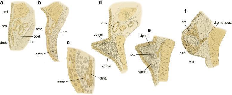
This comparative anatomical diagram series (labeled a-f) illustrates the embryological development of the pectoral fin muscles in the lungfish, providing an evolutionary model for tetrapod limb development. The sequence depicts the progression from Stage 42 to Stage 48. Initial frames (a-c) show the ventral process of the dermomyotome (dmtv) extending across the pronephros (prn) and the subsequent delamination of migratory muscle precursors (MMP). Middle frames (d-e) demonstrate the formation of dorsal and ventral premuscle masses (dpmm and vpmm) within the developing fin bud, surrounding a central precartilage condensation (pcc). The final frame (f) shows the differentiated state with distinct dorsal muscle (dm) and ventral muscle (vm) flanking a central cartilaginous element (cart), alongside the posterior plexus omopterygius (pl.ompt.post). This series highlights key developmental mechanisms such as de-epithelialization and mesenchymal migration, which are foundational concepts in comparative musculoskeletal anatomy and embryology.
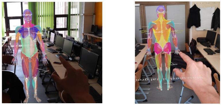
This image depicts an Anatomical Diagram of the human muscular system presented as a three-dimensional holographic projection via Mixed Reality (Microsoft HoloLens). The visual content features two panels showing anterior and posterior views of a life-sized human figure where major muscle groups are color-coded for educational identification. Key anatomical regions are distinguished by vibrant overlays: the pectoral and shoulder muscles are in shades of blue and purple, abdominal and core muscles are represented in green and white, while the lower extremities show thigh muscles in orange/red and lower leg groups in blue/teal. Interactive white labels and leader lines are positioned adjacent to specific muscles (e.g., facial muscles, gluteal muscles, hamstrings) to provide nomenclature context. The image illustrates advanced medical education tools designed for gross anatomy instruction, enabling students to visualize spatial relationships and morphology of the musculoskeletal system within a real-world classroom environment.
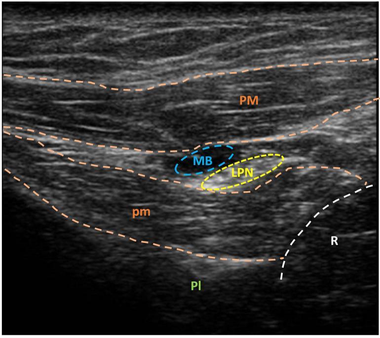
This diagnostic image is a high-frequency ultrasound of the subclavicular region, demonstrating ultrasound-guided needle marking of the lateral pectoral nerve (LPN). The cross-sectional view shows layered musculoskeletal anatomy: the superficial pectoralis major muscle (PM) and the deeper pectoralis minor muscle (pm). The LPN is visualized within the fascial plane between these two muscles, highlighted by a yellow dashed circle. Adjacent to the nerve, a localized hypoechoic area (MB, blue dashed circle) represents injected methylene blue used for intraoperative identification. In the deep field, the pleura (Pl) appears as a hyperechoic line, and a rib (R) is visible as a curved, hyperechoic structure with posterior acoustic shadowing. This visualization is clinically significant for regional anesthesia, spasticity management via selective peripheral neurectomy, and identifying anatomical landmarks for thoracic wall procedures.
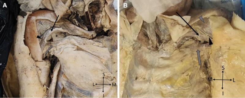
This medical image displays a comparative cadaveric dissection of the anterior thoracoappendicular region in two panels (A: right side; B: left side), illustrating anatomical variations consistent with bilateral partial agenesis of the pectoralis major (PMaj) and pectoralis minor (PMin) muscles. In both panels, the clavicular portion of the PMaj is identified by black arrows, and the sternocostal portion by gray arrows. An asterisk (*) marks the deep-seated PMin, which appears hypoplastic and blends with connective tissue. The deltopectoral sulcus (black arrowheads) is visible as a prominent groove separating the pectoralis major from the deltoid muscle (gray arrowheads in panel B). The dissection highlights the absence of normal muscular bulk in the sternal and abdominal portions of the PMaj, exposing deeper thoracic structures. Anatomical orientation markers (Superior-Inferior, Medial-Lateral) are provided for each panel. This image serves as a clinical reference for studying congenital muscular anomalies, surgical anatomy of the pectoral region, and variations in the thoracoappendicular musculature.
pectoralis major pectoralis minor serratus anterior subclavius anatomy
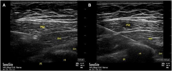
Two side-by-side ultrasound (US) images (A and B) demonstrate the regional anatomy and procedural technique for Pectoralis (PECS) nerve blocks. A high-frequency linear probe reveals the layered musculature of the anterior chest wall. The pectoralis major (PM) and pectoralis minor (Pm) muscles appear as superficial hypoechoic layers with characteristic striated fiber patterns separated by hyperechoic fascial planes. Deep to these lies the serratus anterior muscle (Sm). In the inferior portion of the frame, the fourth rib (r4) is identified as a bright, curvilinear hyperechoic interface with distinct posterior acoustic shadowing. The pleura (Pl) is visible as a smooth, hyperechoic horizontal line located deep to the intercostal space. Image A illustrates the target site for a PECS I block, where local anesthetic is delivered into the fascial plane between the pectoralis major and minor. Image B demonstrates a PECS II block, showing an in-plane needle approach targeting the fascial plane between the pectoralis minor and serratus anterior muscles. This diagnostic ultrasound provides critical guidance for regional anesthesia by identifying vascular and pleural landmarks to prevent complications such as pneumothorax.
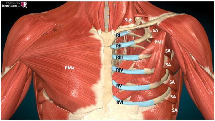
Anatomical diagram of the anterior and lateral thoracic wall muscles and skeletal structures. On the right side of the image (anatomical left), the pectoralis major (PMa) is shown as the most superficial layer. On the left side of the image (anatomical right), the pectoralis major is removed to reveal deeper structures. Key muscles include the pectoralis minor (PMi) originating from ribs III-V, the serratus anterior (SA) with its characteristic digitations on the lateral rib cage, and the subclavius (Sub) muscle inferior to the clavicle. The skeletal system is represented by the sternum and ribs I through VI (RI-RVI). Costal cartilages are highlighted in blue at their junctions with the sternum. The illustration demonstrates the fascial planes and anatomical landmarks relevant for regional anesthesia, specifically for performing pectoral nerve (PECS) and serratus anterior plane (SAP) blocks. This diagram is designed for medical education regarding thoracic wall innervation and ultrasound-guided nerve block techniques.
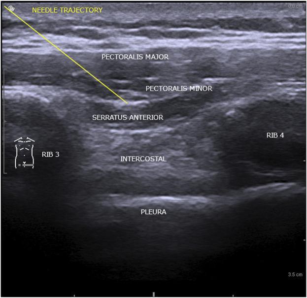
This diagnostic ultrasound image illustrates the regional anatomy involved in the interpectoral (PECS I) and pectoserratus (PECS II) plane blocks. The image displays a cross-sectional view of the anterior chest wall with distinct muscular and osseous landmarks. The Pectoralis Major is the most superficial muscle layer, followed by the Pectoralis Minor. Deep to these is the Serratus Anterior muscle, which sits atop the thoracic cage. Two ribs (Rib 3 and Rib 4) are identified by their hyperechoic anterior surfaces and posterior acoustic shadowing. Between the ribs, the hypoechoic intercostal space is visible, with the pleura appearing as a highly echogenic, horizontal line at the deepest level shown. An annotation line indicates the 'Needle Trajectory,' demonstrating the path through the pectoral muscles toward the fascial plane above the Serratus Anterior. This visual is designed for anesthesia education, specifically targeting ultrasound-guided regional anesthesia for thoracic or breast procedures.
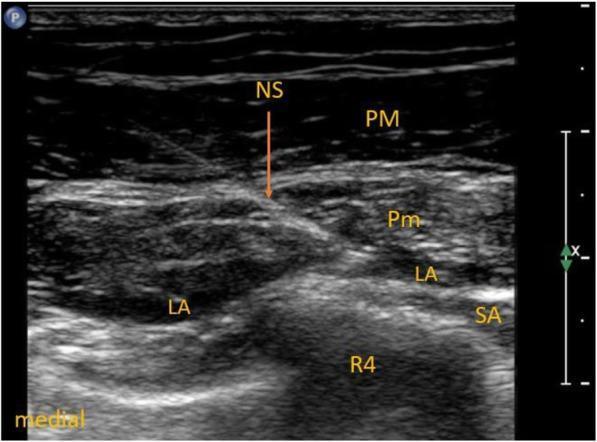
This diagnostic ultrasound image demonstrates a Pectoralis Nerves Block Type II (PECS II) being performed in the thoracic region. The image captures the regional anatomy in a transverse view, showing several muscular and skeletal layers. Visible structures include the pectoralis major (PM) as the most superficial muscle layer, followed by the pectoralis minor (Pm), and the serratus anterior (SA) located deeper and more laterally. The fourth rib (R4) is visible as a hyperechoic structure with posterior acoustic shadowing. A needle shaft (NS) is visualized using an in-plane technique, with its tip positioned to deliver local anesthetic (LA). The image clearly shows the distribution of local anesthetic in two key fascial planes: between the pectoralis major and minor muscles (the PECS I site) and between the pectoralis minor and serratus anterior muscles (the PECS II site). This image serves as an educational reference for ultrasound-guided regional anesthesia, specifically for postoperative analgesia in thoracic or breast surgeries.
Muscles of the Pectoral Region
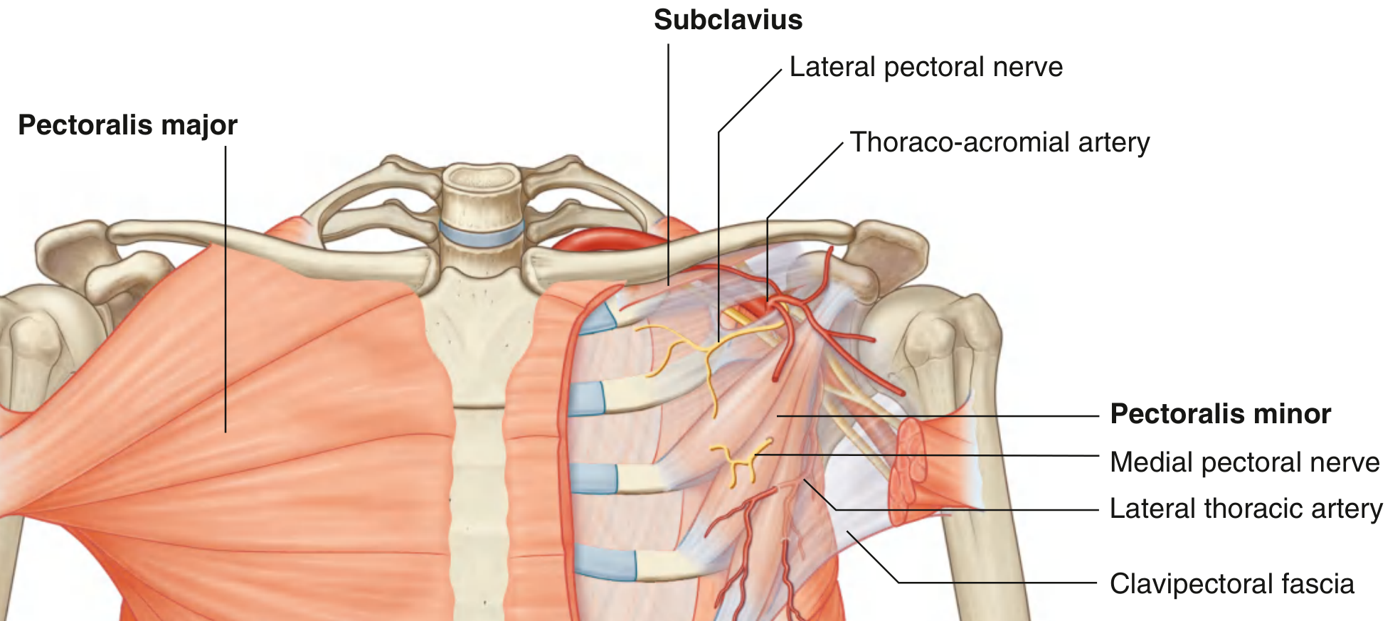
1. Pectoralis Major
| Feature | Details |
|---|---|
| Origin | Medial half of clavicle; anterior surface of sternum; first 7 costal cartilages; aponeurosis of external oblique |
| Insertion | Lateral lip of the intertubercular sulcus (bicipital groove) of the humerus |
| Innervation | Medial and lateral pectoral nerves (C5-T1) |
| Actions | Adduction, medial rotation, and flexion of the humerus at the shoulder joint |
2. Subclavius
| Feature | Details |
|---|---|
| Origin | Rib I at the junction between the rib and its costal cartilage |
| Insertion | Groove on the inferior surface of the middle third of the clavicle |
| Innervation | Nerve to subclavius (C5, C6) |
| Actions | Pulls clavicle medially to stabilize the sternoclavicular joint; depresses the tip of the shoulder |
3. Pectoralis Minor
| Feature | Details |
|---|---|
| Origin | Anterior surfaces of ribs III, IV, and V (and deep fascia of overlying intercostal spaces) |
| Insertion | Coracoid process of the scapula |
| Innervation | Medial pectoral nerve (C8, T1) |
| Actions | Depresses the tip of the shoulder; protracts (draws forward) the scapula |
Clavipectoral Fascia
Clinical Relevance
- The muscles of the pectoral region form the anterior wall of the axilla, the corridor through which all major neurovascular structures pass to and from the upper limb.
- Pectoralis major is used in reconstructive flap surgery (pedicled pectoralis major flap) for head and neck reconstruction.
- Pectoralis minor is a key landmark in axillary dissection - the axillary lymph nodes are divided into levels I, II, and III relative to this muscle.
- Absence or underdevelopment of pectoralis major is a feature of Poland syndrome.
- The lateral pectoral nerve passes through the clavipectoral fascia to supply pectoralis major; the medial pectoral nerve pierces pectoralis minor before entering pectoralis major.