
Elaborate management plan for 28 year old healthy female with acute onset of vertigo . Long answer forr this according to standard ENT textbook in simplified manner
Dix Hallpike test vertigo BPPV nystagmus
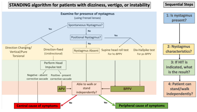
Summary : This figure presents the STANDING algorithm for evaluating patients with dizziness, vertigo, or instability, focusing on the presence and characteristics of nystagmus to differentiate between central and peripheral causes of symptoms.
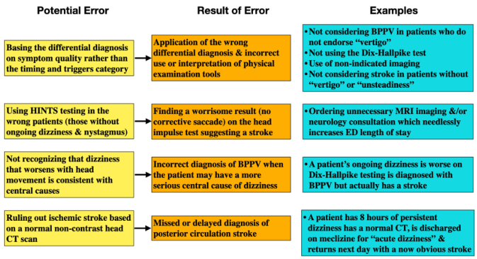
<table><thead><tr><th>Potential Error</th><th>Result of Error</th><th>Examples</th></tr></thead><tbody><tr><td>Basing the differential diagnosis on symptom quality rather than the timing and triggers category</td><td>Application of the wrong differential diagnosis & incorrect use or interpretation of physical examination tools</td><td>• Not considering BPPV in patients who do not endorse "vertigo"<br>• Not using the Dix-Hallpike test<br>• Use of non-indicated imaging<br>• Not considering stroke in patients without "vertigo" or "unsteadiness"</td></tr><tr><td>Using HINTS testing in the wrong patients (those without ongoing dizziness & nystagmus)</td><td>Finding a worrisome result (no corrective saccade) on the head impulse test suggesting a stroke</td><td>• Ordering unnecessary MRI imaging &/or neurology consultation which needlessly increases ED length of stay</td></tr><tr><td>Not recognizing that dizziness that worsens with head movement is consistent with central causes</td><td>Incorrect diagnosis of BPPV when the patient may have a more serious central cause of dizziness</td><td>• A patient's ongoing dizziness is worse on Dix-Hallpike testing is diagnosed with BPPV but actually has a stroke</td></tr><tr><td>Ruling out ischemic stroke based on a normal non-contrast head CT scan</td><td>Missed or delayed diagnosis of posterior circulation stroke</td><td>• A patient has 8 hours of persistent dizziness has a normal CT, is discharged on meclizine for "acute dizziness" & returns next day with a now obvious stroke</td></tr></tbody></table>
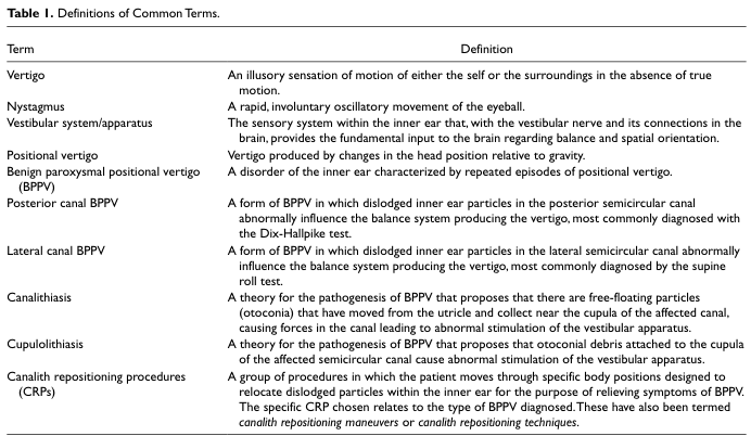
Table 1. Definitions of Common Terms.
Epley maneuver canalith repositioning BPPV treatment steps
Management Plan: Acute Onset Vertigo in a 28-Year-Old Healthy Female
STEP 1: UNDERSTAND WHAT YOU ARE DEALING WITH
STEP 2: INITIAL HISTORY TAKING (Key Questions)
| What to Ask | Why It Matters |
|---|---|
| Describe the dizziness - is the room spinning? | True vertigo vs. presyncope vs. disequilibrium |
| Onset - sudden or gradual? | Acute = vascular/infectious; gradual = tumor |
| Duration - seconds? Minutes to hours? Days? | Key to narrowing diagnosis (see table below) |
| Triggered by head position change? | Suggests BPPV |
| Continuous or episodic? | Episodic = BPPV, Meniere's; Continuous = neuritis |
| Hearing loss? Tinnitus? Ear fullness? | Suggests cochlear/Meniere's involvement |
| Nausea, vomiting? | Severity of acute attack |
| Recent upper respiratory infection? | Suggests vestibular neuritis |
| Neurological symptoms - diplopia, dysphagia, limb weakness? | RED FLAG - central cause |
| Headache, especially occipital? | RED FLAG - posterior fossa lesion |
| Medications - aminoglycosides, loop diuretics, quinine? | Ototoxic drugs |
STEP 3: DURATION-BASED DIFFERENTIAL DIAGNOSIS
| Disorder | Duration of Vertigo | Hearing Loss | Tinnitus | Ear Fullness |
|---|---|---|---|---|
| BPPV | Seconds | No | No | No |
| Meniere's Disease | Minutes to hours | Fluctuant SNHL | Yes | Yes |
| Vestibular Neuritis | Days to weeks | No | No | No |
| Recurrent Vestibulopathy | Minutes to hours | No | No | +/- |
| Acoustic Neuroma | Imbalance | Progressive SNHL | Yes | No |
STEP 4: PHYSICAL EXAMINATION
General Examination
- BP both arms (postural hypotension)
- Pulse, temperature
Ear Examination (Otoscopy)
- Tympanic membrane integrity
- Signs of middle ear disease, cholesteatoma
Neurological Examination - LOOK FOR RED FLAGS
- Cranial nerves: especially V, VII, VIII, IX, X
- Cerebellar signs: finger-nose test, dysdiadochokinesia, heel-shin test
- Gait: ataxic gait suggests cerebellar pathology
Vestibular Examination
- Direction (horizontal/torsional = peripheral; vertical/purely torsional = central)
- Fixation suppression: peripheral nystagmus is suppressed by visual fixation; central nystagmus is NOT suppressed
| Feature | Peripheral | Central |
|---|---|---|
| Direction | Horizontal-torsional | Mixed / purely vertical / torsional |
| Visual fixation suppression | YES | NO |
| CNS symptoms | Absent | Often present |
| Severity | Severe vertigo, vegetative | Vertigo less severe |
- Positive if rotational, geotropic nystagmus appears with:
- Latency (a few seconds before onset)
- Short duration (< 1 minute)
- Fatigues on repeat testing
- Reverses on sitting back up
- Positive (corrective saccade seen) = peripheral lesion (vestibular neuritis)
- Negative = central cause must be considered
- H - Head Impulse: positive HIT suggests peripheral
- I - INtercurrent nystagmus: direction-changing = central
- TS - Test of Skew: skew deviation = central
- Romberg positive = vestibular/proprioceptive dysfunction
- Fukuda: deviation >45° toward affected side = peripheral lesion
STEP 5: INVESTIGATIONS
Bedside / Clinic Tests
- Pure Tone Audiometry (PTA) - mandatory in ALL vertigo patients
- Tympanometry
Blood Tests (to exclude systemic causes)
- Complete Blood Count (CBC) - anemia
- Fasting blood glucose - hypoglycemia
- Thyroid function tests (TFT)
- ESR/CRP - inflammatory/autoimmune
- VDRL/FTA-ABS - syphilis (can cause endolymphatic hydrops)
- In young females specifically: ANA, anti-dsDNA (autoimmune inner ear disease)
Special Vestibular Tests
- Electronystagmography (ENG)/Videonystagmography (VNG): records nystagmus objectively; caloric test is most useful - unilateral caloric weakness identifies the affected side
- Vestibular Evoked Myogenic Potential (VEMP): tests saccule (cVEMP) and utricle (oVEMP)
- Electrocochleography (EcochG): useful in Meniere's disease
Imaging
- MRI brain with gadolinium - ONLY if red flags present (central signs, progressive SNHL, unilateral tinnitus)
- Rules out acoustic neuroma (vestibular schwannoma), posterior fossa tumors, MS plaques
- Normal CT does NOT rule out posterior fossa stroke
- MRI can also show gadolinium enhancement of the vestibular nerve in vestibular neuritis
STEP 6: COMMON DIAGNOSES AND THEIR SPECIFIC MANAGEMENT
A. BENIGN PAROXYSMAL POSITIONAL VERTIGO (BPPV) - Most Common Cause
-
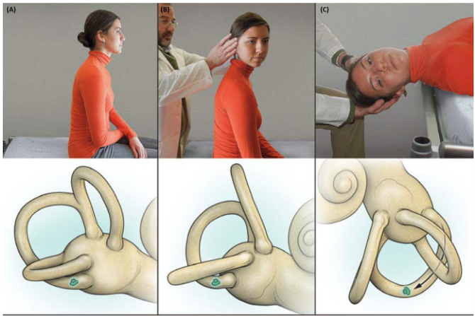
Epley Maneuver - most effective; moves the otoconia out of the canal back into the utricle through a series of 4 head positions held for 30 seconds each. Resolution in >90% of cases.
-
Semont Liberatory Maneuver - rapid side-to-side position changes
-
Brandt-Daroff Exercises - home habituation exercises; patient performs repeated position changes to desensitize the vestibular system
- Posterior canal occlusion
- Singular neurectomy
- Vestibular neurectomy
- Labyrinthectomy (only in deafened ear)
B. VESTIBULAR NEURITIS - Most Common Cause of Acute Prolonged Vertigo
- Acute, severe, persistent vertigo with nausea/vomiting
- Lasts days (acute phase), then gradual improvement over weeks
- Balance complaints may persist for months
- Positive head impulse test toward affected side
- Caloric weakness on the affected side
| Drug | Class | Dose | Notes |
|---|---|---|---|
| Prochlorperazine (Stemetil) | Dopamine antagonist | 5-10 mg TDS oral; buccal/suppository if vomiting | Blocks chemoreceptor trigger zone. More extrapyramidal effects than sedation. Available as buccal (Buccastem) - very useful when patient is vomiting. |
| Cinnarizine (Stugeron) | Antihistamine | 30 mg TDS | Blocks H1 at vomiting centre; also weak calcium channel blocker |
| Cyclizine (Valoid) | Antihistamine | 50 mg TDS oral/IV | Less sedating, useful parenterally |
| Diazepam | Benzodiazepine | 2-5 mg | Reduces vestibular excitability; short-term only |
- Methylprednisolone may improve caloric function
- Benefit limited to caloric test improvement; symptomatic benefit is less clear
- Most beneficial if started within 3 days of onset
- Currently: weak evidence supports use in vestibular neuritis (Cummings Otolaryngology)
- Valacyclovir was NOT found to improve outcomes in controlled trials (Cummings Otolaryngology)
- NOT routinely recommended
- Early ambulation is STRONGLY encouraged
- Physical activity promotes central vestibular compensation (the brainstem "relearns" to compensate for the asymmetric input)
- Started as soon as acute phase settles (usually day 2-3)
- Exercises designed to:
- Promote gaze stabilization
- Improve balance
- Reduce motion sensitivity
- Significantly shortens recovery time
C. MENIERE'S DISEASE - If Recurrent Episodes with Hearing Loss + Tinnitus
- Two or more definitive spontaneous episodes of vertigo lasting at least 20 minutes
- Audiometrically documented hearing loss on at least one occasion
- Tinnitus or aural fullness in the suspected ear
- Other causes excluded
- Low-salt diet (< 2g sodium/day) - reduces endolymph volume
- Avoid caffeine, alcohol, nicotine
- Reduce stress
- Adequate sleep
| Drug | Mechanism | Dose | Notes |
|---|---|---|---|
| Betahistine (Serc) | Improves microcirculation in stria vascularis; inhibits vestibular nuclei | 16 mg TDS initially; maintenance 24-48 mg/day | May reduce vertigo; insufficient RCT evidence. Caution in asthma, peptic ulcer. CI in phaeochromocytoma |
| Diuretics | Reduce endolymph accumulation | Hydrochlorothiazide, co-triamterzide (Dyazide) | Reduces vestibular symptoms; no effect on hearing |
| Steroids | Immunosuppression | Oral prednisolone - 60mg D1, tapering over 3 weeks | Consider if autoimmune etiology suspected |
- Intra-tympanic gentamicin - chemical labyrinthectomy; ablates vestibular function; risk of hearing loss. Used for disabling, medically refractory vertigo with serviceable hearing being acceptable risk.
- Intra-tympanic dexamethasone - preserves hearing while reducing attacks
- Endolymphatic sac surgery (decompression)
- Vestibular nerve section
- Labyrinthectomy (hearing already destroyed)
STEP 7: GENERAL SYMPTOMATIC MANAGEMENT (Regardless of Cause)
Acute Attack Management
- Rest in a quiet, dark room; keep still in position of comfort
- IV fluids if severe vomiting causing dehydration
- Anti-emetics: Prochlorperazine IM/buccal or Cyclizine IV
- Vestibular suppressants: short course only
Vestibular Rehabilitation
- Cawthorne-Cooksey exercises: head movement exercises (looking up and down, side to side, progressively with body movements)
- Improves compensation for all types of peripheral vestibular disorders
Patient Education and Safety
- Avoid driving until vertigo fully resolves
- Avoid heights, ladders, machinery
- Explain the natural history - reassure that most causes in a young healthy person are benign and resolve
- No sudden head movements initially
Lifestyle Advice
- Adequate hydration
- Regular sleep schedule
- Reduce caffeine and salt intake (even outside of Meniere's, these can worsen symptoms)
- Avoid ototoxic drugs (aminoglycosides, furosemide)
STEP 8: RED FLAGS - WHEN TO SUSPECT CENTRAL CAUSE (URGENT MRI)
- Vertical nystagmus or direction-changing nystagmus
- Nystagmus not suppressed by fixation
- Negative head impulse test despite ongoing spontaneous nystagmus
- Skew deviation on eye exam
- Diplopia, dysarthria, dysphagia
- Sudden-onset severe headache ("thunderclap")
- Limb ataxia or truncal ataxia out of proportion to severity of vertigo
- Multiple cranial nerve palsies
- Inability to walk or severe gait ataxia with relatively mild vertigo
STEP 9: FOLLOW-UP PLAN
| Timeframe | Action |
|---|---|
| Day 1-3 | Review response to vestibular suppressants; ensure antiemetics working; stop suppressants as soon as tolerable |
| Week 1 | PTA, tympanometry results; start vestibular rehabilitation |
| Week 2-4 | Review recovery; if no improvement, consider VNG/ENG, specialist referral |
| 3 months | If recurrent episodes - full Meniere's workup; MRI if not done |
| 6 months | Repeat PTA to document hearing trajectory |
Summary Flowchart (Simplified)
Acute Vertigo in 28-yr female
         |
    HISTORY + EXAMINATION
         |
  ┌──────┴──────┐
Central signs?  No central signs
(vertical nystagmus,   |
no HIT correction,    Peripheral
neurological sx)       |
  |              ┌─────┴──────────┐
Urgent MRI    Position-    Continuous/
              triggered    Days/weeks
                 |              |
               BPPV         ±Hearing loss?
                 |              |
           Dix-Hallpike    No HL → Vestibular Neuritis
           positive        HL+Tinnitus → Consider Meniere's
                 |
          Epley Maneuver
- Cummings Otolaryngology Head and Neck Surgery - Vestibular Neuritis, Meniere's Disease chapters
- K.J. Lee's Essential Otolaryngology, 11th Ed. - BPPV, Differential Diagnosis table
- Scott-Brown's Otorhinolaryngology Head & Neck Surgery Vol. 1 - Pharmacological management of vertigo