
Brachial fistula and sinus
branchial cleft fistula sinus neck anatomy diagram
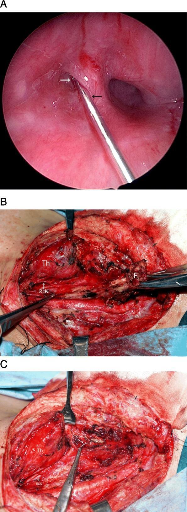
This composite of three images illustrates a surgical resection of a branchial pouch sinus (fourth branchial cleft fistula). Panel A is an endoscopic view of the piriform fossa, showing a metal probe (black arrow) inserted into the internal opening (white arrow) of the fistula tract. Panels B and C are intraoperative clinical photographs of an open neck dissection. Panel B shows the surgical field with labeled anatomical structures: the thyroid gland (Th), the fistula (F) being retracted, the recurrent laryngeal nerve (RLN) visualized as a thin white strand, and the carotid artery (CA) deeper in the field. Panel C demonstrates the completed tracing and excision of the fistula (F) using the metal probe as a guide. The image highlights the proximity of the fistula to critical neurovascular structures and the thyroid gland, emphasizing the importance of careful dissection to preserve the RLN and CA during the removal of the tract and associated scar tissue.
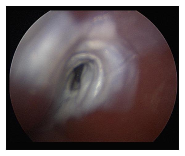
This clinical diagnostic image shows a direct laryngoscopic view of the right pyriform sinus apex in a patient with a suspected branchial cleft cyst. Centrally, the image demonstrates a small, dark, oval fistulous tract opening. The immediate perifistular tissue exhibits a blanched, white-to-light-gray appearance, characteristic of chemical cauterization following the application of 40% trichloroacetic acid (TCA). The surrounding pharyngeal mucosa is reddish-pink with a moist, glistening texture. This visual represents the intraoperative endpoint of chemocauterization for the management of a branchial cleft fistula, where the goal is to induce mucosal inflammation and subsequent fibrosis to achieve tract closure. The anatomical context is critical for otolaryngologists in identifying and treating congenital neck anomalies through endoscopic approaches.
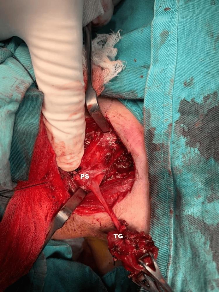
This clinical photograph provides an intraoperative view of the surgical excision of a branchial fistula tract in the right neck. The operative field is exposed through a surgical incision, with drapes and retractors maintaining visibility. A gloved finger and surgical forceps are seen manipulating the tissues. Two key structures are labeled: 'TG' (Thyroid Gland) and 'PS' (Pyriform Sinus). A distinct, elongated, fibrous fistula tract is clearly visible, extending from the superior pole of the right thyroid gland toward the right pyriform sinus. The thyroid tissue appears hemorrhagic and partially dissected, reflecting the surgical manipulation required to isolate the tract. The image illustrates the complex anatomical relationship between the branchial fistula and adjacent endocrine and pharyngeal structures, specifically highlighting the pathway where a fourth branchial pouch sinus typically traverses. This material is educationally relevant for otorhinolaryngology and endocrine surgery trainees to understand the surgical anatomy of congenital neck fistulas.
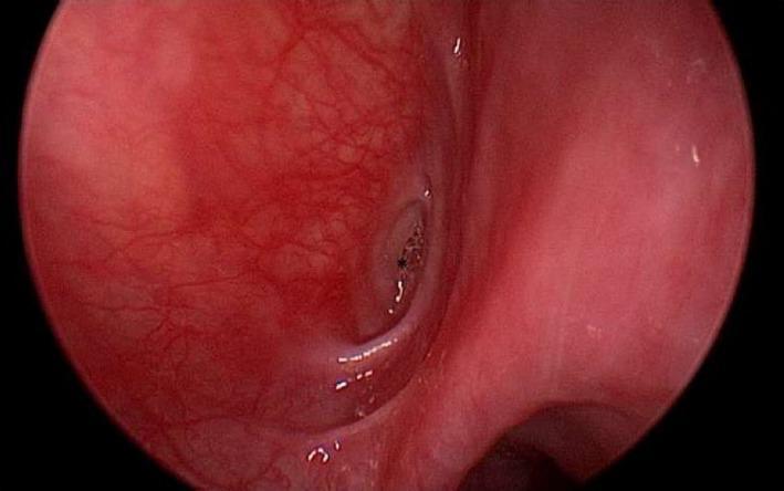
This clinical diagnostic image shows an endoscopic view of the left piriform sinus, located within the hypopharynx. The mucosa is predominantly reddish-pink and demonstrates prominent superficial vascularization. At the apex of the piriform sinus, a distinct, abnormal opening is visible, representing a branchial cleft fistula or sinus tract. The orifice of the fistula appears as a dark, circular indentation with irregular, slightly elevated, and erythematous margins, suggesting localized chronic inflammation or tissue remodeling. The surrounding mucosal texture is uneven, transitioning from smooth areas to more granular surfaces near the pathological opening. This visual finding is characteristic of a congenital branchial anomaly, specifically a third or fourth branchial cleft remnant, which typically presents with recurrent neck infections or abscesses. The image serves as a key educational reference for identifying anatomical landmarks and pathological findings during direct laryngoscopy in Otolaryngology (ENT).
Branchial Fistula and Sinus
Note: The correct term is branchial (from the branchial/pharyngeal arch apparatus), not "brachial" (which refers to the arm).
Definitions
| Term | Definition |
|---|---|
| Branchial fistula | A congenital skin-lined tract with two openings - an internal opening in the pharynx and an external opening on the neck skin |
| Branchial sinus | Has only one opening - either internal (into the pharynx) or more commonly external (on the neck skin), with no communication at the other end |
| Branchial cyst | A closed remnant with no opening at all |
Embryological Basis
- 2nd branchial pouch - in the tonsillar fossa (most common)
- 3rd branchial pouch - in the pyriform sinus/larynx area
- 4th branchial pouch - in the lower pyriform fossa
- Scott-Brown's Otorhinolaryngology Head & Neck Surgery, Vol 2
- KJ Lee's Essential Otolaryngology
Classification by Arch of Origin
Second Branchial Anomalies (most common ~95%)
- Deep to second arch derivatives (CN VII, external carotid artery)
- Superficial to third arch derivatives (stylopharyngeus, CN IX, internal carotid artery)
- Superficial to CN XII
- In close relation with but superficial to the carotid sheath
- Pierces the middle constrictor muscle, deep to the stylohyoid ligament
- Internal opening: Anterior aspect of the tonsillar fossa (slit-like)
Third Branchial Anomalies (rare)
- External opening: Anterior to SCM, low in the neck
- Deep to the internal carotid artery and CN IX
- Pierces the thyrohyoid membrane above the internal branch of the superior laryngeal nerve
- Opens into the pyriform fossa
Fourth Branchial Anomalies (rare)
- Also opens into the lower pyriform sinus or larynx
- Third and fourth anomalies are difficult to distinguish clinically; often present with recurrent thyroid infections
First Branchial Anomalies
-
Work Type I: Preauricular, parallel to the EAC
-
Work Type II: Through the parotid gland, ending at the bony-cartilaginous EAC junction (angle of mandible)
-
Variable relation to the facial nerve (deep, superficial, or between branches) - making surgery hazardous
-
Schwartz's Principles of Surgery, 11th Ed.
-
KJ Lee's Essential Otolaryngology
Clinical Features
- Most patients are young infants or children presenting with a discharging sinus in the neck
- The external opening appears just in front of the lower third of the anterior border of the sternomastoid
- Discharge is mucoid or mucopurulent
- The tract may intermittently become infected, forming a neck abscess
- Some patients notice increased discharge during upper respiratory tract infections
- A complete fistula extends all the way to the supra-tonsillar fossa
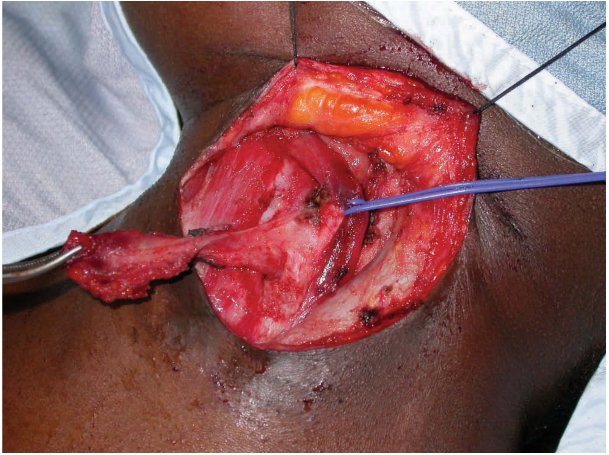
Investigations
- Clinical diagnosis - position and nature of the opening is often diagnostic
- Fistulogram / CT fistulogram - inject radio-opaque dye (e.g., Uropac) into the external opening to delineate the tract length and differentiate sinus from fistula; a complete fistula will extend to the supra-tonsillar fossa
- Contrast swallow - useful for 3rd/4th arch anomalies with pyriform sinus involvement
- Endoscopy - can visualize internal opening in the pyriform sinus for 3rd/4th arch lesions
- Ultrasound + CT/MRI - for surgical planning in complex cases
Surgical Treatment
- An elliptical incision is made around the external opening at the skin
- The tract is followed upward as high as possible
- A second (stepladder) incision is made higher up in a skin crease to allow further dissection
- The entire tract is excised, including the internal opening if accessible
- Facial nerve (particularly for 1st arch lesions - the tract may be deep, superficial, or between branches of the nerve; intraoperative nerve monitoring is advised)
- Internal carotid artery and external carotid artery
- Hypoglossal nerve (CN XII), Glossopharyngeal nerve (CN IX)
- Recurrent laryngeal nerve (for 3rd/4th arch lesions)
Summary Table
| Feature | 2nd Arch Fistula | 3rd Arch Fistula | 4th Arch Fistula |
|---|---|---|---|
| Frequency | Most common (~95%) | Rare | Rare |
| External opening | Ant. SCM (mid-lower junction) | Low in neck | Low in neck |
| Internal opening | Tonsillar fossa | Pyriform sinus | Lower pyriform/larynx |
| Key nerve risk | CN IX, XII | CN IX, SLN | RLN |
| Passes relative to ICA | Superficial | Deep to ICA | Deep to ICA |
| Typical presentation | Neck discharge from infancy | Recurrent neck abscess | Recurrent thyroiditis |
- Scott-Brown's Otorhinolaryngology Head & Neck Surgery, Vol 2 - Branchial Fistulae and Sinuses section
- KJ Lee's Essential Otolaryngology - Embryology and Derivatives of the Pouches
- Schwartz's Principles of Surgery, 11th Ed. - Benign Neck Masses
- S. Das - A Manual on Clinical Surgery, 13th Ed. - Physical Examination of the Neck
Branchial cyst
branchial cyst neck clinical presentation CT scan
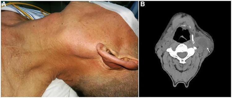
This composite image consists of a clinical photograph and a diagnostic CT scan illustrating a branchial cleft cyst in the neck. (A) A clinical photograph showing the left side of a patient's neck, where a large, prominent mass is visibly protruding from the skin in the upper cervical region. The overlying skin appears intact without obvious erythema. (B) An axial non-contrast CT scan of the neck at the level of the cervical vertebrae. It reveals a well-circumscribed, hypodense, ovoid mass (indicated by a white arrow) located on the left lateral aspect of the neck, deep to the platysma and lateral to the carotid sheath. The low-density center is characteristic of fluid-filled cystic structures. The lesion is located inferior to the parotid gland and anterior to the sternocleidomastoid muscle. The case illustrates the classic presentation of a branchial cleft cyst, which in this clinical context was diagnosed as a primary branchial cleft carcinoma (squamous cell carcinoma).
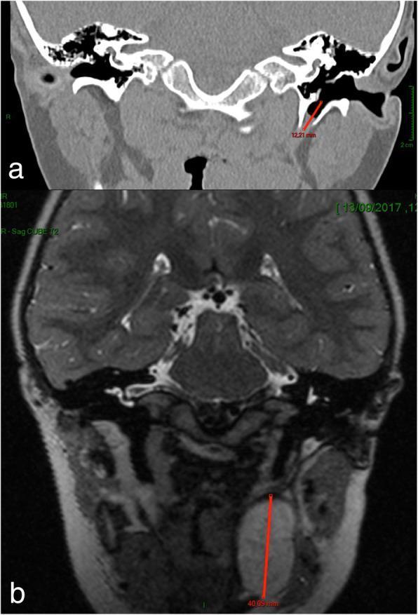
This dual-panel diagnostic image presents clinical imaging of the head and neck, specifically evaluating a suspected first branchial cleft cyst. Image (a) is a coronal CT scan of the temporal bones and skull base, highlighting a congenital anomaly: a bony canal (fistulous tract) measuring approximately 12.21 mm. This canal is situated in the deep portion of the tympanic bone, medial to the facial nerve and originating from the pre-tympanic region of the external auditory canal. Image (b) is a coronal T2-weighted MRI scan of the same patient, revealing a large, hyperintense, well-circumscribed parapharyngeal cyst in the left peri-tonsillar region. The cyst measures approximately 40.09 mm in its craniocaudal dimension. Together, these images illustrate the characteristic multi-component presentation of branchial cleft anomalies, involving both a cartilaginous/bony tract near the ear and a deeper soft tissue cystic component in the parapharyngeal space. The imaging modality and precise measurements are essential for pre-operative planning in otolaryngology to avoid injury to the adjacent facial nerve and carotid vasculature.
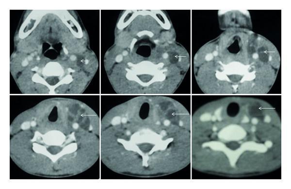
Diagnostic axial CT scan of the neck with contrast demonstrating a large, multiloculated cystic lesion in the left parapharyngeal and cervical space. The image series shows a hypoattenuating (fluid-density) mass indicated by white arrows, characterized by thin, internal enhancing septae. Anatomically, the lesion extends vertically from the level of the mandible down to the C7 vertebral level and the superior mediastinum. It is situated posteromedial to the left sternocleidomastoid muscle, displacing adjacent vascular structures. The clinical presentation is highly suggestive of a branchial cleft cyst or an ectopic cervical thymic cyst. This imaging highlights key diagnostic features of deep neck cystic masses, including internal architecture, density, and spatial relationship to cervical fascia and major vessels, essential for surgical planning and differential diagnosis in otolaryngology and radiology.
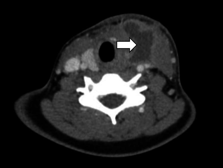
This diagnostic axial CT scan of the neck with contrast demonstrates an ill-defined, heterogeneous lesion in the left paratracheal soft tissue. A white arrow points to the primary pathology: a fourth branchial cleft cyst showing as a hypodense, fluid-filled area with rim enhancement, suggesting inflammation or infection. Key anatomical landmarks visible include the central air-filled trachea (dark circular structure) and the cervical vertebra (bright white structure posterior to the airway). The image captures the characteristic fistulous tract associated with this congenital anomaly, which is seen extending from the paratracheal region toward the left pyriform sinus. The lesion is located medial to the vascular structures of the carotid sheath and lateral to the trachea, illustrating the typical course of a fourth branchial arch remnant. This image is used to teach medical students and radiology residents the diagnostic presentation of a rare branchial cleft anomaly and its anatomical relationship to the upper aerodigestive tract.
Branchial Cyst
Definition
Aetiology / Theories of Origin
| Theory | Basis | Argument Against |
|---|---|---|
| Branchial apparatus theory | Remnants of pharyngeal pouches or branchial clefts (3rd-8th week of gestation) | Cysts should present at birth; they don't |
| Cervical sinus theory | Remnants of the cervical sinus of His (formed when 2nd arch grows down to cover 3rd/4th clefts) | Most widely accepted |
| Thymopharyngeal duct theory | Remnants of thymus-3rd pouch connection | No cyst ever found deep to thyroid; duct never identified |
| Inclusion theory | Epithelial inclusions within lymph nodes | Explains why cysts have lymphoid walls, absent in neonates, peak in adults - most favoured |
- Scott-Brown's Otorhinolaryngology Head & Neck Surgery, Vol 2
Pathology
| Feature | Detail |
|---|---|
| Lining | Stratified squamous epithelium (90%); pseudostratified columnar / respiratory epithelium (10%) |
| Wall | Fibrous with lymphoid tissue and prominent germinal centres (80% of cases) |
| Contents | Straw-coloured / golden-yellow fluid containing cholesterol crystals, fat globules, and desquamated epithelial cells |
| Size | Usually 2-10 cm in diameter; typically 5-10 cm |
| Malignant transformation | Rarely occurs; most cystic SCCs in the neck are metastases from upper aerodigestive tract cancers |
- Robbins & Cotran Pathologic Basis of Disease
- S. Das - Manual on Clinical Surgery, 13th Ed.
Site and Presentation
- 60% are in the upper third of the neck, at the anterior border of the sternocleidomastoid muscle (specifically at the junction of its upper and middle thirds, where the posterior belly of digastric crosses)
- Classically described as being half in front and half deep to the sternomastoid
- Long axis runs forwards and downwards
- Presents as a smooth, fluctuant, non-tender swelling - ovoid in shape
- 80% present as a persistent swelling; 20% are intermittent
- 70% feel cystic; up to 30% may feel solid (tense cyst)
- ~40% have a prior history of URTI before noticing the mass
- Transillumination is usually negative (content is thick/turbid)
- Does NOT move on swallowing (not attached to larynx/trachea)
- Regional lymph nodes are NOT enlarged in an uncomplicated cyst
- Bailey and Love's Short Practice of Surgery, 28th Ed.
Imaging
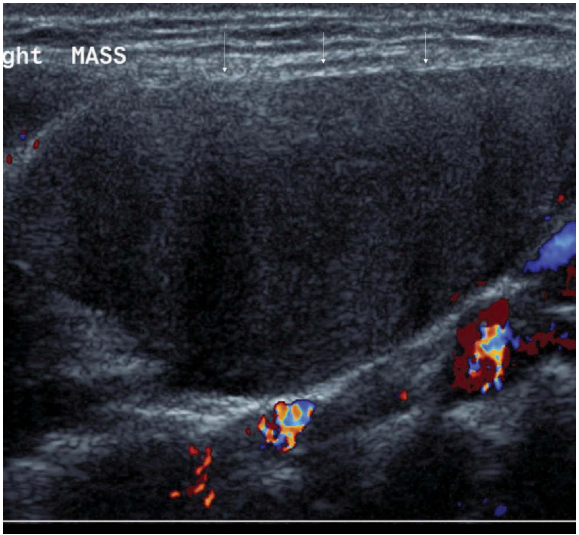
- Range from anechoic simple cyst with posterior enhancement to pseudosolid / heterogeneous with debris and septae
- If a medial beak is seen pointing toward the pharynx, CT/MRI is warranted to exclude a sinus or fistula
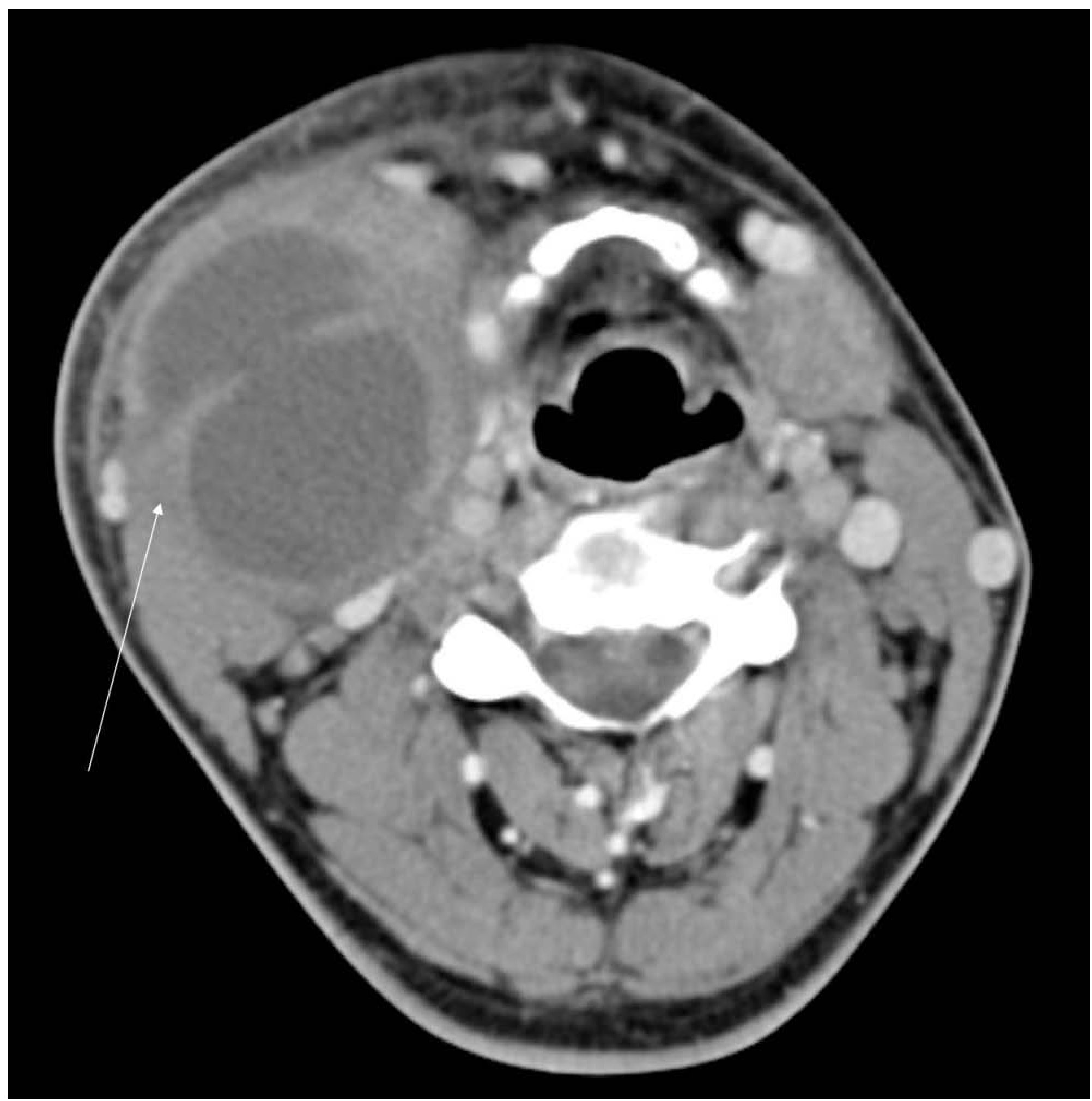
-
Classic: homogeneous low-attenuation mass with thin wall
-
Infected: ill-defined, heterogeneous, thick-walled with debris and septae
-
MRI: high T2, low T1 signal
-
Scott-Brown's Otorhinolaryngology Head & Neck Surgery
Diagnosis / Investigations
- Clinical diagnosis - characteristic site, age, and feel
- FBC (baseline)
- Ultrasound + FNAC - acellular fluid with cholesterol crystals on microscopy; this is diagnostic. (Caution: squamous debris may be misread as malignancy)
- CT / MRI - for large cysts, surgical planning, or when a medial beak is seen
- PET-CT - if malignancy (metastatic SCC) cannot be excluded, especially in patients over 40 who are smokers/drinkers
Key clinical rule: In any patient over 35 years of age, a cystic neck mass must be treated as metastatic SCC until proven otherwise. HPV-related oropharyngeal SCC commonly presents as a cystic nodal metastasis mimicking a branchial cyst.
Differential Diagnosis
| Condition | Differentiating feature |
|---|---|
| Cervical lymphadenopathy (TB cold abscess) | Palpable regional nodes, systemic features |
| Cystic hygroma (lymphangioma) | Brilliantly transilluminant, multilocular, infants |
| Carotid body tumour | Transmitted pulsation, bruit, at carotid bifurcation |
| Metastatic cystic SCC | Age >35, smoker, find primary in oropharynx |
| Thyroglossal cyst | Midline, moves on swallowing and tongue protrusion |
| Dermoid cyst | Midline, doughy |
Surgical Treatment
- Tendency to become infected
- Increasing size causing discomfort or cosmetic deformity
- Pressure symptoms
- Transverse cervical skin crease incision over the cyst
- Platysma incised; sub-platysmal flaps elevated
- Cervical fascia incised along the anterior border of SCM, which is retracted laterally
- Cyst identified and dissected from fascial attachments without rupturing the wall
- Carotid sheath and contents identified and protected
- Cyst passes superficial to CN IX and CN XII (hypoglossal), deep to the posterior belly of digastric
- Complete excision - the "tail" of the cyst may need dissection from the parotid gland or parapharyngeal space
- Marginal branch of the facial nerve (CN VII)
- Hypoglossal nerve (CN XII)
- Vagus nerve (CN X)
- Spinal accessory nerve (CN XI)
- Glossopharyngeal nerve (CN IX)
Branchiogenic Carcinoma (rare)
- Scott-Brown's Otorhinolaryngology Head & Neck Surgery, Vol 2 - Branchial Cysts section
- Bailey and Love's Short Practice of Surgery, 28th Ed.
- Robbins & Cotran Pathologic Basis of Disease
- S. Das - A Manual on Clinical Surgery, 13th Ed.
- KJ Lee's Essential Otolaryngology
Midline neck swelling
midline neck swelling thyroglossal cyst clinical anatomy diagram
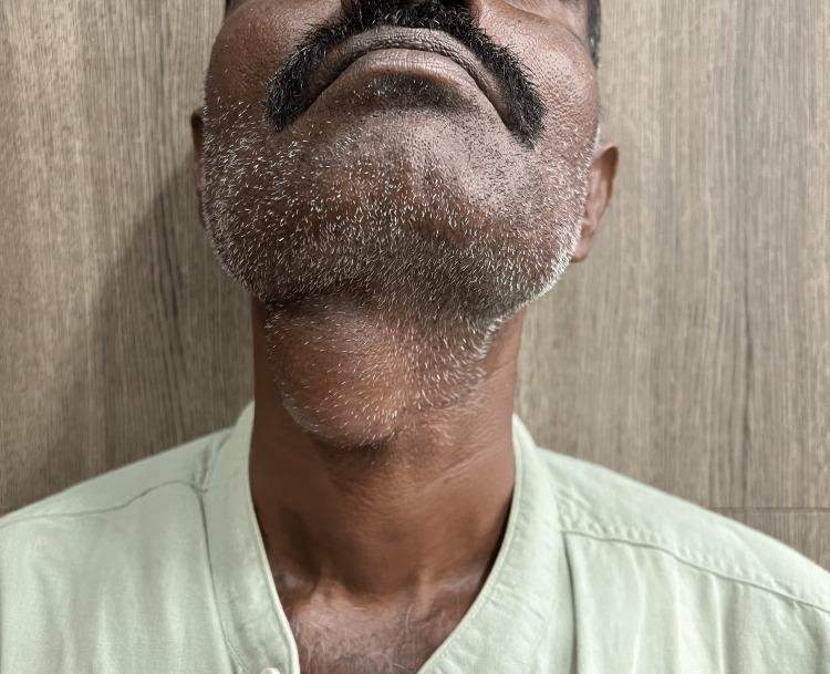
A clinical photograph of an adult male depicting a prominent midline neck mass, consistent with a thyroglossal duct cyst. The patient's neck is slightly extended, revealing a well-circumscribed, ovoid, and globular swelling located in the anterior midline of the neck, specifically at the level of the hyoid bone and inferior to the submental region. The overlying skin is intact and appears unremarkable, without signs of erythema, inflammation, ulceration, or fistulous tracts. This visual presentation is characteristic of a congenital thyroglossal duct anomaly, which typically manifests as a painless, mobile midline mass that often moves superiorly with tongue protrusion or swallowing. The image serves as a clinical example for Otolaryngology and General Surgery education, emphasizing the anatomical localization and physical characteristics necessary for the differential diagnosis of midline neck swellings, such as dermoid cysts or ectopic thyroid tissue.
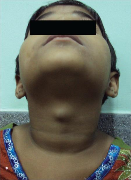
Clinical photograph of a pediatric patient's neck, viewed from an inferior-anterior perspective (looking up toward the chin). The image demonstrates a prominent, well-circumscribed, midline cystic swelling in the anterior neck, located superior to the thyroid cartilage. The overlying skin appears normal with no visible erythema, inflammation, or ulceration. The morphology is characteristic of a thyroglossal duct cyst, a common congenital abnormality occurring along the descent pathway of the thyroid gland. In this clinical context, such a mass often contains ectopic functional thyroid tissue and typically moves upward upon protrusion of the tongue or swallowing. The visual findings illustrate a classic presentation of a midline neck mass in childhood, serving as an educational example for differential diagnosis in pediatric otolaryngology and endocrinology, specifically regarding ectopic thyroid tissue and thyroglossal duct remnants.
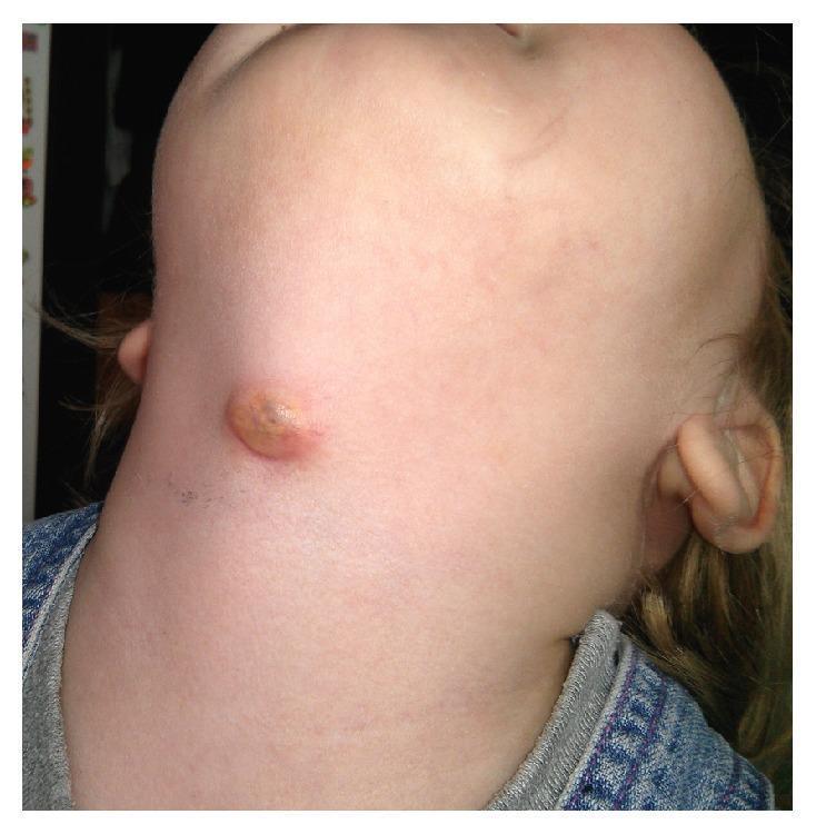
This clinical photograph displays a 2-year-old female patient with a prominent anterior neck swelling, characteristic of an infected thyroglossal duct cyst (TGDC). The lesion is located near the midline in the thyrohyoid region, slightly below the mandibular level. The swelling is approximately 1-2 cm in diameter, presenting as a raised, nodular mass with a circular to oval morphology. The surface exhibits a yellowish-brown crusting and erythema of the surrounding skin, suggesting an inflammatory or infectious process, such as an abscess or a draining fistula. This image serves as a clinical example of a common congenital midline neck mass in pediatric otolaryngology, illustrating the typical presentation of a thyroglossal duct cyst when secondary infection occurs. It is an essential visual reference for differential diagnosis of pediatric neck masses, distinguishing TGDC from other entities like dermoid cysts or lymphadenopathy based on its characteristic anatomical position.
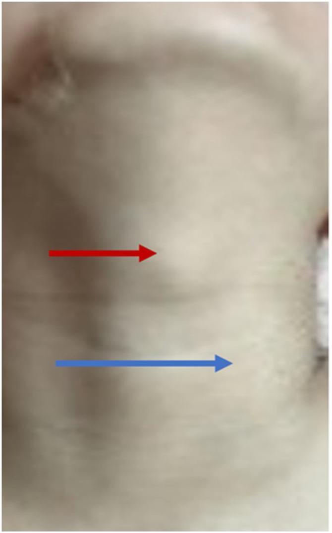
This clinical photograph displays the anterior and lateral neck of a 7-year-old patient, highlighting two distinct areas of subcutaneous swelling. A red arrow indicates a midline mass located in the infrahyoid region, specifically between the thyroid and cricoid cartilages; clinically, this represents a thyroglossal duct cyst. A blue arrow points to a second, more lateral protrusion situated anterior to the left carotid artery, which represents ectopic thymic tissue with cyst formation (thymic ectasia). The overlying skin appears normal, with no evidence of erythema, inflammation, or ulceration. This image is an educational example of concurrent congenital neck masses, illustrating the clinical presentation of a common midline lesion (thyroglossal cyst) alongside a rarer lateral developmental anomaly (ectopic thymus). It is intended for pediatric surgery and otolaryngology training to aid in the differential diagnosis of pediatric neck masses.
Midline Neck Swellings
Classification: From Above Downwards
| Level | Swelling |
|---|---|
| Submental | Ludwig's angina, submental lymph nodes, sublingual dermoid, lipoma |
| Hyoid level | Thyroglossal cyst, subhyoid bursitis, dermoid cyst |
| Below hyoid / thyroid level | Goitre (isthmus/pyramidal lobe), thyroid adenoma/carcinoma |
| Suprasternal | Lymph nodes, lipoma (space of Burns), retrosternal goitre, thymic swelling |
- S. Das - A Manual on Clinical Surgery, 13th Ed.
The Key Clinical Tests
| Test | Positive in |
|---|---|
| Moves on swallowing | Thyroid, thyroglossal cyst, subhyoid bursitis, pretracheal lymph nodes fixed to trachea |
| Moves on tongue protrusion | Thyroglossal cyst only (specific sign) |
| Does NOT move on tongue protrusion | Dermoid cyst, subhyoid bursitis |
| Transilluminates brilliantly | Cystic hygroma (not midline) |
1. Thyroglossal Duct Cyst (TDC)
Embryology
- The thyroid gland originates from the floor of the primitive pharynx between the 1st and 2nd pharyngeal pouches (foramen caecum at the base of the tongue)
- It descends to its final position via the thyroglossal duct, which normally involutes by the 6th-8th week of gestation
- Failure of involution leaves epithelial remnants that can form cysts, sinuses, or fistulae along the tract
Sites
- 90% in the midline; 10% are slightly off-midline (95% of these on the left)
- 75% are pre-hyoid (infrahyoid); 15% at the level of the hyoid bone; 10% above the hyoid (suprahyoid or intralingual)
- Rare locations: mediastinum, base of tongue (lingual thyroid)
Clinical Features
- Mean age of presentation: 5 years (range: 4 months to old age); no sex preponderance
- Painless, smooth, cystic midline swelling at or just below the hyoid bone
- Moves upward on swallowing AND on tongue protrusion - the pathognomonic sign
- Usually asymptomatic; ~5% present with acute infection
- 15% have an associated discharging fistula (usually after spontaneous drainage or inadequate surgery)
- The relationship to the hyoid bone is intimate - the tract passes through, in front of, or behind the hyoid
Investigations
- Thyroid function tests (TFTs) - rule out ectopic thyroid as the only functional thyroid tissue
- Ultrasound + FNAC - confirms cystic mass; FNAC shows cyst contents with colloid; US confirms presence of normal thyroid gland in its anatomical position
- CT / MRI - for large or atypical cysts
- Isotope scan (Tc99 or I123) - when cyst is above the hyoid or in posterior tongue, to exclude the cyst being the only thyroid tissue (lingual thyroid)
- Imaging note: 1% incidence of papillary thyroid carcinoma (PTC) within a TDC
Treatment: Sistrunk Procedure (1920)
-
Simple cystectomy alone has a recurrence rate of ~46% (vs 5.8% with Sistrunk procedure)
-
Modified Sistrunk operation is the current standard:
- Transverse midline neck incision below the cyst
- Dissection of the cyst from infrahyoid strap muscles and laryngeal cartilages
- At the hyoid: detach suprahyoid muscles (mylohyoid, geniohyoid, genioglossus)
- Resect the middle third of the hyoid bone between the lesser cornu, en bloc with the cyst
- Continue dissection upward into the tongue base - include a core of tissue incorporating the tract/raphe between mylohyoid muscles and a portion of each genioglossus up to the foramen caecum
-
Recurrence rate after adequate Sistrunk: ~8%
-
Previously leaving the hyoid resulted in recurrence rates up to 50%
-
Scott-Brown's Otorhinolaryngology Head & Neck Surgery, Vol 2
-
Sabiston Textbook of Surgery
-
Cummings Otolaryngology Head and Neck Surgery
2. Dermoid Cyst
-
Midline or paramedian swelling anywhere in the neck; also seen submentally (sublingual dermoid)
-
A sequestration dermoid - trapped epithelium at embryonic fusion lines
-
Contains keratin, sebaceous material, hair follicles (unlike thyroglossal cyst which contains colloid)
-
Does NOT move on tongue protrusion - key differentiating feature from thyroglossal cyst
-
Doughy in consistency (keratin-filled); does not transilluminate
-
Treatment: simple cystectomy (no need to remove hyoid bone)
-
KJ Lee's Essential Otolaryngology
3. Subhyoid Bursitis
- A bursa between the hyoid bone and the thyrohyoid membrane
- Presents as a smooth midline swelling at or just below the hyoid
- Moves on swallowing (attached to laryngeal structures) but does NOT move on tongue protrusion
- Can be confused with a thyroglossal cyst
- Treatment: excision
4. Thyroid Swellings (Midline Component)
- Enlargement of the isthmus or pyramidal lobe of the thyroid
- Moves upward on swallowing (tethered to the larynx/trachea)
- Does NOT move on tongue protrusion
- Includes: multinodular goitre, thyroid adenoma, thyroid carcinoma, Hashimoto's thyroiditis, de Quervain's thyroiditis
5. Ludwig's Angina
- Severe, rapidly spreading cellulitis of the floor of the mouth and submental region
- Produces a diffuse, brawny, tender swelling beneath the jaw
- Fever, trismus, drooling, voice changes
- Life-threatening: can cause oedema glottis and mediastinitis
- Not truly a cystic swelling - an inflammatory condition
6. Lymph Node Swellings (Submental / Pretracheal)
- Submental nodes - just above the hyoid; drain the floor of the mouth, lower lip, chin
- Pretracheal/prelaryngeal nodes - can become fixed to the trachea and may move on swallowing
- Usually associated with inflammatory or neoplastic primary lesions
7. Retrosternal Goitre and Thymic Swelling
- Found at the suprasternal level (space of Burns)
- Retrosternal goitre may cause tracheal deviation and compression (stridor, dysphagia)
- Thymic swelling: rare; in children
8. Lingual Thyroid / Ectopic Thyroid
- Lingual thyroid: the most common form of ectopic thyroid (~90% of ectopic thyroid tissue)
- Thyroid tissue fails to descend - remains as a midline mass at the base of the tongue (foramen caecum level)
- Appears as a reddish, smooth, submucosal midline mass at the posterior tongue
- May be the only functional thyroid tissue - isotope scan is mandatory before any intervention
- Presents with dysphagia, dysphonia, or dyspnea
Differential Diagnosis Summary Table
| Swelling | Age | Site | Movement on swallowing | Movement on tongue protrusion | Consistency | Key feature |
|---|---|---|---|---|---|---|
| Thyroglossal cyst | Children/young adults | Hyoid level, midline | Yes | Yes (pathognomonic) | Soft, cystic | Colloid on FNAC |
| Dermoid cyst | Any | Midline, anywhere | No | No | Doughy | Keratin-filled |
| Subhyoid bursitis | Adults | Just below hyoid | Yes | No | Soft | Between hyoid and thyrohyoid |
| Thyroid (isthmus) | Any | Below hyoid | Yes | No | Variable | TFTs, USS |
| Submental nodes | Any | Above hyoid | No | No | Firm/hard | Reactive/infective |
| Ludwig's angina | Adults | Submental/floor mouth | No | No | Brawny, indurated | Septic, life-threatening |
| Lingual thyroid | Young females | Base of tongue | - | - | Soft-firm | Only thyroid tissue |
- S. Das - A Manual on Clinical Surgery, 13th Ed.
- Scott-Brown's Otorhinolaryngology Head & Neck Surgery, Vol 2
- Sabiston Textbook of Surgery
- KJ Lee's Essential Otolaryngology
- Cummings Otolaryngology Head and Neck Surgery
- Mulholland and Greenfield's Surgery, 7th Ed.