
Hypertension 1. Definition 1.types 2.etiology 3. Pathophysiology 4. Clinical manifestation 5. Complications 6. Non Pharmacological management 7. Pharmacological management 8. Risk factors
hypertension pathophysiology diagram blood pressure regulation
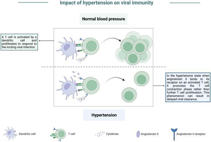
A pathophysiology diagram titled 'Impact of hypertension on viral immunity' comparing cellular immune responses under normal and hypertensive conditions. The top panel, 'Normal blood pressure,' illustrates a dendritic cell presenting antigens to a T cell via receptor-ligand interactions and cytokine release. This stimulation leads to robust T-cell activation and proliferation, resulting in a large cluster of effector T cells essential for viral clearance. The bottom panel, 'Hypertension,' depicts the same initial dendritic cell-T cell interaction but highlights the presence of elevated Angiotensin II. Angiotensin II binds to Angiotensin II receptors expressed on the activated T cell. This signaling inhibits sustained proliferation and instead promotes a T-cell contraction phase, resulting in a significantly reduced population of T cells. The diagram indicates that this hypertensive state leads to an impaired effector response and delayed viral clearance. Key components labeled include dendritic cells, T cells, cytokines, Angiotensin II, and Angiotensin II receptors.
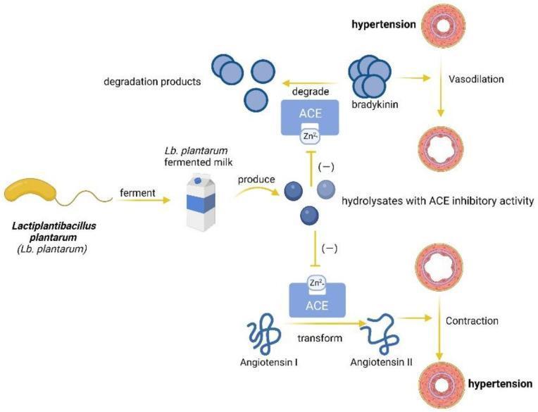
This pathophysiology diagram illustrates the anti-hypertensive mechanism of Lactiplantibacillus plantarum (Lb. plantarum) in functional foods. The process begins with Lb. plantarum fermenting milk, which produces bioactive hydrolysates. These hydrolysates function as Angiotensin-Converting Enzyme (ACE) inhibitors, targeting the zinc-binding (Zn2+) site of the ACE protein. The diagram depicts two physiological pathways influenced by this inhibition: 1) The Bradykinin Pathway: Typically, ACE degrades the vasodilator bradykinin into inactive products. By inhibiting ACE, hydrolysates prevent bradykinin degradation, promoting vasodilation and relieving hypertension. 2) The Renin-Angiotensin Pathway: Normally, ACE transforms Angiotensin I into the potent vasoconstrictor Angiotensin II, leading to vascular contraction and hypertension. The hydrolysates inhibit this transformation, preventing pathological vasoconstriction. The visual contrast emphasizes the transition from constricted, hypertensive blood vessel states to relaxed, dilated states through the modulation of enzymatic activity. This diagram serves as a model for how probiotic-derived peptides can mitigate cardiovascular risk factors by interfering with the biochemical pathways responsible for systemic blood pressure regulation.
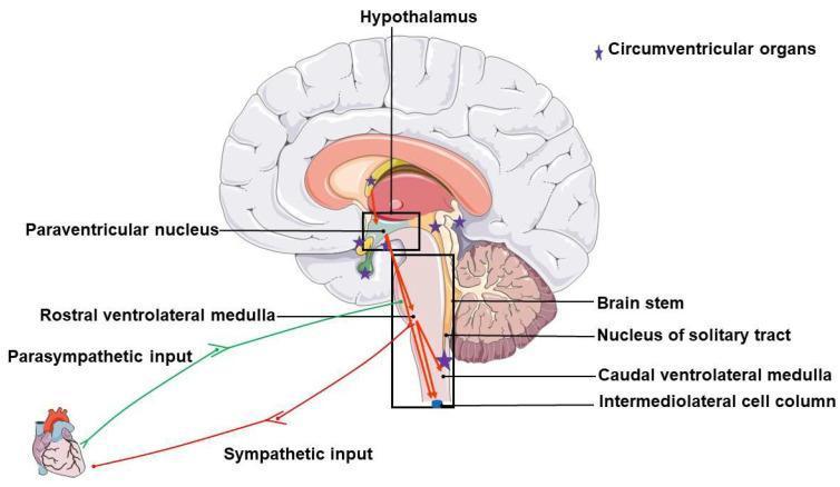
An anatomical and pathophysiology diagram illustrating the brain regions and neural pathways involved in neurogenic hypertension. The illustration features a sagittal view of the human brain with key regulatory centers labeled, including the hypothalamus, paraventricular nucleus (PVN), and components of the brainstem: the rostral ventrolateral medulla (RVLM), nucleus of the solitary tract (NTS), caudal ventrolateral medulla (CVLM), and the intermediolateral cell column (IML). Purple stars denote the locations of circumventricular organs (CVOs) distributed near the hypothalamus and brainstem. The diagram maps the functional connectivity between these regions and the heart. A green line represents the parasympathetic input originating from the RVLM/brainstem area, while a red line represents the sympathetic input originating from the IML, both descending to the cardiac target. This illustration highlights the neuroregulatory network responsible for blood pressure control and the autonomic imbalance (increased sympathetic and decreased parasympathetic tone) characteristic of neurogenic hypertension.
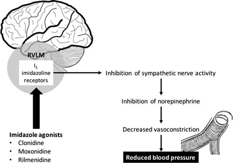
This pathophysiology diagram illustrates the mechanism of action of imidazoline receptor agonists on blood pressure regulation. The visual is divided into a sagittal brain anatomical diagram and a secondary physiological flowchart. On the left, the diagram highlights the 'I1 imidazoline receptors' located within the Rostral Ventrolateral Medulla (RVLM) of the brainstem, situated inferior to the cerebrum and anterior to the cerebellum. Below this, three pharmacological agents—Clonidine, Moxonidine, and Rilmenidine—are listed as imidazole agonists that target these receptors. To the right, a downward flowchart depicts the systemic consequences of receptor activation: stimulation of the RVLM leads to the 'Inhibition of sympathetic nerve activity,' followed by the 'Inhibition of norepinephrine' release. This progression results in 'Decreased vasoconstriction,' visually represented by an illustration of a dilated blood vessel with reduced smooth muscle tension. The final clinical outcome is identified as 'Reduced blood pressure.' The content is designed for intermediate medical education, focusing on neuropharmacology and cardiovascular physiology.
hypertension complications stroke heart failure kidney damage retinopathy
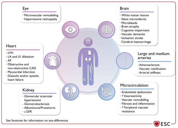
Summary : This figure illustrates the organ-specific and vascular effects of hypertension, detailing pathological changes in the eye, heart, kidney, brain, large and medium arteries, and microcirculation. Each organ system is listed with its associated hypertensive complications. diagram: # Organ Systems and Hypertensive Effects : ## Eye : • Microvascular remodelling • Hypertensive retinopathy ## Heart : • LVH (left ventricular hypertrophy) • LA and LV dilatation (left atrial and left ventricular) • AF (atrial fibrillation) • Obstructive and non-obstructive CAD (coronary artery disease) • Myocardial infarction • Diastolic and/or systolic heart failure ## Kidney : • Glomerular arteriolar hypertension • Glomerulosclerosis • Albuminuria/Proteinuria • ↓ GFR (glomerular filtration rate) ## Brain : • White matter lesions • Silent microinfarcts • Microbleeds • Brain atrophy • Cognitive impairment • Vascular dementia • Ischaemic stroke • Cerebral haemorrhage ## Large and Medium Arteries : • Atherosclerosis • Vascular calcification • Arterial stiffness ## Microcirculation : • Endothelial dysfunction • ↑ Vasoreactivity • Vascular remodelling • Fibrosis and inflammation • ↑ Peripheral vascular resistance # Layout : • Central human figure with icons representing each organ system. • Each organ system is connected to a text box listing its hypertensive complications. • Colour-coded circles and boxes for each organ/vascular system. # Additional Information : • Footnote mentions sex-differences (not detailed in the figure). • ESC (European Society of Cardiology) logo at the bottom right. # Analysis : • The figure demonstrates that hypertension affects multiple organ systems, with distinct pathological changes in each. • Both macrovascular (large arteries) and microvascular (small vessels, microcirculation) complications are highlighted. • The heart, brain, and kidney are shown to be particularly vulnerable, with a wide range of structural and functional consequences. • The diagram visually emphasises the systemic nature of hypertensive disease and its multi-organ impact.
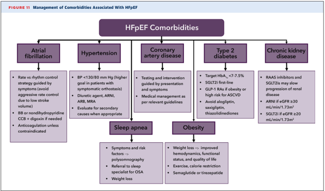
Summary : This figure presents a flowchart for the management of comorbidities associated with Heart Failure with Preserved Ejection Fraction (HFpEF), detailing recommended strategies for atrial fibrillation, hypertension, coronary artery disease, type 2 diabetes, chronic kidney disease, sleep apnea, and obesity. flowchart: # Nodes : • HFpEF Comorbidities (central header, rectangle) • Atrial fibrillation (rectangle) – Rate vs rhythm control strategy guided by symptoms (avoid aggressive rate control due to low stroke volume) – BB or nondihydropyridine CCB + digoxin if needed – Anticoagulation unless contraindicated • Hypertension (rectangle) – BP <130/80 mm Hg (higher goal in patients with symptomatic orthostasis) – Diuretic agent, ARNI, ARB, MRA – Evaluate for secondary causes when appropriate • Coronary artery disease (rectangle) – Testing and intervention guided by presentation and symptoms – Medical management as per relevant guidelines • Type 2 diabetes (rectangle) – Target HbA1c <7-7.5% – SGLT2i first-line – GLP-1 RAs if obesity or high risk for ASCVD – Avoid alogliptin, saxagliptin, thiazolidinediones • Chronic kidney disease (rectangle) – RAAS inhibitors and SGLT2is may slow progression of renal disease – ARNI if eGFR ≥30 mL/min/1.73m² – SGLT2i if eGFR ≥20 mL/min/1.73m² • Sleep apnea (rectangle, below hypertension/coronary artery disease) – Symptoms and risk factors → polysomnography – Referral to sleep specialist for OSA – Weight loss • Obesity (rectangle, below type 2 diabetes/chronic kidney disease) – Weight loss → improved hemodynamics, functional status, and quality of life – Exercise, calorie restriction – Semaglutide or tirzepatide # Connectors : • Central node “HFpEF Comorbidities” branches to five main comorbidity nodes (atrial fibrillation, hypertension, coronary artery disease, type 2 diabetes, chronic kidney disease) in a horizontal row. • Sleep apnea and obesity nodes are placed below and connected to the main row, indicating additional comorbidities. • No explicit decision diamonds; all nodes are rectangles with bulleted management strategies. # Layout : • Horizontal top row: five main comorbidity nodes. • Central header above. • Two additional comorbidity nodes (sleep apnea, obesity) below the main row. • All nodes contain bulleted lists of management recommendations. # Analysis : • The flowchart provides a comprehensive, organized approach to managing common comorbidities in HFpEF, emphasizing tailored strategies for each condition. • There is a focus on evidence-based pharmacologic and non-pharmacologic interventions, with specific drug classes and targets listed for each comorbidity. • The inclusion of sleep apnea and obesity highlights the importance of addressing lifestyle and secondary risk factors in HFpEF management. • The structure allows clinicians to quickly reference recommended actions for each comorbidity, supporting integrated care.
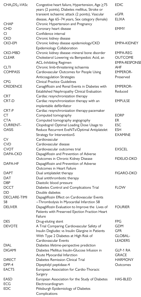
<table><tbody><tr><td>CHA₂DS₂-VASc</td><td>Congestive heart failure, Hypertension, Age ≥75 years (2 points), Diabetes mellitus, Stroke or transient ischaemic attack (2 points), Vascular disease, Age 65–74 years, Sex category (female)</td><td>EDIC</td></tr><tr><td></td><td></td><td>eGFR</td></tr><tr><td></td><td></td><td>ELIXA</td></tr><tr><td>CHAP</td><td>Chronic Hypertension and Pregnancy</td><td></td></tr><tr><td>CHD</td><td>Coronary heart disease</td><td>EMMY</td></tr><tr><td>CI</td><td>Confidence interval</td><td></td></tr><tr><td>CKD</td><td>Chronic kidney disease</td><td></td></tr><tr><td>CKD-EPI</td><td>Chronic kidney disease epidemiology/CKD Epidemiology Collaboration</td><td>EMPA-KIDNEY</td></tr><tr><td>CKD-MBD</td><td>Chronic kidney disease–mineral bone disorder</td><td>EMPA-REG</td></tr><tr><td>CLEAR</td><td>Cholesterol Lowering via Bempedoic Acid, an ACL-Inhibiting Regimen</td><td>OUTCOME</td></tr><tr><td></td><td></td><td>EMPA-RESPONSE</td></tr><tr><td>CLTI</td><td>Chronic limb-threatening ischaemia</td><td>AHF</td></tr><tr><td>COMPASS</td><td>Cardiovascular Outcomes for People Using Anticoagulation Strategies</td><td>EMPEROR-Preserved</td></tr><tr><td>CPG</td><td>Clinical Practice Guidelines</td><td></td></tr><tr><td>CREDENCE</td><td>Canagliflozin and Renal Events in Diabetes with Established Nephropathy Clinical Evaluation</td><td>EMPEROR-Reduced</td></tr><tr><td>CRT</td><td>Cardiac resynchronization therapy</td><td></td></tr><tr><td>CRT-D</td><td>Cardiac resynchronization therapy with an implantable defibrillator</td><td>EMPULSE</td></tr><tr><td>CRT-P</td><td>Cardiac resynchronization therapy-pacemaker</td><td></td></tr><tr><td>CT</td><td>Computed tomography</td><td>EORP</td></tr><tr><td>CTA</td><td>Computed tomography angiography</td><td>ER</td></tr><tr><td>CURRENT-OASIS</td><td>Clopidogrel Optimal Loading Dose Usage to Reduce Recurrent EveNTs/Optimal Antiplatelet Strategy for InterventionS</td><td>ESC</td></tr><tr><td></td><td></td><td>ESH</td></tr><tr><td></td><td></td><td>EXAMINE</td></tr><tr><td>CV</td><td>Cardiovascular</td><td></td></tr><tr><td>CVD</td><td>Cardiovascular disease</td><td></td></tr><tr><td>CVOT</td><td>Cardiovascular outcomes trial</td><td>EXSCEL</td></tr><tr><td>DAPA-CKD</td><td>Dapagliflozin and Prevention of Adverse Outcomes in Chronic Kidney Disease</td><td>FIDELIO-DKD</td></tr><tr><td>DAPA-HF</td><td>Dapagliflozin and Prevention of Adverse Outcomes in Heart Failure</td><td></td></tr><tr><td>DAPT</td><td>Dual antiplatelet therapy</td><td>FIGARO-DKD</td></tr><tr><td>DAT</td><td>Dual antithrombotic therapy</td><td></td></tr><tr><td>DBP</td><td>Diastolic blood pressure</td><td></td></tr><tr><td>DCCT</td><td>Diabetes Control and Complications Trial</td><td>FLOW</td></tr><tr><td>DD</td><td>Double diabetes</td><td></td></tr><tr><td>DECLARE-TIMI 58</td><td>Dapagliflozin Effect on Cardiovascular Events –Thrombolysis In Myocardial Infarction 58</td><td></td></tr><tr><td>DELIVER</td><td>Dapagliflozin Evaluation to Improve the Lives of Patients with Preserved Ejection Fraction Heart Failure</td><td>FOURIER</td></tr><tr><td>DES</td><td>Drug-eluting stent</td><td>FPG</td></tr><tr><td>DEVOTE</td><td>A Trial Comparing Cardiovascular Safety of Insulin Degludec vs Insulin Glargine in Patients With Type 2 Diabetes at High Risk of Cardiovascular Events</td><td>GDM</td></tr><tr><td></td><td></td><td>GFR</td></tr><tr><td></td><td></td><td>GLOBAL-LEADERS</td></tr><tr><td>DIAL</td><td>Diabetes lifetime-perspective prediction</td><td></td></tr><tr><td>DIGAMI</td><td>Diabetes Mellitus Insulin-Glucose Infusion in Acute Myocardial Infarction</td><td>GLP-1 RA</td></tr><tr><td></td><td></td><td>GRACE</td></tr><tr><td>DiRECT</td><td>Diabetes Remission Clinical Trial</td><td>HARMONY</td></tr><tr><td>DPP-4</td><td>Dipeptidyl peptidase-4</td><td>Outcomes</td></tr><tr><td>EACTS</td><td>European Association for Cardio-Thoracic Surgery</td><td></td></tr><tr><td>EASD</td><td>European Association for the Study of Diabetes</td><td>HAS-BLED</td></tr><tr><td>ECG</td><td>Electrocardiogram</td><td></td></tr><tr><td>EDC</td><td>Pittsburgh Epidemiology of Diabetes Complications</td><td></td></tr></tbody></table>
antihypertensive drug classes mechanism of action ACE inhibitor calcium channel blocker

Clinical photography of a generalized morbilliform drug eruption on the skin illustrating an exanthematous eruption due to fosinopril, an ACE inhibitor prescribed for hypertension. The image depicts numerous erythematous macules and papules, some coalescing into diffuse patches on the trunk and proximal limbs, including the exposed upper arm. Lesions are blanching on pressure, non-vesicular, slightly raised, and pruritic in many patients. There is no mucosal involvement evident in this view. Morphology is stereotypical for a morbilliform eruption: symmetric distribution, rapid onset after drug exposure, and a proximal-to-distal spread pattern. The clinical context supports a cutaneous adverse drug reaction triggered by fosinopril, with timing typically 5–14 days after initiation or dose escalation, consistent with a type IV hypersensitivity mechanism. Diagnostic significance lies in recognizing drug-induced exanthem to prevent progression, guide drug withdrawal, and prompt substitution with an alternative antihypertensive (e.g., an ARB). Differential diagnoses include viral exanthem, allergic contact dermatitis, and pityriasis rosea; clinical correlation with medication history is essential. If biopsy is performed, histology would commonly show superficial perivascular lymphocytic infiltrate with eosinophils and mild spongiosis. This image is useful for clinical education, pharmacovigilance, and dermatology training on ACE inhibitor–related rashes and drug eruption management. Prompt action improves patient outcomes.
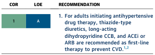
Summary : This figure presents a clinical guideline recommendation for first-line antihypertensive drug therapy in adults, including the class of recommendation (COR) and level of evidence (LOE). table: COR & LOE : • COR: 1 (Class of Recommendation 1, indicating strong recommendation). • LOE: A (Level of Evidence A, indicating high-quality evidence). Recommendation : • For adults initiating antihypertensive drug therapy, the following are recommended as first-line therapy to prevent cardiovascular disease (CVD): – Thiazide-type diuretics – Long-acting dihydropyridine calcium channel blockers (CCB) – Angiotensin-converting enzyme inhibitors (ACEi) or angiotensin receptor blockers (ARB) • Recommendation is supported by references 1 and 2. Analysis : • The figure provides a clear, evidence-based recommendation for initial antihypertensive therapy, prioritizing specific drug classes based on strong evidence for CVD prevention. • The use of COR 1 and LOE A signals that these recommendations are both strongly endorsed and supported by robust clinical data.
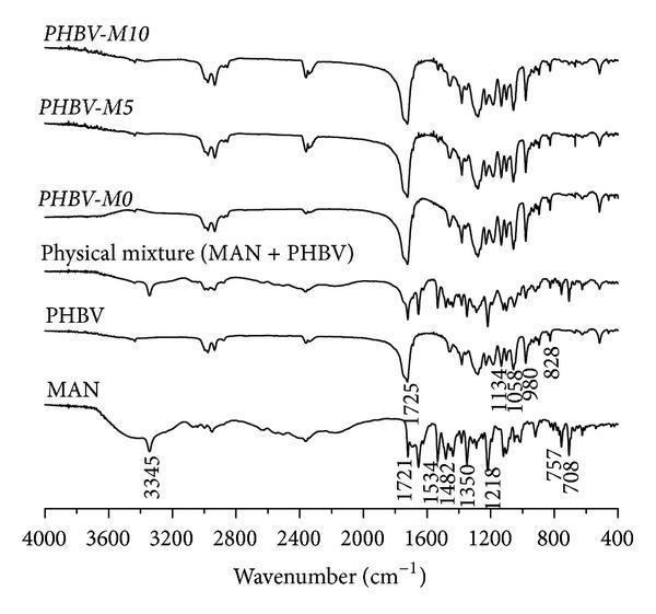
This diagnostic image displays the Fourier-transform infrared (FTIR) spectra used in pharmaceutical research for drug delivery systems. The chart compares the chemical fingerprints of Manidipine (MAN), a third-generation calcium channel blocker used for hypertension, and poly(3-hydroxybutyrate-co-3-hydroxyvalerate) (PHBV), a biodegradable polymer used for microencapsulation. The x-axis represents the wavenumber in cm⁻¹ (ranging from 4000 to 400), while the y-axis shows transmittance. The MAN spectrum reveals characteristic peaks at 3345 cm⁻¹ (N-H stretching) and 1721 cm⁻¹ (C=O stretching). The PHBV and microparticle formulation spectra (PHBV-M0, M5, M10) demonstrate a dominant carbonyl peak at approximately 1725 cm⁻¹. The comparison between the physical mixture (MAN + PHBV) and the microparticle formulations is used to evaluate drug-polymer interactions and the successful incorporation of the antihypertensive agent into the porous polymer matrix, which is clinically relevant for developing sustained-release medications.
Hypertension: A Complete Clinical Overview
1. Definition
| Category | Systolic BP (mmHg) | Diastolic BP (mmHg) |
|---|---|---|
| Normal | < 120 | < 80 |
| Elevated | 120-129 | < 80 |
| Stage 1 Hypertension | 130-139 | 80-89 |
| Stage 2 Hypertension | ≥ 140 | ≥ 90 |
| Hypertensive Crisis | > 180 | > 120 |
2. Types of Hypertension
A. Primary (Essential) Hypertension
- Accounts for 90-95% of all hypertension cases
- No identifiable single underlying cause; multifactorial
- Related to excess weight gain, sedentary lifestyle, genetics, and neurohormonal dysregulation
- Also called idiopathic hypertension
B. Secondary Hypertension
- Accounts for 5-10% of cases; has an identifiable, potentially reversible cause
- Major causes include:
- Renal parenchymal disease (chronic glomerulonephritis, diabetic nephropathy, polycystic kidney disease)
- Renovascular hypertension (renal artery stenosis - Goldblatt hypertension; most common in young women with fibromuscular dysplasia and older adults with atherosclerosis)
- Primary hyperaldosteronism (Conn's syndrome)
- Pheochromocytoma (adrenal medullary tumor)
- Cushing's syndrome (glucocorticoid excess)
- Coarctation of the aorta
- Obstructive sleep apnea
- Thyroid disorders (hypothyroidism, hyperthyroidism)
- Drug-induced (NSAIDs, oral contraceptives, sympathomimetics, cocaine)
C. Special Types
| Type | Description |
|---|---|
| Isolated systolic hypertension | SBP ≥ 140, DBP < 90; common in elderly due to arterial stiffness |
| White-coat hypertension | Elevated in clinic, normal outside; risk of true hypertension over time |
| Masked hypertension | Normal in clinic, elevated outside; higher CV risk than white-coat |
| Resistant hypertension | BP uncontrolled on ≥ 3 agents including a diuretic |
| Malignant/accelerated hypertension | Severe hypertension with acute target organ damage (papilledema, encephalopathy) |
| Hypertensive urgency | SBP > 180 or DBP > 120 without acute organ damage |
| Hypertensive emergency | Severe elevation with acute target organ damage requiring immediate controlled lowering |
3. Etiology
Primary Hypertension - Key Causes
-
Genetic factors: Multiple gene mutations affecting renal sodium handling (monogenic hypertension accounts for < 1%; polygenic contribution is the norm). Familial aggregation is well established.
-
Obesity and excess weight gain: Adiposity may account for 65-75% of primary hypertension risk. Excess adipose tissue increases cardiac output, activates the sympathetic nervous system (especially renal sympathetics), stimulates the renin-angiotensin-aldosterone system (RAAS), and raises leptin - which stimulates hypothalamic sympathetic centers.
-
Increased sympathetic nervous system activity: Sympathetic overactivation raises heart rate, cardiac output, and peripheral vascular resistance. Renal sympathetic nerves impair pressure-natriuresis.
-
RAAS activation: Elevated angiotensin II causes vasoconstriction and promotes renal sodium and water retention via direct tubular effects and aldosterone secretion.
-
Dietary sodium excess: High salt intake leads to volume expansion and pressure natriuresis reset.
-
Reduced kidney function / nephron loss: Reduced renal filtration surface impairs pressure natriuresis; glomerular arteriolar resistance increases.
-
Insulin resistance and hyperinsulinemia: Stimulates sympathetic nervous system, promotes sodium retention.
-
Endothelial dysfunction: Reduced nitric oxide bioavailability increases peripheral vascular resistance.
- Guyton and Hall Textbook of Medical Physiology; National Kidney Foundation Primer on Kidney Diseases, 8e
4. Pathophysiology
A. Renin-Angiotensin-Aldosterone System (RAAS) - Central Mechanism
- Renin (from juxtaglomerular cells of kidney) cleaves angiotensinogen → Angiotensin I
- ACE converts Angiotensin I → Angiotensin II (in lungs and tissues)
- Angiotensin II:
- Causes direct vasoconstriction (rapid pressor effect, within minutes)
- Stimulates aldosterone from adrenal cortex → Na+ and water retention → volume expansion
- Has direct renal tubular effects - increases proximal tubule Na+ reabsorption
- Constricts efferent glomerular arterioles, reducing renal blood flow and driving up tubular reabsorption
- Resets pressure-natriuresis to a higher BP set-point
B. Neurogenic Mechanisms
- Increased sympathetic outflow from rostral ventrolateral medulla (RVLM) → increased heart rate, contractility, and peripheral vasoconstriction
- Renal sympathetic innervation: stimulates renin release and direct tubular sodium reabsorption
- Reduced baroreceptor sensitivity in obesity blunts reflex buffering of BP rises
C. Renal-Fluid Volume Mechanism
- The kidney's pressure-natriuresis curve is reset rightward in hypertension: a higher BP is required to achieve normal sodium excretion
- Volume expansion raises filling pressures → increased CO → over time, autoregulation raises TPR
D. Structural Vascular Remodeling
- Prolonged hypertension causes medial hypertrophy, reduced arterial compliance, increased arterial stiffness
- Increased pulse wave velocity → isolated systolic hypertension in the elderly
- Endothelial dysfunction reduces NO production and vasodilatory reserve
E. Obesity-Related Hypertension (Key Pathway)
- Excess adipose tissue → increased metabolic demand → increased CO
- Leptin hypersecretion → hypothalamic sympathetic activation
- Reduced baroreceptor sensitivity
- Activation of RAAS (adipose tissue is a source of angiotensinogen)
- Obstructive sleep apnea → episodic hypoxia → chemoreceptor activation → sympathetic surges
5. Clinical Manifestations
"The Silent Killer"
Symptoms When Present
| System | Manifestations |
|---|---|
| General | Morning occipital headache (throbbing), dizziness, fatigue |
| Neurological | Headache, blurred vision, tinnitus, epistaxis (nosebleeds) |
| Cardiovascular | Palpitations, exertional dyspnea (from LVH/HF) |
| Renal | Nocturia (early sign of renal involvement) |
| Eye | Visual disturbances (from hypertensive retinopathy) |
Signs on Examination
| Finding | Significance |
|---|---|
| Elevated BP (repeatedly) | Diagnostic |
| Forceful apex beat | Left ventricular hypertrophy (LVH) |
| S4 gallop | Reduced LV compliance from LVH |
| AV nicking, copper/silver wiring | Hypertensive retinopathy on fundoscopy |
| Papilledema | Malignant hypertension / hypertensive encephalopathy |
| Bruits (renal or carotid) | Suggests renovascular or atherosclerotic disease |
| Radio-femoral delay | Coarctation of the aorta (secondary cause) |
Hypertensive Emergency Features
- Headache, confusion, altered consciousness (hypertensive encephalopathy)
- Acute chest pain (aortic dissection, MI)
- Acute dyspnea (pulmonary edema)
- Neurological deficits (hemorrhagic/ischemic stroke)
- Oliguria, hematuria (acute kidney injury)
6. Complications
A. Cardiovascular Complications
- Left ventricular hypertrophy (LVH): Compensatory concentric LVH → diastolic dysfunction → eventually systolic heart failure (HFrEF or HFpEF)
- Coronary artery disease: Accelerated atherosclerosis → angina, myocardial infarction
- Heart failure: Both diastolic and systolic types
- Atrial fibrillation: From LA enlargement and fibrosis
- Aortic dissection: High wall stress from sustained hypertension
B. Cerebrovascular Complications
- Ischemic stroke (most common)
- Hemorrhagic stroke / intracerebral hemorrhage: From rupture of Charcot-Bouchard microaneurysms in small penetrating arteries
- Lacunar infarcts / white matter lesions (silent microinfarcts)
- Vascular dementia
- Hypertensive encephalopathy: Acute syndrome of headache, confusion, seizures, papilledema from breakthrough cerebral vasodilation (BP > 180/120)
C. Renal Complications
- Hypertensive nephrosclerosis: Benign arteriolar nephrosclerosis → progressive GFR decline
- Malignant nephrosclerosis: Rapidly progressive renal failure in malignant hypertension
- Glomerulosclerosis: Glomerular capillary hypertension → focal segmental sclerosis
- Albuminuria/proteinuria: Early marker of renal damage
- Hypertension and CKD form a vicious cycle: CKD worsens hypertension
D. Ophthalmic Complications (Keith-Wagener-Barker Classification)
| Grade | Features |
|---|---|
| I | Mild arteriolar narrowing / increased light reflex (copper wiring) |
| II | AV nipping/nicking + Grade I changes |
| III | Flame-shaped hemorrhages, cotton-wool spots, hard exudates + Grade II |
| IV | Papilledema (indicates malignant hypertension) |
E. Peripheral Vascular Disease
- Accelerated atherosclerosis → peripheral artery disease (PAD), claudication
- Hypertension confers a 2.5-fold (men) to 3.9-fold (women) increased risk of PAD
F. Metabolic
- Associated with metabolic syndrome, insulin resistance, dyslipidemia
7. Non-Pharmacological Management
| Intervention | Expected BP Reduction | Details |
|---|---|---|
| Weight loss | ~1 mmHg per kg lost | Target BMI < 25; most impactful single intervention |
| DASH diet | 8-14 mmHg | Rich in fruits, vegetables, low-fat dairy; low saturated fat; high K+, Mg2+, Ca2+ |
| Sodium restriction | 2-8 mmHg | Reduce to < 2,300 mg/day (ideally < 1,500 mg/day) |
| Physical activity | Aerobic: 3.8 mmHg SBP / 2.6 mmHg DBP | 150 min/week moderate aerobic exercise; resistance and isometric training also effective 3-5×/week |
| Limit alcohol | 2-4 mmHg | ≤ 2 drinks/day men, ≤ 1 drink/day women |
| Smoking cessation | Indirect CV risk reduction | Smoking raises BP acutely and amplifies atherosclerotic risk |
| Stress reduction | Variable | Relaxation techniques, biofeedback, mindfulness |
| Sleep hygiene | Addresses masked hypertension | Treat obstructive sleep apnea (CPAP reduces nocturnal and daytime BP) |
8. Pharmacological Management
First-Line Drug Classes (ACC/AHA Recommendation: COR 1, LOE A)
Drug Class Summary
| Drug Class | Examples | Mechanism | Key Indications / Notes |
|---|---|---|---|
| Thiazide diuretics | Hydrochlorothiazide, chlorthalidone, indapamide | Block Na-Cl cotransporter in DCT → initial volume depletion, then reduced TPR | First-line; especially effective in Black patients; SE: hypokalemia, hyperuricemia, hyperglycemia |
| Loop diuretics | Furosemide, bumetanide, torsemide | Block Na-K-2Cl cotransporter in loop of Henle | Preferred with heart failure or severe CKD (GFR < 30); SE: hypokalemia, ototoxicity |
| K+-sparing diuretics / MRAs | Spironolactone, eplerenone, amiloride, triamterene | Aldosterone antagonism or direct ENaC blockade | Useful in primary aldosteronism, HF, resistant hypertension; SE: hyperkalemia, gynecomastia (spiro) |
| ACE inhibitors (ACEi) | Enalapril, lisinopril, ramipril, captopril, perindopril | Block conversion of Ang I → Ang II; reduce vasoconstriction and aldosterone | First-line; preferred in CKD with proteinuria, diabetes, HF, post-MI; SE: dry cough (10-15%), angioedema (rare); contraindicated in pregnancy |
| Angiotensin receptor blockers (ARBs) | Losartan, valsartan, irbesartan, olmesartan | Block AT1 receptor; same hemodynamic effects as ACEi | Use when ACEi not tolerated (no cough); SE: angioedema (rare); contraindicated in pregnancy |
| Dihydropyridine CCBs | Amlodipine, nifedipine (ER), felodipine | Block L-type Ca2+ channels in vascular smooth muscle → vasodilation | First-line; effective in elderly, Black patients, angina; SE: peripheral edema, flushing, reflex tachycardia |
| Non-dihydropyridine CCBs | Verapamil, diltiazem | Block cardiac and vascular L-type Ca2+ channels → reduced HR, CO, and vasodilation | Useful in angina, supraventricular tachyarrhythmias; avoid with beta-blockers (AV block risk) |
| Beta-blockers (β-blockers) | Metoprolol (ER), atenolol, bisoprolol, nebivolol, carvedilol, labetalol | Reduce HR and CO; decrease renin secretion; nebivolol also releases NO | No longer first-line for uncomplicated hypertension; preferred post-MI, angina, HF, tachyarrhythmias; SE: fatigue, bradycardia, bronchospasm (avoid in asthma) |
| Alpha-1 blockers | Prazosin, doxazosin, terazosin | Block α1-adrenoceptors → vasodilation | Used with BPH; not first-line monotherapy (fluid retention); SE: first-dose orthostatic hypotension |
| Central sympatholytics | Clonidine, methyldopa, moxonidine | Stimulate α2 or imidazoline I1 receptors in RVLM → reduce sympathetic outflow | Methyldopa: safe in pregnancy; clonidine: SE = rebound hypertension if stopped abruptly |
| Direct vasodilators | Hydralazine, minoxidil | Directly relax arteriolar smooth muscle | Reserved for resistant hypertension; hydralazine used in pregnancy; SE: reflex tachycardia, fluid retention |
| Nitroprusside | Sodium nitroprusside | Releases NO → arterial and venous dilation | IV only; hypertensive emergencies; SE: cyanide toxicity with prolonged use |
Treatment Principles
- Goal BP: < 130/80 mmHg for most adults (ACC/AHA 2017). < 140/90 mmHg by JNC-8 / older guidelines. In elderly, avoid DBP < 70 mmHg in those with CAD.
- Start low, go slow in the elderly; usual single-agent response: SBP ↓ 12-15 mmHg, DBP ↓ 8-10 mmHg.
- Combination therapy: Preferred when BP is > 20/10 mmHg above goal; shortens time to control and reduces side effects vs. high-dose monotherapy.
- Once-daily long-acting agents improve adherence.
- Compelling indications guide drug choice:
| Comorbidity | Preferred Drug Class |
|---|---|
| CKD + proteinuria | ACEi or ARB (mandatory) |
| Diabetes | ACEi or ARB (first-line) |
| Heart failure (reduced EF) | ACEi/ARB + beta-blocker + MRA |
| Post-MI | Beta-blocker + ACEi |
| Angina pectoris | Beta-blocker or CCB |
| Atrial fibrillation (rate control) | Beta-blocker or non-DHP CCB |
| Primary aldosteronism | MRA (spironolactone) |
| Pregnancy | Methyldopa, nifedipine, labetalol |
| Black patients | CCB + thiazide diuretic (ACEi less effective as monotherapy) |
| BPH + hypertension | Alpha-1 blocker |
| Resistant hypertension | Add MRA (spironolactone) as 4th agent |
Hypertensive Emergency Management
-
Admit to ICU; IV antihypertensives
-
Target: reduce MAP by no more than 25% in the first hour, then gradually to 160/100 over 2-6 hours
-
Avoid excessive rapid lowering (risk of ischemic stroke, MI, renal failure from reduced perfusion)
-
IV agents used: labetalol, nicardipine, sodium nitroprusside, hydralazine, clevidipine, esmolol
-
Goodman & Gilman's Pharmacological Basis of Therapeutics, 14e; National Kidney Foundation Primer on Kidney Diseases, 8e
9. Risk Factors
Non-Modifiable Risk Factors
| Factor | Details |
|---|---|
| Age | BP rises progressively; isolated systolic hypertension predominates after age 60 |
| Sex | Men develop hypertension earlier; post-menopausal women catch up; women at higher CV risk per given BP level |
| Race / Ethnicity | Black individuals have higher prevalence, earlier onset, more severe hypertension, and greater target organ damage; APOL1 genetic variants contribute to higher rates of nephrosclerosis |
| Family history / Genetics | Strong hereditary component; polygenic; monogenic forms account for < 1% |
Modifiable Risk Factors
| Factor | Mechanism |
|---|---|
| Obesity / Overweight | Adiposity accounts for 65-75% of primary hypertension risk; activates SNS and RAAS |
| Sedentary lifestyle | Reduces vascular compliance and cardiorespiratory fitness |
| Excess dietary sodium | Volume expansion, resetting of pressure natriuresis |
| Low dietary potassium | Reduces natriuresis; increases vascular tone |
| Excess alcohol | Raises BP acutely and chronically; activates SNS |
| Tobacco/Smoking | Acute pressor effect; accelerates atherosclerosis |
| Dyslipidemia | Promotes endothelial dysfunction and atherosclerosis |
| Diabetes mellitus | Insulin resistance activates SNS; combined risk greatly magnifies CV events |
| Obstructive sleep apnea | Intermittent hypoxia → chemoreceptor activation → sympathetic surges → sustained hypertension |
| Chronic stress / Psychological factors | Sustained sympathetic activation |
| NSAIDs / Certain drugs | Blunt prostaglandin-mediated vasodilation; reduce antihypertensive drug efficacy |
| Chronic kidney disease | Impaired sodium excretion and RAAS activation form a bidirectional relationship with hypertension |
Summary Table
| Aspect | Key Points |
|---|---|
| Definition | SBP ≥ 130/DBP ≥ 80 (ACC/AHA); ≥ 140/90 (JNC-7/WHO) |
| Most common type | Primary (essential) - 90-95% |
| Core pathophysiology | RAAS activation, sympathetic overactivity, impaired renal natriuresis |
| Key symptom | Usually none ("silent killer"); headache when severe |
| Major complications | Stroke, MI, heart failure, CKD, retinopathy |
| First-line lifestyle | DASH diet, weight loss, sodium restriction, exercise |
| First-line drugs | Thiazide diuretics, ACEi/ARB, dihydropyridine CCBs |
| Hypertensive emergency | Lower MAP ≤ 25% in first hour; IV agents |