
Okay what you have given is great Yes. Given what I know about your radiology seminar style and your goal, I would not make this a routine “textbook” presentation. I would make it a teaching seminar where even first-year residents and clinicians can understand the pathology simply by looking at the imaging. Your topic Diffuse Sclerotic Diseases can easily become confusing because there are many diseases with overlapping appearances. The secret is pattern recognition. Ultimate Seminar Strategy (Radiology PG Level) Presentation duration: 35–45 minutes Goal: By the end of the seminar, anyone in the room should be able to look at a diffusely sclerotic skeleton and narrow it down to 3–4 likely diagnoses within seconds. That should be the theme throughout. ⸻ Overall Flow Instead of Definition Classification Disease 1 Disease 2 Disease 3 (which everyone does) We’ll do PART 1 Introduction PART 2 Approach to diffuse skeletal sclerosis PART 3 Disease-wise discussion PART 4 Pattern recognition tables PART 5 Rapid diagnosis algorithm PART 6 Important viva points PART 7 Recent advances ⸻ Approximate Slide Distribution Section Slides Introduction 3 Bone physiology 3 Causes & Classification 3 Radiological approach 4 Diseases 25–30 Comparison tables 5 Diagnostic algorithm 2 Key take-home points 2 Total 45–50 slides This is ideal. ⸻ Every Disease Should Follow SAME Template Never randomly explain diseases. Each disease should have identical headings. Example ⸻ Osteopetrosis Definition (one slide) Image Small box with definition. ⸻ Etiology & Genetics Simple flowchart Mutation ↓ Osteoclast dysfunction ↓ Bone resorption ↓ ↓ Dense brittle bone ⸻ Pathophysiology Instead of paragraph Use animation Normal bone ↓ Osteoclast resorption ↓ Marrow cavity ↓ Normal bone VS Osteopetrosis ↓ Failed osteoclast ↓ Dense bone ↓ Poor marrow ↓ Fracture ⸻ Clinical Features Table Age Symptoms Complications ⸻ Radiology This is MOST important. One slide for each modality. Radiograph CT MRI Bone scan (if useful) ⸻ Classical Imaging Findings Image occupies 70% Beside image Bullet points Example ✔ Bone within bone ✔ Sandwich vertebra ✔ Erlenmeyer flask deformity ✔ Diffuse sclerosis ✔ Narrow medullary cavity ⸻ Why these findings occur? This is where examiners get impressed. Example Sandwich vertebra ↓ Failure of normal endplate remodeling ↓ Dense endplates ↓ Central lucency Instead of memorizing. Understand. ⸻ Differential Diagnosis Table Disease How to differentiate ⸻ Pearls One slide “Never miss” ⸻ Same format for ALL diseases ⸻ Images This is where your seminar becomes outstanding. For every disease Include Radiograph ↓ CT ↓ MRI ↓ Nuclear scan (if applicable) ↓ Pathology correlation ↓ Clinical photo (only if important) ⸻ Beside Every Image Never simply put image. Example Image occupies left. Right side Arrow Circle Short annotation Red arrow → Bone within bone Yellow arrow → Narrow marrow cavity Blue arrow → Dense pelvis Audience immediately understands. ⸻ Before Disease Discussion Need one VERY IMPORTANT SECTION Radiological Approach This is what professors love. Patient has diffuse sclerosis. Now what? Step 1 Age? Child Adult ⸻ Step 2 Localized Diffuse ⸻ Step 3 Symmetric Asymmetric ⸻ Step 4 Axial Appendicular ⸻ Step 5 Marrow involved? Yes No ⸻ Step 6 Associated findings Fracture Anemia Renal disease Metastasis ⸻ Make algorithm. This becomes the backbone of seminar. ⸻ Diseases to Cover I suggest this order ⸻ Genetic Osteopetrosis Pyknodysostosis Melorheostosis (mention focal vs diffuse involvement) Osteopoikilosis (brief differentiation) ⸻ Metabolic Renal osteodystrophy Fluorosis Hypervitaminosis D Hyperparathyroidism after treatment Paget disease ⸻ Hematological Myelofibrosis Sickle cell (late sclerosis) Mastocytosis ⸻ Neoplastic Osteoblastic metastases Prostate Breast Carcinoid Lymphoma ⸻ Drugs Bisphosphonates Vitamin A Fluoride therapy ⸻ Miscellaneous Hepatitis C osteosclerosis Camurati–Engelmann disease Intramedullary osteosclerosis ⸻ Every Disease Should Have Definition Etiology Genetics (if applicable) Pathophysiology Clinical features Radiograph CT MRI Bone scan Complications Differential diagnosis Treatment (one slide) Key imaging pearls Exam points ⸻ Add Histopathology Correlation Professors LOVE this. Example Normal trabecula ↓ Histology ↓ Radiograph Explain correlation. ⸻ Comparison Tables These will make your seminar unforgettable. Example Disease Marrow Cortex Vertebra Skull Bone scan Another Diffuse sclerosis in child vs Adult ⸻ Another Benign vs Malignant ⸻ Another Metabolic vs Metastatic ⸻ Include Diagnostic Algorithm Example Diffuse sclerosis ↓ Age <20 ↓ Bone within bone? ↓ Yes ↓ Osteopetrosis No ↓ Short stature? ↓ Pyknodysostosis ⸻ Adult ↓ Known malignancy? ↓ Yes ↓ Metastasis No ↓ Renal disease? ↓ Renal osteodystrophy etc. ⸻ High Yield Radiology Signs Every sign should have image. Bone within bone Rugger jersey spine Sandwich vertebra Ivory vertebra Erlenmeyer flask Cotton wool skull Picture frame vertebra Superscan Bone island Flask deformity These should each have an image with one-line explanation. ⸻ Recent Advances (2–3 slides) Don’t spend too much time, but include: * Role of whole-body MRI in marrow infiltration and occult neoplastic disease. * FDG PET/CT and bone scintigraphy/SPECT-CT in differentiating diffuse metastatic osteoblastic disease from metabolic causes. * Dual-energy CT and quantitative CT techniques for assessing bone density in selected conditions. * Emerging use of AI-assisted pattern recognition for diffuse skeletal abnormalities (brief mention only). ⸻ End With Take Home Message Instead of memorizing diseases Remember Age ↓ Distribution ↓ Pattern ↓ Associated findings ↓ Diagnosis ⸻ Presentation Style For every disease, use this sequence: 1. Clinical vignette (1–2 lines): “A 12-year-old child with recurrent fractures and anemia…” 2. Show the radiograph first and ask the audience, “What do you notice?” 3. Reveal the annotated findings. 4. Explain why those findings occur (pathophysiology). 5. Summarize with a “Radiology Pearl” and key differentials before moving to the next disease. This keeps the audience engaged instead of passively reading slides. My proposed workflow Rather than creating all 50 slides at once, let’s build this as a high-quality, textbook-level seminar. We’ll proceed in sessions: 1. Session 1: Introduction, bone remodeling, definition, classification, and the radiologist’s approach to diffuse sclerosis. 2. Session 2: Genetic sclerosing dysplasias (Osteopetrosis, Pyknodysostosis, etc.). 3. Session 3: Metabolic and endocrine causes. 4. Session 4: Hematologic and neoplastic causes. 5. Session 5: Infectious, toxic, and miscellaneous causes. 6. Session 6: Differential diagnosis tables, diagnostic algorithms, viva questions, and the final polished PPT structure. This approach will let us go into real depth for every disease while ensuring the seminar is coherent, visually rich, and at the level expected of a radiology postgraduate presentation. Can you check this pattern and give this way as you given above as professional manner in depth refer from related articles, journls, textbooks of radiology
"diffuse osteosclerosis" OR "skeletal sclerosis" radiology imaging
bone within bone osteopetrosis radiograph
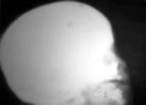
This lateral skull radiograph demonstrates a marked, generalized increase in bone density, characterized by a homogenous radio-opacity throughout the cranial vault and facial skeleton. This 'bone-within-bone' or 'chalk-like' appearance is a classic hallmark of osteopetrosis (Albers-Schönberg disease). The diffuse sclerosis obscures normal anatomical landmarks, including the distinction between the inner and outer tables of the skull, the diploic space, and the paranasal sinuses. The maxilla and mandible also exhibit significant thickening and hyperdensity, which contributes to the poor visualization of developing dental structures and an apparent limitation in alveolar ridge development. Clinically, such findings in a pediatric patient are significant for impaired bone remodeling, which can lead to complications such as cranial nerve compression (resulting in vision or hearing loss) and a predisposition to osteomyelitis due to reduced vascularity.
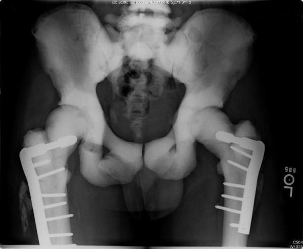
Anteroposterior (AP) radiograph of the pelvis and bilateral proximal femurs demonstrating characteristic features of osteopetrosis, including generalized increased bone density (sclerosis) and the 'bone-within-a-bone' (endostoma) appearance. The radiograph shows bilateral subtrochanteric femur fractures managed with orthopedic internal fixation. On both sides, lateral locking plates are secured with multiple cortical screws; the hardware remains intact without signs of loosening or failure. On the right side (anatomical right), the subtrochanteric fracture line appears attenuated and poorly defined, indicating chronic healing progression. Adjacent to the right proximal femur, there is evidence of mature heterotopic ossification. The left-sided fracture site shows a higher degree of healing with no clearly visible residual fracture lines. The iliac wings and sacrum exhibit significant opacification, and the visualized lumbar vertebrae demonstrate 'sandwich vertebrae' end-plate thickening typical of marble bone disease.
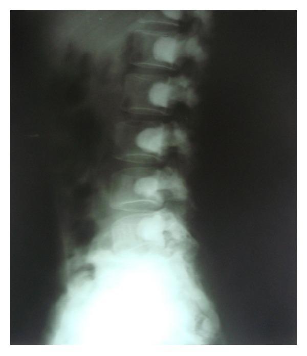
This lateral lumbar spine radiograph demonstrates a significant, generalized increase in bone density (osteosclerosis) across all visible vertebral levels. A hallmark radiographic feature known as the 'bone-within-bone appearance' (endobone) is present, where a dense, well-defined silhouette of a smaller vertebral body appears nested within the outer cortical margins of each vertebra. This phenomenon is caused by systemic failure of normal bone remodeling, resulting in the persistence of primary spongiosa. Despite the marked sclerosis and loss of normal trabecular detail, the overall morphology, height, and alignment of the vertebral bodies, as well as the spinous processes, appear relatively preserved. Such findings are characteristic of sclerosing bone dysplasias, most notably osteopetrosis (Albers-Schönberg disease). This diagnostic image serves as an educational example of how systemic mineralization disorders manifest in the axial skeleton, emphasizing the distinction between increased radiographic density and actual structural integrity.
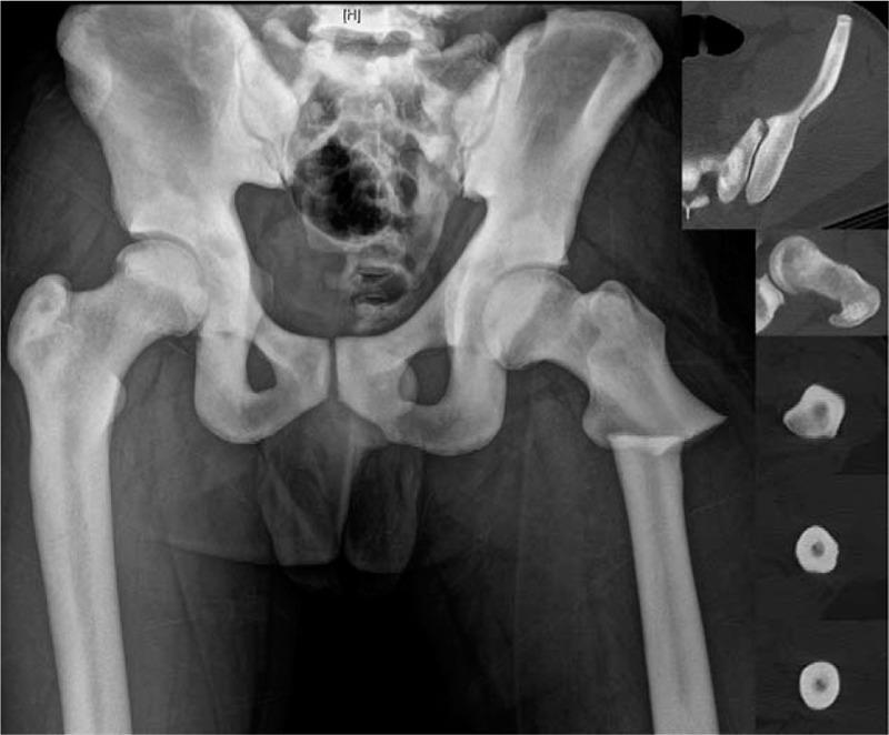
This composite diagnostic image features an anteroposterior (AP) radiograph of the pelvis and bilateral femurs, supplemented by axial computed tomography (CT) insets. The primary finding is diffuse, generalized osteosclerosis characteristic of autosomal dominant type II osteopetrosis (Albers-Schönberg disease). The bones exhibit markedly increased radiodensity, resulting in a 'chalk-like' appearance and loss of normal corticomedullary differentiation. A distinctive 'bone-within-bone' (endobone) appearance is visible in the pelvic bones and vertebral elements. The medullary canals of the femurs are severely narrowed or obliterated by dense sclerotic bone. A displaced subtrochanteric fracture is evident in the left femur. The axial CT insets at the level of the femoral shaft confirm the increased cortical thickness and high-density central marrow space, illustrating the extreme bone hardness associated with this metabolic condition. These images provide critical educational context regarding the challenges of intramedullary nail placement in patients with osteopetrosis due to the lack of a patent medullary canal.
"osteopetrosis" imaging radiology
rugger jersey spine renal osteodystrophy fluorosis vertebra
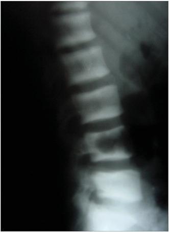
A lateral view X-ray of the lumbar spine illustrating the 'rugger jersey spine' sign. The radiograph reveals a characteristic pattern of alternating radiopacity within the vertebral bodies. Specifically, there are prominent horizontal bands of increased bone density (osteosclerosis) located at the superior and inferior endplates of each vertebra. These sclerotic bands contrast with the relatively radiolucent (normal to osteopenic) central portion of the vertebral bodies, creating a striped appearance reminiscent of a rugby jersey. This finding is highly indicative of renal osteodystrophy secondary to chronic kidney disease or hyperparathyroidism. The image demonstrates the structural skeletal manifestations of systemic metabolic bone disease, focusing on the lumbar region's vertebral architecture and density distribution.
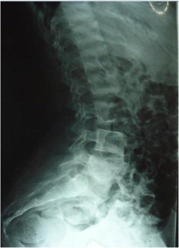
This lateral thoracolumbar X-ray displays a classic 'rugger jersey spine' appearance, a characteristic radiographic sign of osteosclerosis associated with hyperparathyroidism or renal osteodystrophy. The vertebral bodies demonstrate prominent, horizontal bands of increased radiodensity (sclerosis) at the superior and inferior endplates, contrasting with relatively radiolucent (osteopenic) central vertebral bodies. This alternating horizontal pattern mimics the striped design of a rugby jersey. The anatomical region covered includes the lower thoracic spine, lumbar spine, and sacral region. While intervertebral disc spaces appear preserved in height, the pelvic bones visible at the base of the image show heterogeneous bone density. The finding is a critical clinical marker for metabolic bone disease, illustrating the pathological bone remodeling and increased osteoid production that occurs in response to elevated parathyroid hormone levels.
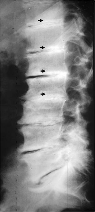
This lateral plain X-ray of the spine demonstrates diffuse osteosclerosis characteristic of renal osteodystrophy. The most prominent finding is the presence of dense, sclerotic horizontal bands located at the superior and inferior sub-endplate regions of multiple contiguous vertebral bodies. These radiopaque bands contrast with the relatively radiolucent (normal density) central portion of the vertebral bodies, creating a striped appearance. This classic radiographic sign is known as 'rugger-jersey spine.' This pattern is a key indicator of secondary hyperparathyroidism, often seen in patients with chronic renal failure or end-stage renal disease undergoing long-term hemodialysis. The image serves as a diagnostic hallmark for systemic metabolic bone disease where bone resorption and disordered bone formation occur simultaneously.
Erlenmeyer flask deformity femur distal metaphysis
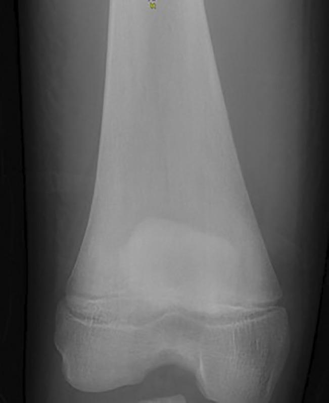
This diagnostic radiograph of the distal femur and knee joint demonstrates a classic 'Erlenmeyer flask deformity.' The image shows an abnormal widening and expansion of the distal femoral metaphysis, where the cortical bone fails to undergo normal modeling, resulting in a bulbous, flared configuration instead of the typical tapered diaphysis-metaphysis transition. Key features include thinning of the cortical margins in the expanded region and heterogeneous bone density with an altered trabecular pattern within the metaphysis. The epiphysis and the knee joint space appear relatively preserved in morphology. This radiographic sign is highly characteristic of marrow infiltrative or metabolic bone disorders, most notably Gaucher disease, where the accumulation of glucocerebroside-laden histiocytes (Gaucher cells) in the bone marrow interferes with normal osteoclast-mediated remodeling. The image is a critical educational tool for identifying skeletal manifestations of lysosomal storage diseases and skeletal dysplasias.
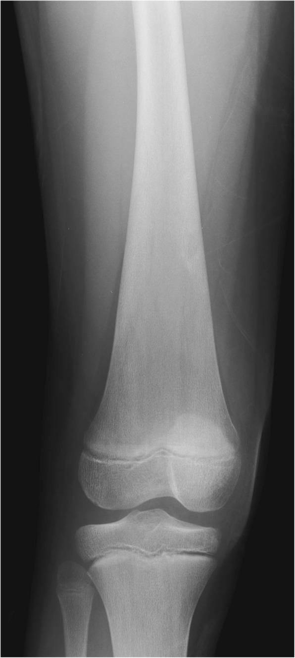
This diagnostic image is a frontal (anteroposterior) radiograph of the right knee of a 12-year-old male. The primary clinical finding is a prominent 'Erlenmeyer flask deformity' of the distal femur. This morphology is characterized by significant widening and expansion of the distal femoral diaphysis and metaphysis, where the bone fails to undergo normal cortical remodeling and tapering. The expanded metaphysis appears disproportionately large relative to the femoral epiphysis. The radiodensity of the distal femur is comparable to the proximal tibia, though the fibula shows slightly higher radiodensity. This characteristic bony deformity is a classic skeletal manifestation of Gaucher disease, resulting from the accumulation of Gaucher cells within the marrow cavity causing medullary expansion. The image serves as a key educational example of metabolic bone disease and storage disorders affecting the appendicular skeleton in pediatric patients.
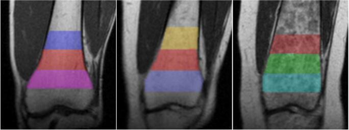
This diagnostic image consists of three coronal MRI slices of the distal femur, illustrating the spectrum of Erlenmeyer flask deformity (EFD) severity. The panels show varying degrees of metaphyseal remodeling: 'No EFD' (left), 'Mild EFD' (center), and 'Severe EFD' (right). Each image features three superimposed, color-coded Regions of Interest (ROIs) used for volumetric and morphometric analysis. The ROIs are segmented based on distance from the physis: 0–2 cm, 2–4 cm, and 4–6 cm proximally. Visually, the progress from left to right demonstrates a loss of the normal concave 'waist' transition from the metaphysis to the diaphysis. In the 'Severe EFD' case, there is pronounced metaphyseal flaring and cortical thinning, resulting in a characteristic bulbous shape associated with skeletal dysplasias or metabolic bone diseases such as Gaucher disease or osteopetrosis. The image serves as a comparison chart for diagnostic staging and quantitative assessment of bone morphology using medical imaging software.
"osteoblastic metastasis" imaging diagnosis prostate breast
ivory vertebra osteoblastic metastasis prostate lymphoma spine
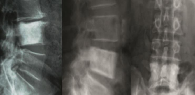
This diagnostic imaging composite displays anteroposterior (AP) and lateral x-ray radiographs of the lumbar spine. The primary finding is a classic 'ivory vertebra' involving the L4 level. In both views, the L4 vertebral body exhibits diffuse, uniform sclerosis, appearing significantly more radiopaque (whiter) than the adjacent levels. The lateral views (left and center panels) demonstrate the preservation of the vertebral height and cortical margins despite the intense internal hyperdensity. The AP view (right panel) shows the midline spinous processes, pedicles, and transverse processes, with L4 standing out as a densely mineralized rectangular structure. Clinically, this radiographic sign is characteristic of osteoblastic metastases, most commonly from prostate cancer, though it can also be seen in Paget's disease or lymphoma. The image serves as an educational example of sclerotic bone lesions in the axial skeleton.
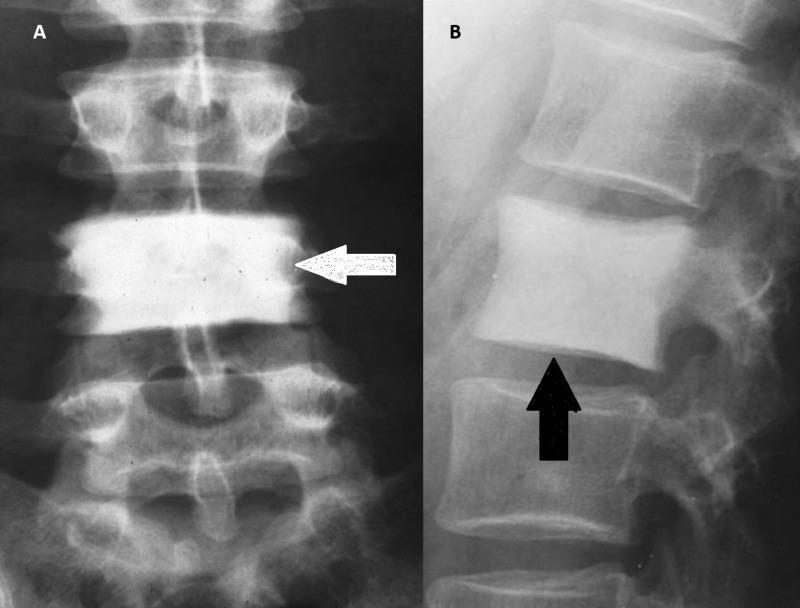
This diagnostic image consists of two radiographic views of the human spine demonstrating the 'ivory vertebra' sign. Image A is an anteroposterior (AP) X-ray showing a single vertebral body (indicated by a white arrow) with a diffuse, homogeneous increase in radiopacity, appearing significantly denser and whiter than the adjacent vertebrae above and below. Image B is a lateral X-ray of the lumbar spine where a black arrow points to a vertebral body exhibiting similar dense, white sclerosis throughout its structure. The affected vertebra in both views maintains its basic height and shape, though it shows a slight squared-off appearance of the endplates. This radiological finding is an important diagnostic indicator for several conditions, including osteoblastic metastases (commonly from prostate or breast cancer), Paget's disease of the bone, Hodgkin lymphoma, and occasionally chronic osteomyelitis. The preservation of the vertebral size and the uniform nature of the density are key educational features for differentiating ivory vertebra from compression fractures or other sclerotic bone lesions.
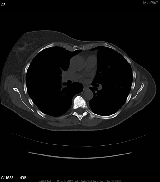
**Imaging Modality:** Axial computed tomography (CT) scan. **Anatomical Region:** Mid-thoracic spine at the level of the mediastinum and pulmonary hilum. **Observed Pathology:** The image demonstrates a classic "ivory vertebra" sign. A single thoracic vertebral body exhibits diffuse, homogeneous, and intense sclerosis (increased bone density) throughout the entire marrow space. **Characteristic Visual Features:** * **Hyperdensity:** Markedly increased radiopacity of the vertebral body compared to adjacent skeletal structures and posterior elements. * **Morphology:** The vertebral body maintains its normal height, shape, and cortical margins without evidence of expansion, fragmentation, or cortical destruction. * **Distribution:** Isolated involvement of the vertebral body, with the posterior elements (laminae and spinous process) appearing relatively spared or less densely sclerotic. * **Surrounding Structures:** Visible ribs, sternum, and soft tissues appear within normal limits for this window setting. **Clinical Significance:** This radiologic finding is a key diagnostic cue for osteoblastic metastases (e.g., prostate or breast cancer), Paget’s disease (though usually associated with osseous expansion), or lymphoma (specifically Hodgkin lymphoma). It is differentiated from other sclerotic vertebral patterns by its uniform, high-density "ivory" appearance.
Paget disease skull cotton wool calvarium radiograph
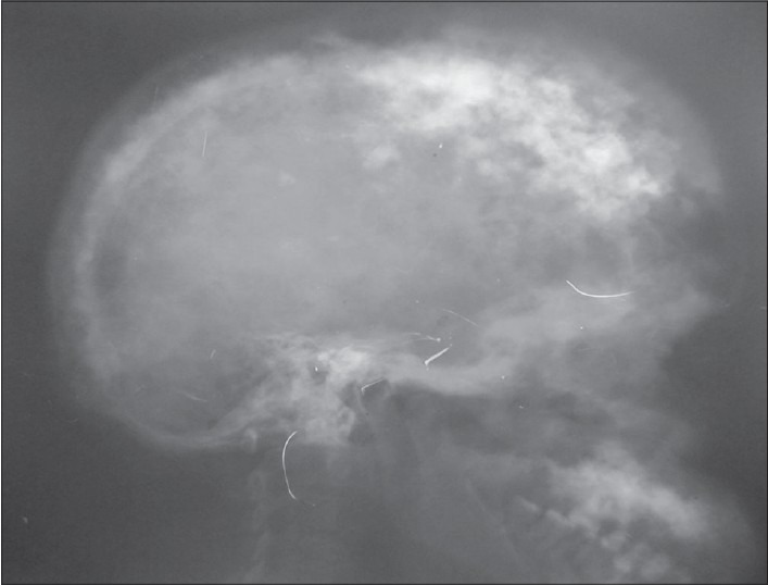
**Imaging Modality:** Lateral plain film radiograph (X-ray). **Anatomical Region:** Skull (cranium). **Observed Pathology:** Advanced Paget's disease of bone (osteitis deformans). **Characteristic Visual Features:** The image demonstrates a classic "cotton wool appearance" characterized by multifocal, ill-defined sclerotic patches interspersed with radiolucent areas throughout the calvarium. There is marked thickening of the diploic space and expansion of the outer table of the skull. The normal cortical margins of the inner and outer tables are blurred due to disorganized bone remodeling. **Key Diagnostic Features:** - **Cotton wool spots:** Focal areas of opacification representing the sclerotic phase of Paget's disease. - **Diploic thickening:** Significant widening of the cranial vault. - **Bone expansion:** Generalized enlargement of the skull landmarks compared to normal anatomical proportions. **Clinical Significance:** These radiographic findings are pathognomonic for the mixed lytic and sclerotic stages of Paget’s disease involving the axial skeleton, specifically the calvarium.
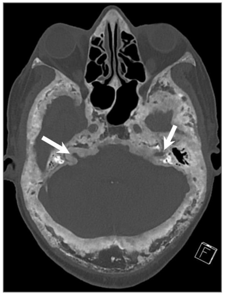
This axial CT image of the skull illustrates advanced Paget’s disease of the bone. The calvarium and skull base exhibit a characteristic 'cotton wool' appearance, defined by heterogeneous, mixed lytic and sclerotic areas. Notable pathological features include diffuse bone expansion, prominent cortical thickening, and coarsened, thickened trabeculae. White arrows specifically highlight the narrowing (stenosis) of the bilateral internal auditory canals (IACs) resulting from significant bony overgrowth and cortical expansion. The image demonstrates the progressive osseous deformity and remodeling typical of the mixed and sclerotic phases of Paget’s disease, which can lead to neurological complications such as cranial nerve entrapment or hearing loss due to foraminal and canal narrowing.
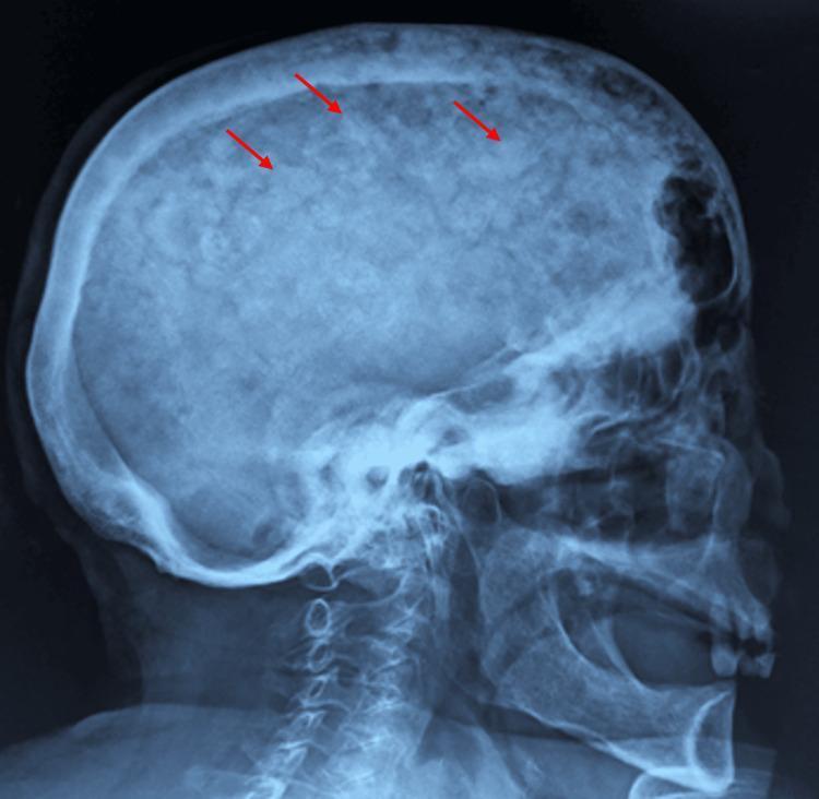
This lateral skull X-ray demonstrates a classic 'cotton wool' appearance of the cranium, a hallmark radiographic finding of Paget's disease of bone (osteitis deformans). The image shows diffuse thickening of the calvarium with a characteristic mottled texture. Multiple, ill-defined sclerotic patches are visible throughout the skull (highlighted by red arrows), representing areas of disordered, high-density bone formation interspersed with radiolucent areas of osteolysis. There is a noticeable widening of the diploic space and a loss of distinction between the inner and outer tables of the skull. The irregular mineralization replaces the normally smooth, homogeneous trabecular pattern of the cranial vault. These visual features are clinically significant as they reflect the late sclerotic phase of Paget's disease, where excessive and disorganized bone remodeling leads to structural deformities and potential neurological complications due to foraminal narrowing or cranial vault thickening.
fluorosis skeletal sclerosis spine chalk bones radiograph
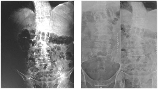
This diagnostic image set consists of conventional anteroposterior (AP) X-ray radiographs of the lumbar spine, pelvis, and abdomen. The left image demonstrates a marked increase in bone density (osteosclerosis) throughout the visualized axial skeleton, including the lumbar vertebrae and pelvic bones, resulting in a 'chalk-like' appearance where the trabecular patterns are obscured. The right set provides comparative views of the lumbar spine and pelvic brim. The prominent radiographic finding of diffuse sclerosis is a hallmark characteristic of skeletal fluorosis, particularly in the context of chronic high fluoride exposure. Visible anatomical structures include the lumbar vertebral bodies, transverse processes, sacroiliac joints, and the iliac crests. These images illustrate the skeletal manifestations of fluoride toxicity, which can lead to bone pain and increased fracture risk despite the apparent increase in density. The clinical relevance focuses on environmental health and metabolic bone diseases, emphasizing the use of radiography in identifying chronic fluoride intoxication.
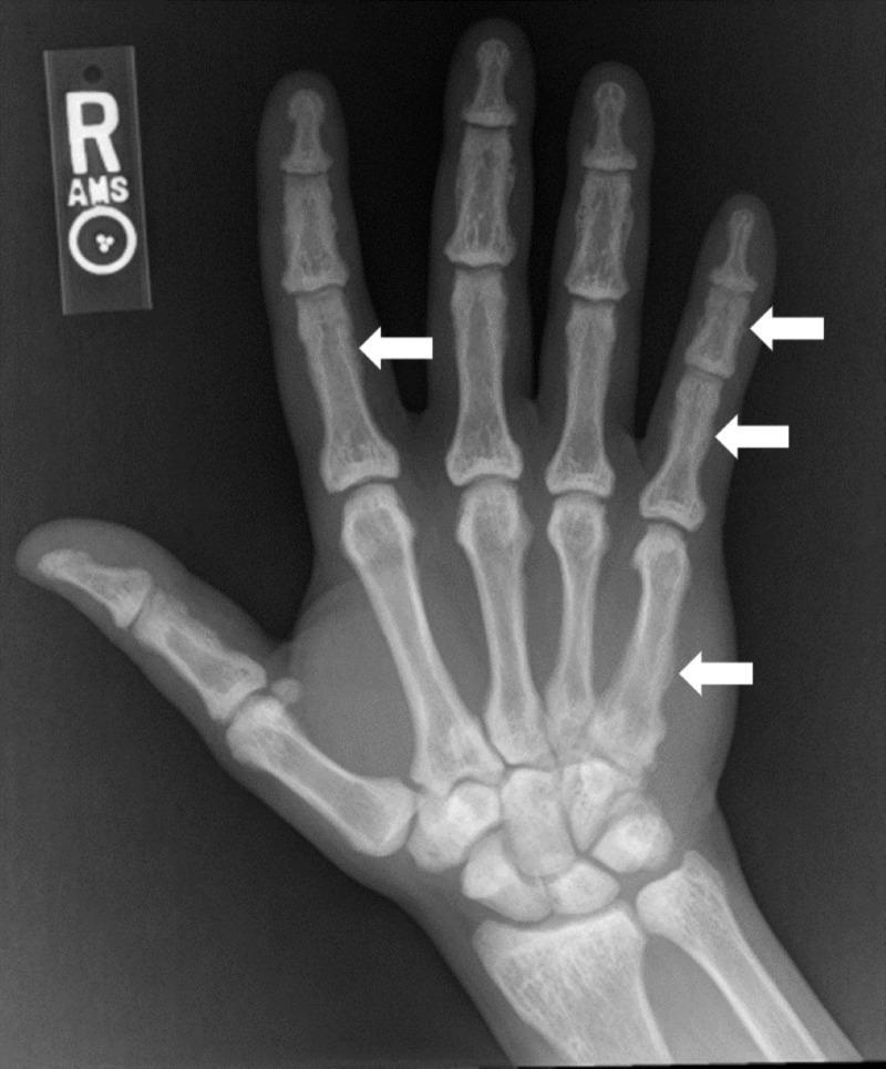
This anteroposterior (AP) radiograph of the right hand and wrist demonstrates diffuse skeletal sclerosis, characterized by a generalized increase in bone density across the metacarpals, phalanges, and carpal bones. The osseous structures appear hyper-opaque with a significant loss of normal corticomedullary differentiation; the medullary cavities are nearly obliterated by dense trabecular bone. Several white arrows highlight focal abnormalities along the diaphyses, including the second proximal phalanx, the fifth metacarpal, and the fifth proximal and intermediate phalanges. These areas exhibit irregular, indistinct cortical margins and subtle periosteal reactions, giving the bone surfaces a smudged appearance. Such widespread osteosclerosis in the small bones of the hand is a key radiological finding for systemic metabolic or toxic bone conditions, such as skeletal fluorosis. The image serves as a clinical example of secondary osteosclerosis and its impact on peripheral skeletal architecture.
myelofibrosis bone marrow superscan bone scan nuclear medicine
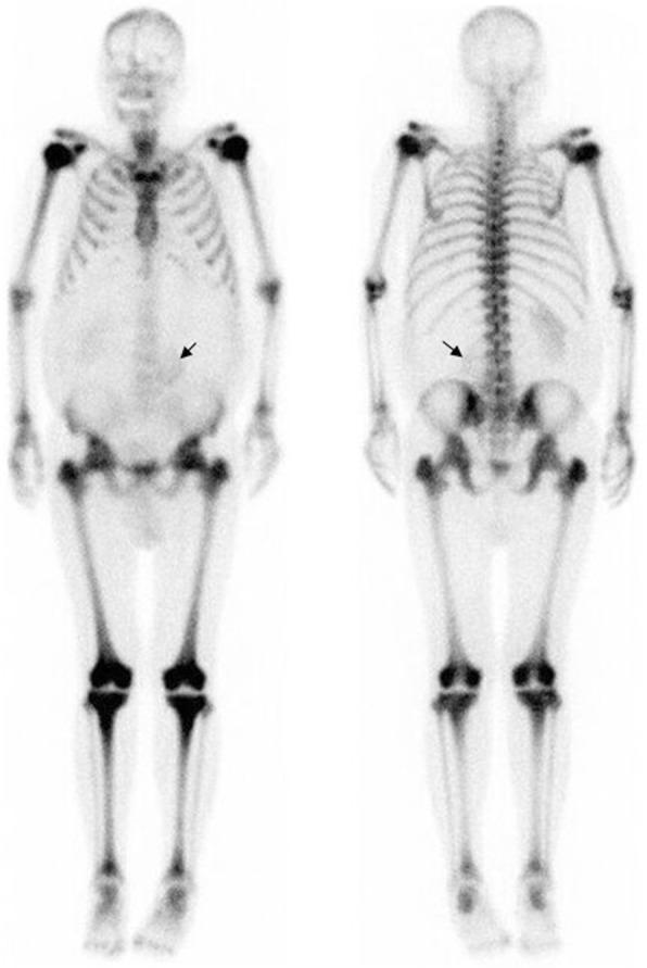
This diagnostic image is a whole-body nuclear medicine bone scan using 99mTc-hydroxydiphosphonate (HDP), showing both anterior and posterior projections. The image demonstrates a classic 'superscan' appearance, characterized by diffuse, intensely increased radiotracer uptake throughout the entire axial and appendicular skeleton. This intense skeletal uptake is accompanied by a near-total absence of normal physiological tracer distribution in the soft tissues and a lack of tracer excretion by the kidneys. In the abdominal region, a black arrow indicates the inferior displacement of the left kidney, caused by mass effect from massive splenomegaly. This constellation of findings—diffuse osteosclerosis reflected as a bone superscan combined with massive splenomegaly—is highly suggestive of a myeloproliferative neoplasm such as primary myelofibrosis, which can also involve extramedullary hematopoiesis. The visual focus is on the extreme metabolic activity of the bone marrow space across all visualized skeletal structures including the skull, spine, pelvis, and long bones.
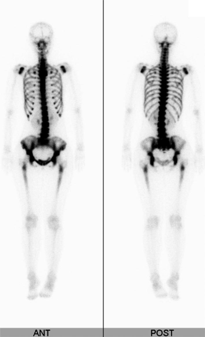
This diagnostic image shows anterior (ANT) and posterior (POST) whole-body views of a nuclear medicine bone scintigraphy (bone scan) using technetium-99m methylene diphosphonate (Tc-99m MDP). The scan demonstrates a classic 'superscan' pattern, often referred to as the 'beautiful bone sign' in specific clinical contexts. Visually, there is intense, diffuse, and symmetrical radiotracer uptake throughout the axial skeleton, including the entire vertebral column, ribs, and sternum. The pelvic girdle, particularly the iliac alae and sacrum, shows markedly increased activity. A key diagnostic feature is the complete absence of radiotracer visualization in the kidneys and urinary bladder ('absent kidney sign'), occurring because the skeletal system is sequestering the majority of the tracer due to high metabolic turnover. The appendicular skeleton shows uptake primarily in the proximal humeri and femora, with significantly reduced visualization of the distal extremities and soft tissues. This pattern is highly suggestive of diffuse osteoblastic metastatic disease, commonly associated with disseminated carcinomatosis of the bone marrow from primary malignancies like gastric or breast adenocarcinoma.
bone remodeling osteoclast osteoblast physiology
🦴 DIFFUSE SCLEROTIC DISEASES OF THE SKELETON
A Systematic Radiology Seminar for Postgraduate Residents
Session 1 of 6 — Introduction · Bone Physiology · Classification · Radiologist's Approach
Seminar Theme (announce this at the start): "By the end of this session, you will be able to look at any diffusely sclerotic skeleton and narrow it down to 3-4 diagnoses within seconds - using a structured, reproducible radiological approach."
PART 1 — INTRODUCTION
Slides 1–3
SLIDE 1 — Opening Clinical Vignette
"A 35-year-old man presents with progressive bone pain, recurrent fractures since childhood, and severe anemia requiring repeated transfusions. His family history reveals a sibling with the same problem. On examination, hepatosplenomegaly is present."
"What is going on in his bones?"
- 🔴 Red arrow → "Bone within bone" (endobone) sign in pelvis and femora
- 🟡 Yellow arrow → Complete obliteration of the medullary canal
- 🟢 Green arrow → Sandwich vertebra at lower lumbar levels
- 🔵 Blue arrow → Left subtrochanteric fracture - the "chalk bone that breaks like glass"
"This is not just dense bone. This is bone that cannot remodel, cannot house marrow, and is paradoxically brittle despite being radiographically the densest skeleton you will ever see. That is the essence of diffuse sclerotic disease - increased density does NOT mean increased strength."
SLIDE 2 — Why This Topic Matters in Your Clinical Practice
| Clinical Question | Why It Matters |
|---|---|
| Is this benign or malignant? | Determines urgency |
| Is this metabolic or genetic? | Determines workup |
| Is the marrow compromised? | Determines transfusion, transplant |
| Is there an underlying treatable cause? | Determines management |
| What is the prognosis? | Determines counselling |
"Radiology is not just a reporting tool here. In many of these conditions, the radiograph is the FIRST clue - sometimes the only clue before biopsy."
- Osteosclerosis literally means "hard bone" (Greek: osteon = bone, skleros = hard)
- Normal bone density on plain radiography reflects a balance between bone formation (osteoblasts) and resorption (osteoclasts)
- Any condition that tips this balance toward formation OR prevents resorption produces sclerosis
- Diffuse sclerosis involves the majority of the skeleton - as opposed to focal lesions
SLIDE 3 — The Spectrum of Diffuse Skeletal Sclerosis
| Category | Diseases |
|---|---|
| Genetic / Dysplastic | Osteopetrosis, Pyknodysostosis, Melorheostosis, Osteopoikilosis, Camurati-Engelmann |
| Metabolic / Endocrine | Renal osteodystrophy, Fluorosis, Hypervitaminosis D, Paget disease |
| Hematological | Myelofibrosis, Sickle cell disease (late), Mastocytosis |
| Neoplastic | Osteoblastic metastases (prostate, breast, carcinoid), Lymphoma |
| Drugs / Toxic | Bisphosphonates, Fluoride therapy, Vitamin A toxicity |
| Miscellaneous | Hepatitis C osteosclerosis, Intramedullary osteosclerosis |
"Each category has a different patient, a different age, a different clinical context, and a different radiological fingerprint. Our job today is to learn those fingerprints."
PART 2 — BONE PHYSIOLOGY FOR THE RADIOLOGIST
Slides 4–6
"You cannot understand WHY a bone looks sclerotic until you understand HOW normal bone remodels."
SLIDE 4 — Bone Remodeling: The Basic Unit
NORMAL BONE REMODELING CYCLE
─────────────────────────────────────────────────────
① RESORPTION PHASE  (2–4 weeks)
   Osteoclasts (multi-nucleated, from monocyte lineage)
   ↓
   Secrete H+ ions + lysosomal enzymes (cathepsin K)
   ↓
   Dissolve mineral → erode Howship's lacuna

② REVERSAL PHASE  (1–2 weeks)
   Mononuclear cells prepare surface
   ↓
   Release coupling signals (IGF-1, TGF-β)

③ FORMATION PHASE  (2–3 months)
   Osteoblasts (from mesenchymal stem cells)
   ↓
   Deposit osteoid (collagen matrix)
   ↓
   Mineralization with hydroxyapatite

④ QUIESCENCE
   Osteoblasts → become osteocytes (embedded) or lining cells
─────────────────────────────────────────────────────
- RANK / RANKL / OPG axis:
- Osteoblasts express RANKL → activates osteoclasts
- Osteoblasts also express Osteoprotegerin (OPG) → decoy receptor, inhibits osteoclasts
- Balance of RANKL:OPG ratio determines net remodeling direction
"Any condition that impairs osteoclast function (like osteopetrosis where TCIRG1 mutation blocks the H+ pump) shifts this cycle toward net bone accumulation → dense bone." "Any condition with excess osteoblast stimulation (like osteoblastic metastases from prostate cancer) → also produces dense bone but through a different mechanism."
SLIDE 5 — Why Does Bone Appear White on Radiograph?
| Component | Contribution to Radiodensity |
|---|---|
| Hydroxyapatite [Ca₁₀(PO₄)₆(OH)₂] | PRIMARY - absorbs X-rays strongly |
| Cortical bone thickness | Increases apparent density |
| Trabecular compaction | Increases density when marrow space reduces |
| Water/soft tissue | Negligible |
- Calcium phosphate content per unit volume
- Trabecular architecture
- Cortical thickness
MECHANISM OF SCLEROSIS
──────────────────────────────────────────────────────────
RESORPTION ↓     →  More bone remains  →  SCLEROSIS
  (Osteopetrosis, Pyknodysostosis, bisphosphonates)

FORMATION ↑      →  More bone deposited →  SCLEROSIS
  (Osteoblastic metastasis, Paget sclerotic phase)

MINERAL ↑ in bone → Denser per volume  →  SCLEROSIS
  (Fluorosis - fluorapatite replaces hydroxyapatite)

MARROW FIBROSIS  →  Reactive bone forms →  SCLEROSIS
  (Myelofibrosis)
──────────────────────────────────────────────────────────
Teaching Pearl: "Not all sclerosis looks the same. Osteopetrosis gives you chalk-white uniformity. Paget gives you coarse disorganized density. Osteoblastic metastasis gives you patchy sclerosis. This difference in TEXTURE is your first discriminator."
SLIDE 6 — Bone Compartments: What Is Affected in Each Disease?
| Compartment | Normal Function | Diseased In |
|---|---|---|
| Cortex | Structural support | Osteopetrosis, Pyknodysostosis, Fluorosis |
| Medullary cavity (marrow) | Hematopoiesis + fat | Myelofibrosis, Osteopetrosis |
| Trabeculae | Load distribution | Paget, Metastasis, Renal osteodystrophy |
- Cortical changes → look at long bone diaphysis
- Medullary changes → look at corticomedullary differentiation
- Trabecular changes → look at vertebral body texture
- Periosteal changes → look at bone outline
PART 3 — CAUSES AND CLASSIFICATION
Slides 7–9
SLIDE 7 — Etiological Classification (Master List)
GENERALIZED OSTEOSCLEROSIS
├── CONGENITAL / GENETIC
│   ├── Osteopetrosis (autosomal dominant & recessive)
│   ├── Pyknodysostosis
│   ├── Camurati-Engelmann disease (progressive diaphyseal dysplasia)
│   ├── Melorheostosis (flowing hyperostosis)
│   └── Osteopoikilosis (bone islands)
│
├── METABOLIC
│   ├── Renal osteodystrophy (2° hyperparathyroidism)
│   ├── Fluorosis (endemic / industrial / iatrogenic)
│   ├── Hypervitaminosis D
│   └── Paget disease of bone (osteitis deformans)
│
├── HEMATOLOGICAL
│   ├── Primary myelofibrosis
│   ├── Sickle cell disease (late stage)
│   └── Mastocytosis (systemic)
│
├── NEOPLASTIC
│   ├── Osteoblastic metastases
│   │   ├── Prostate carcinoma (MC in males)
│   │   ├── Breast carcinoma (MC in females)
│   │   ├── Carcinoid tumour
│   │   ├── Medulloblastoma
│   │   └── Bladder, GI primaries
│   └── Lymphoma (Hodgkin > Non-Hodgkin)
│
├── DRUGS & TOXIC
│   ├── Bisphosphonate therapy (long-term)
│   ├── Fluoride therapy (osteoporosis treatment)
│   └── Hypervitaminosis A
│
└── MISCELLANEOUS
    ├── Hepatitis C-related osteosclerosis
    ├── Intramedullary osteosclerosis
    └── Heavy metal poisoning (lead, phosphorus)
Bone islands, osteoma, fibrous dysplasia (ground-glass, not true sclerosis), healing fractures, osteoid osteoma nidus
SLIDE 8 — Age-Based Classification (Clinical Discriminator #1)
"In diffuse skeletal sclerosis, the age of the patient is the single most powerful discriminator."
| Age Group | Most Likely Diagnoses |
|---|---|
| Neonate / infant | Autosomal recessive (malignant) osteopetrosis |
| Child (1–10 yrs) | Osteopetrosis (AD or AR), Camurati-Engelmann |
| Adolescent (10–20 yrs) | Pyknodysostosis, Sickle cell, Fluorosis (endemic) |
| Young adult (20–40 yrs) | Mastocytosis, Myelofibrosis, Fluorosis |
| Middle-aged adult (40–60 yrs) | Paget disease, Renal osteodystrophy, early metastasis |
| Elderly (>60 yrs) | Osteoblastic metastasis, Paget, Myelofibrosis |
"A diffusely sclerotic skeleton in a 2-month-old infant - this is a radiology emergency. Malignant osteopetrosis without bone marrow transplant is potentially fatal."
"A diffusely sclerotic skeleton in a 65-year-old man with back pain - until proven otherwise, think prostate metastases."
SLIDE 9 — Sex and Distribution-Based Classification
| Feature | Discriminating Value |
|---|---|
| Male, >60 yrs, axial sclerosis | Prostate metastases |
| Female, >50 yrs, known breast Ca | Breast metastases (mixed lytic/sclerotic) |
| Child, short stature, fractures | Osteopetrosis, Pyknodysostosis |
| Fluoride-endemic area | Fluorosis |
| Chronic renal failure | Renal osteodystrophy |
| Splenomegaly + leukoerythroblastic picture | Myelofibrosis |
| Urticaria pigmentosa on skin | Mastocytosis |
PART 4 — THE RADIOLOGIST'S APPROACH TO DIFFUSE SCLEROSIS
Slides 10–13
"This is the section that separates a good presentation from a great one. Give the audience a tool they can use tomorrow morning."
SLIDE 10 — Step-by-Step Algorithm (The Core Framework)
STEP 1: CONFIRM IT IS TRULY DIFFUSE
────────────────────────────────────
Ask: Does it involve axial + appendicular skeleton?
  If FOCAL → not diffuse; pursue focal differential
  If DIFFUSE → proceed to Step 2

STEP 2: ASSESS THE PATIENT AGE
────────────────────────────────────
< 20 years → Genetic / Metabolic causes predominate
  → Osteopetrosis, Pyknodysostosis, Fluorosis, Sickle cell

20–50 years → Metabolic + Hematological causes
  → Renal osteodystrophy, Myelofibrosis, Mastocytosis

> 50 years → Neoplastic + Paget + Metabolic
  → Osteoblastic metastases, Paget, Myelofibrosis

STEP 3: LOOK AT BONE TEXTURE
────────────────────────────────────
UNIFORM chalk-white, no cortical-medullary distinction
  → Osteopetrosis, Pyknodysostosis

COARSE disorganized trabeculae with bone expansion
  → Paget disease

PATCHY or nodular density
  → Metastases, Mastocytosis, Myelofibrosis

TRABECULAR THICKENING, ligament calcification
  → Fluorosis

STEP 4: LOOK AT SPECIFIC BONES
────────────────────────────────────
SKULL:
  ├── Cotton wool → Paget disease
  ├── Dense base → Osteopetrosis, Paget
  └── Widened diploë → Paget, hemolytic anemias

SPINE:
  ├── Sandwich vertebra → Osteopetrosis
  ├── Rugger jersey → Renal osteodystrophy
  ├── Ivory vertebra → Paget (single), Metastasis, Lymphoma
  ├── Picture frame vertebra → Paget
  └── Bamboo spine-like ligament calcification → Fluorosis

PELVIS:
  ├── Dense iliac wings → Myelofibrosis, Fluorosis
  └── Sclerotic pubic symphysis → Paget, Metastasis

LONG BONES:
  ├── Erlenmeyer flask → Osteopetrosis, Gaucher
  ├── Transverse bands → Osteopetrosis, heavy metal
  └── Periosteal reaction → Fluorosis, Camurati-Engelmann

STEP 5: LOOK AT MARROW CAVITY
────────────────────────────────────
OBLITERATED medullary canal
  → Osteopetrosis (hallmark), Pyknodysostosis

EXPANDED marrow (fibrotic tissue)
  → Myelofibrosis

PRESERVED corticomedullary differentiation
  → Metabolic causes (fluorosis early, renal OD)

STEP 6: LOOK FOR ASSOCIATED FINDINGS
────────────────────────────────────
Fractures + anemia in child → Osteopetrosis
Known malignancy → Metastases
Renal failure → Renal osteodystrophy
Skull thickening + bowing of long bones → Paget
Splenomegaly + tear-drop cells → Myelofibrosis
Skin lesions (urticaria pigmentosa) → Mastocytosis
Dental anomalies + short stature → Pyknodysostosis
SLIDE 11 — Visual Algorithm (Flowchart for Rapid Diagnosis)
DIFFUSE SKELETAL SCLEROSIS
           │
    ┌──────┴──────┐
  Age <20       Age >20
    │               │
Genetic/          ┌──────┴──────┐
Metabolic      Known          No known
    │          malignancy?    malignancy
    │            │               │
  ┌─┴─┐        YES             ┌─┴────────┐
Bone   Short    ↓           Renal     Metabolic/
within  stature Osteoblastic disease?  Hematological
bone?    ↓      metastasis   │           │
  │   Pykno-               YES         ┌─┴───┐
YES    dysostosis          ↓         Marrow  Spine
↓      or C-E          Renal OD    disease? pattern?
Osteo-               (Rugger          │       │
petrosis              jersey)       MF/SM  Paget/
                                    Fluorosis
"Tape this algorithm inside your reporting room. It won't give you the diagnosis - it will give you a framework."
SLIDE 12 — Multimodality Approach to Diffuse Sclerosis
"Not every patient needs every modality. Choose wisely."
| Modality | Best For | Key Findings |
|---|---|---|
| Plain Radiograph | Initial detection, pattern recognition, comparison | Density, distribution, specific signs (bone within bone, rugger jersey) |
| CT | Bone detail, corticomedullary differentiation, narrow canal, ligament ossification | Density measurements, cortical thickness |
| MRI | Marrow involvement, early disease, nerve compression | T1 low signal (sclerotic), T2 variable; whole-body MRI for marrow infiltration |
| Bone Scintigraphy (Tc-99m MDP) | Metabolic activity, extent of disease, superscan | Diffuse uptake = metastases/metabolic; photopenic areas = lack of vascularity |
| SPECT-CT | Precise anatomical localization of scintigraphy findings | Differentiates Paget vs metastasis at specific site |
| FDG PET/CT | Active malignant marrow disease | FDG avid = malignant/metabolically active |
| DEXA / QCT | Quantitative bone density | Not for diagnosis but for monitoring |
"Always start with a radiograph. It costs the least and teaches the most in diffuse skeletal sclerosis."
SLIDE 13 — The Superscan — A Special Mention
A bone scintigraphy scan with diffuse, intensely increased skeletal uptake with absent or reduced renal and soft tissue activity - because the skeleton is consuming almost all the tracer.
- 🔴 Red arrows → Intense, symmetrical axial and appendicular skeletal uptake
- 🟡 Yellow arrow → Absent kidney visualization (tracer all consumed by bone)
- 🔵 Blue arrow → Displaced left kidney (splenic enlargement in myelofibrosis)
| Cause | Pattern |
|---|---|
| Diffuse osteoblastic metastases (prostate, breast) | Axial > appendicular; patchy |
| Primary myelofibrosis | Axial + appendicular; uniform |
| Paget disease (diffuse) | Heterogeneous, expands bones |
| Hyperparathyroidism / renal OD | Axial predominant; "metabolic" pattern |
| Mastocytosis | Patchy; appendicular also involved |
Examiner Pearl: "In a superscan from metastases, you often see a photopenic area (cold spot) at the site of a fracture, while in metabolic causes the uptake is more uniform."
PART 5 — HIGH-YIELD RADIOLOGY SIGNS ATLAS
Pre-disease reference (Each sign with image + 1-line explanation)
SIGN 1 — Bone Within Bone (Endobone Sign)
SIGN 2 — Rugger Jersey Spine
SIGN 3 — Sandwich Vertebra
- Rugger jersey: central body OSTEOPENIC (looks empty)
- Sandwich: central body NORMAL density (the endplates are simply abnormally dense)
SIGN 4 — Ivory Vertebra
- P = Paget disease (body enlarged + dense)
- L = Lymphoma (Hodgkin - body normal size or expanded)
- O = Osteoblastic metastasis (body normal or compressed)
- T = Tuberculosis / chronic infection (rare)
SIGN 5 — Cotton Wool Skull (Paget Disease)
- Lytic phase → Osteoporosis circumscripta (V-shaped frontal resorption)
- Mixed phase → Cotton wool appearance
- Sclerotic phase → Thick, dense calvarium
SIGN 6 — Erlenmeyer Flask Deformity
- Grade 0 → No deformity
- Grade 1 → Mild flaring
- Grade 2 → Moderate (loss of waist)
- Grade 3 → Severe (bulbous, cortical thinning)
SIGN 7 — Picture Frame Vertebra (Paget)
SIGN 8 — Fluorosis Ligament Calcification
- Ligament ossification (not seen in osteopetrosis)
- Calcification of interosseous membrane (tibia-fibula, radius-ulna)
- Dense pelvis with calcified ligaments
- Normal skull (unlike osteopetrosis where skull is very dense)
- Endemic geography clue (India - Punjab, Rajasthan; Africa; China)
COMPARISON TABLE — Session 1 Master Reference
Table 1: Key Differentiating Features at a Glance
| Disease | Skull | Spine Sign | Long Bone | Medullary Canal | Key Clue |
|---|---|---|---|---|---|
| Osteopetrosis | Dense, "chalk" | Sandwich vertebra | Erlenmeyer flask; bone within bone | Obliterated | Child, anemia, fractures |
| Paget disease | Cotton wool, thickened | Ivory / Picture frame | Bowing, cortical thickening | Enlarged | Enlarged bones, older adult |
| Renal OD | Normal / dense | Rugger jersey | Periosteal resorption at middle phalanx | Normal | Chronic renal failure |
| Fluorosis | Normal | Dense all vertebrae + ligament calcification | Dense, ligament ossification | Normal/narrowed | Endemic area, spine stiffness |
| Osteoblastic mets | Patchy sclerosis | Multiple ivory or patchy | Patchy metaphyseal | Variable | Known Ca prostate/breast |
| Myelofibrosis | Normal | Diffuse sclerosis | Dense cortex | Fibrosed | Splenomegaly, leukoerythroblastic |
| Mastocytosis | Patchy | Patchy sclerosis | Osteoporosis + focal sclerosis | Normal | Urticaria pigmentosa |
Table 2: Bone Scan (Tc-99m MDP) Patterns
| Diagnosis | Bone Scan Pattern |
|---|---|
| Osteopetrosis | Low or normal uptake (poor vascularity, no remodeling) |
| Paget disease | Markedly increased focal uptake ("hot" in Paget bone) |
| Osteoblastic metastases | Multiple foci of increased uptake; superscan |
| Myelofibrosis | Superscan pattern |
| Renal osteodystrophy | Metabolic superscan; absent kidney sign |
| Fluorosis | Diffuse increased axial uptake |
VIVA QUESTIONS — Session 1
A: Osteoblastic metastases - most commonly prostate carcinoma in males, breast carcinoma in females.
A: Cyclical arrest of bone remodeling (during illness or therapy), leaving behind unresorbed primary spongiosa as a ghost vertebra or ghost bone. Seen classically in osteopetrosis due to failure of TCIRG1-dependent osteoclast proton pump.
A: In sandwich vertebra, the ENTIRE vertebra is denser than normal with prominent endplates but the central band is relatively less dense (not truly osteopenic). In rugger jersey spine, the CENTRAL body is genuinely osteopenic (reduced density) and only the endplate bands are sclerotic. Clinical context (child with anemia vs. renal failure patient) confirms.
A: PLOTL mnemonic - Paget disease, Lymphoma (Hodgkin), Osteoblastic metastasis, Tuberculosis (sclerotic healing phase), Lymphoma-associated sclerosis.
A: Superscan = whole-body bone scan with diffuse intense skeletal uptake, absent renal activity. Causes: diffuse osteoblastic metastases, myelofibrosis, Paget disease (polyostotic), renal osteodystrophy, mastocytosis.
A: Conditions with osteoclast dysfunction or marrow crowding: osteopetrosis, Gaucher disease, Niemann-Pick disease, thalassemia major, lead poisoning. All involve failure of normal metaphyseal remodeling.
A: Fluorosis. The replacement of hydroxyl group by fluoride → fluorapatite formation, plus direct periosteal stimulation → calcification of paraspinal ligaments, interosseous membranes, entheses. Also differentiates from osteopetrosis (which does not calcify ligaments).
RECENT ADVANCES PREVIEW (Full Session 7)
-
Whole-body MRI (WB-MRI): Now the preferred modality for assessing bone marrow infiltration in conditions like myeloma, myelofibrosis, and diffuse osteoblastic disease. Detects marrow changes before plain radiograph abnormalities appear. (PMID: 36116759 - Calder et al., Bone 2022)
-
FDG PET/CT vs. SPECT-CT: PET/CT better delineates metabolically active malignant disease from metabolic bone disease in cases of mixed sclerotic patterns. SPECT-CT provides better anatomical co-registration for spinal lesions. (PMID: 41433193 - Mastocytosis Imaging Spectrum, RadioGraphics 2026)
-
RANK/RANKL pathway therapeutics: Denosumab (anti-RANKL monoclonal antibody) understanding has deepened our knowledge of how targeted manipulation of the remodeling cycle can create iatrogenic osteosclerosis. Important for drug-induced sclerosis differential.
-
Genetic testing + imaging correlation: With TCIRG1, CLCN7, and other mutation panels now available, radiology reports in childhood diffuse sclerosis should suggest genetic correlation. (PMID: 36360203 - Spinnato et al., Genes 2022)
-
AI pattern recognition: Early systems now trained on diffuse skeletal sclerosis patterns for automated flagging - not yet clinical standard but emerging.
TAKE-HOME MESSAGE — SESSION 1
"There is no need to memorize 20 diseases separately. Learn the APPROACH."
AGE        →  Narrows your differential from 20 to 5
TEXTURE    →  Narrows from 5 to 3
SITE       →  Narrows from 3 to 2
ASSOCIATED →  Gives you the diagnosis
- How old is this patient?
- Is the density uniform or heterogeneous?
- Which specific bones are most affected?
- What is happening in the medullary cavity?
📚 References for Session 1
- Goldman-Cecil Medicine, 26th Edition - Paget Disease of Bone (Chapter by Ralston SH) - definition, epidemiology, pathophysiology
- Robbins & Kumar Pathologic Basis of Disease - Bone remodeling, osteoclast-osteoblast coupling, RANK-RANKL-OPG axis
- Grainger & Allison's Diagnostic Radiology - Sclerosing bone disorders, approach to diffuse sclerosis
- Kildal D, Braunschweig R, Schaal M. Sclerosing hyperostotic bone disorders. Radiologe. 2021 Dec. [PMID: 34735583]
- Calder AD, Arulkumaran S, D'Arco F. Imaging in osteopetrosis. Bone. 2022 Dec. [PMID: 36116759]
- Spinnato P, Pedrini E, Petrera MR, et al. Spectrum of Skeletal Imaging Features in Osteopetrosis: Inheritance Pattern and Radiological Associations. Genes (Basel). 2022 Oct. [PMID: 36360203]
- Khot R, Diab M, Jing JM. Mastocytosis: Imaging Spectrum and Diagnostic Insights. RadioGraphics. 2026 Jan. [PMID: 41433193]
- Delgado-Calle J, Bellido T. The osteocyte as a signaling cell. Physiol Rev. 2022. [PMID: 34337974]
SESSION ROADMAP — What Is Coming
| Session | Topic | Status |
|---|---|---|
| Session 1 | Introduction · Bone Physiology · Classification · Approach | ✅ COMPLETE |
| Session 2 | Genetic Sclerosing Dysplasias - Osteopetrosis, Pyknodysostosis, Melorheostosis, Camurati-Engelmann | 🔜 Next |
| Session 3 | Metabolic & Endocrine - Renal OD, Fluorosis, Paget, Hypervitaminosis D | ⏳ |
| Session 4 | Hematological & Neoplastic - Myelofibrosis, Mastocytosis, Osteoblastic Metastases | ⏳ |
| Session 5 | Drugs, Toxic, Miscellaneous causes | ⏳ |
| Session 6 | Comparison Tables · Diagnostic Algorithm · Viva Q&A · Final PPT polish | ⏳ |
No need of sessions generate a complete pdf all together
pyknodysostosis dense bones short stature mandible radiograph
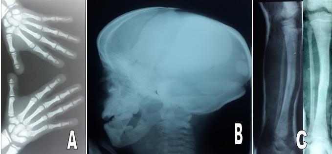
This composite diagnostic image features three radiographs illustrating skeletal manifestations consistent with a systemic bone disorder such as pyknodysostosis. Panel A is an anteroposterior (AP) X-ray of both hands, showing short, stubby phalanges with notable acro-osteolysis (resorption of the distal tufts) and associated soft tissue changes indicating nail dysplasia. Panel B presents a lateral skull radiograph demonstrating a large, prominent cranial vault with delayed closure of the fontanelles and sutures, multiple intra-sutural Wormian bones, hypoplasia of the facial bones, and a small, underdeveloped mandible with an increased (obtuse) mandibular angle. Panel C includes AP and lateral views of the lower leg, identifying a transverse fracture of the mid-tibial shaft. Collectively, these images illustrate key diagnostic features of osteosclerosis and skeletal dysplasia, highlighting both cranial and peripheral bone abnormalities along with a predisposition to pathological fractures.
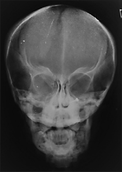
**Imaging Modality:** Posteroanterior (PA) conventional radiograph of the skull. **Anatomical Region:** Craniofacial skeleton, including the calvarium, orbits, nasal cavity, maxilla, and mandible. **Observed Pathology and Visual Features:** * **Cranial Vault:** The skull demonstrates a brachycephalic configuration with a noticeably widened transverse diameter. There is evidence of a persistent, open anterior fontanel and widened cranial sutures. The calvarium appears diffusely thickened with increased bone density. * **Facial Skeleton:** Marked generalized osteosclerosis is present throughout the facial bones. There is significant hypoplasia of the maxilla and mandible, contributing to a diminished midface and lower face height. * **Sinuses:** There is a lack of visible pneumatization of the paranasal sinuses, consistent with sinus aplasia or high-grade hypoplasia. * **Dental/Mandibular:** The mandible appears small and dense, with crowded or unerupted permanent dentition visible. **Diagnostic Context:** These radiographic findings—specifically the combination of delayed suture closure, osteosclerosis, and midface hypoplasia—are highly characteristic of pyknodysostosis. **Key Features for Retrieval:** Brachycephaly, open fontanelle, osteosclerosis, maxillary hypoplasia, nonpneumatized sinuses, pyknodysostosis, skeletal dysplasia.
mastocytosis bone sclerosis pelvis radiograph systemic
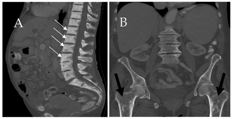
This dual-panel figure displays computed tomography (CT) scans of the skeletal system in a patient with systemic mastocytosis. Image A is a sagittal CT of the lumbar and sacral spine, demonstrating diffuse osteosclerosis of the axial skeleton. White arrows indicate multiple vertebral bodies showing pathologically increased bone density with a patchy, sclerotic appearance. Image B is a coronal CT of the pelvis and proximal femurs, where black arrows highlight a diffuse loss of normal trabecular architecture. The proximal femurs exhibit a heterogeneous, osteolytic, and somewhat 'mottled' internal structure, indicative of bone marrow infiltration. The images collectively illustrate the varied osseous manifestations of mast cell infiltration, ranging from lytic-appearing trabecular loss to profound focal or diffuse sclerosis. These findings are clinically significant for the radiological evaluation of hematologic malignancies and metabolic bone diseases.
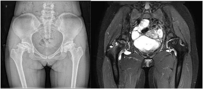
This dual-modality comparison presents an anteroposterior (AP) radiograph and a coronal MRI T2-weighted short tau inversion recovery (STIR) sequence of the pelvis in a 35-year-old patient with systemic lupus erythematosus (SLE). The visual illustrates advanced femoral head osteonecrosis (FHON). On the left (radiograph), bilateral femoral head abnormalities are visible, more severe on the patient's right side, characterized by significant flattening, subchondral sclerosis, and loss of the normal spherical articular contour. On the right (MRI), the STIR sequence highlights extensive bone marrow edema and cystic changes within both femoral heads. High signal intensity (bright areas) indicates fluid accumulation and active inflammatory processes, while heterogeneous low-signal areas correspond to bone death and collapse. The images demonstrate a comparison between the sensitivity of plain films for detecting structural bone changes versus the MRI's ability to visualize intramedullary pathology and joint effusion. This serves as an educational example of secondary osteonecrosis associated with systemic autoimmune disease and corticosteroid therapy.
sickle cell disease H-shaped vertebra bone infarct sclerosis spine
This diagnostic image displays two plain radiographs of the lumbar spine: an anteroposterior (AP) view (a) and a lateral view (b). The radiographs demonstrate classic skeletal manifestations of sickle cell disease. Image (a) shows 'H-shaped vertebrae' characterized by sharp, step-like depressions in the central portion of the vertebral endplates, secondary to microvascular end-plate infarction (Reynold's sign). Image (b) illustrates 'fish mouth vertebrae,' showing smooth, biconcave depressions of the endplates caused by bone softening and chronic osteopenia. Beyond the spine, the AP view reveals patchy sclerosis of the pelvic bones, suggesting chronic medullary bone infarction, and radiopaque cholecystectomy clips in the right upper quadrant, indicating prior surgical removal of the gallbladder for pigment stones. The images provide high-yield educational examples of musculoskeletal complications in sickle cell anemia, including avascular necrosis of the vertebral endplates and changes in bone density.
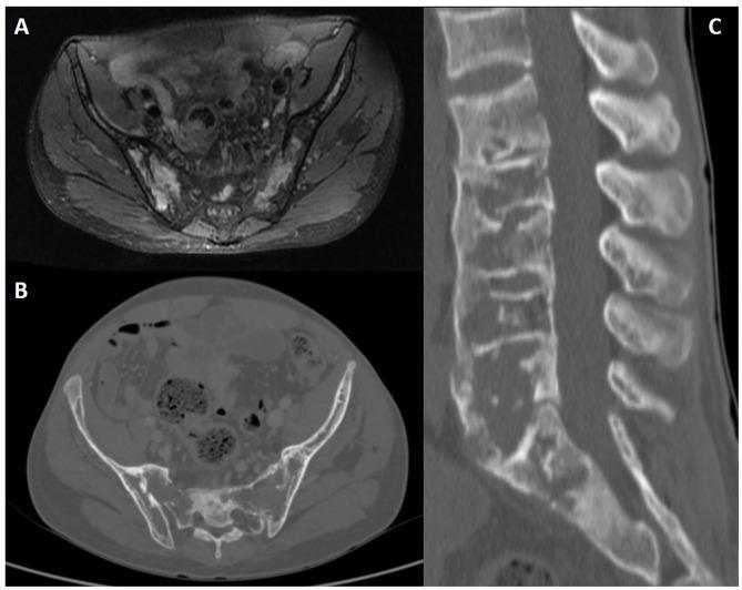
Multi-modal diagnostic imaging of the lumbosacral region in a 31-year-old male with sickle cell disease. (A) Axial fat-saturated proton density (PD) MRI shows bilateral sacroiliac joints with prominent, high-signal intensity areas indicative of bone marrow edema and surrounding soft tissue inflammation. (B) Axial CT slice of the pelvis demonstrates thinning of the iliac cortical bone and mottled medullary density. (C) Sagittal lumbar spine CT reconstruction reveals extensive skeletal changes including widened medullary spaces, thinning of the cortical bone, and characteristic 'fish-mouth' or 'H-shaped' vertebral deformities due to central endplate depressions. These findings collectively illustrate musculoskeletal complications of sickle cell disease, specifically bone marrow hyperplasia and secondary ischemic changes (infarctions) leading to structural vertebral remodeling and sacroiliac joint involvement.
myelofibrosis sclerotic pelvis spine diffuse osteosclerosis radiograph
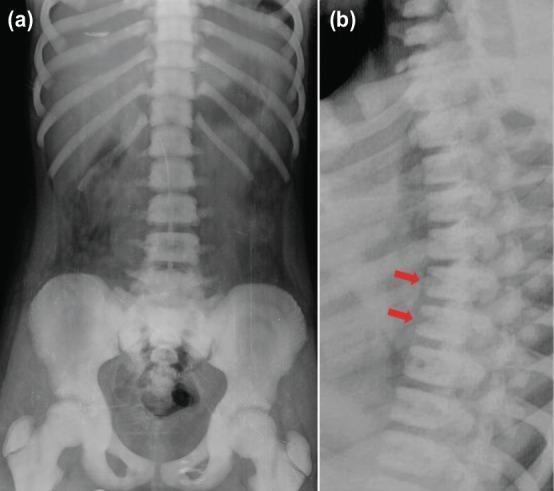
Diagnostic imaging series comprising (a) an anteroposterior and (b) a lateral radiograph of the thoracolumbar spinal column and pelvis. The images demonstrate diffuse, generalized osteosclerosis, marked by a significant increase in bone density throughout the axial skeleton, ribs, and proximal femurs. In the lateral view (b), red arrows highlight the 'sandwich vertebrae' sign, characterized by dense, sclerotic bands at the superior and inferior endplates of the vertebral bodies with relatively lucent central regions. This radiological appearance is also known as 'rugger jersey spine' and is a hallmark of hereditary metabolic bone diseases such as osteopetrosis (Albers-Schönberg disease). The AP view confirms the loss of normal corticomedullary differentiation in the pelvis and long bones. These findings illustrate impaired osteoclast function resulting in failure of normal bone resorption and remodeling.
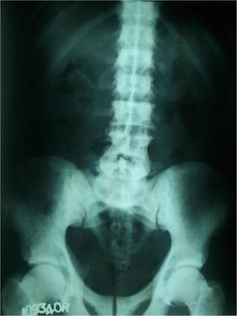
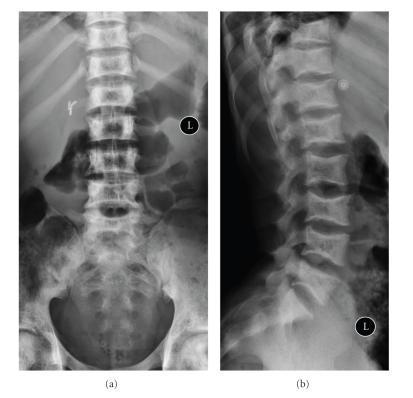
This diagnostic image is an anteroposterior (AP) X-ray of the lumbosacral spine and pelvis. The radiograph demonstrates a diffuse and marked increase in bone density (osteosclerosis) involving the lumbar vertebrae, sacrum, and pelvic bones, including the iliac wings and proximal femora. The affected bony structures exhibit a characteristic hyper-radiopaque or 'whitened' appearance with a significant loss of the normal trabecular pattern and corticomedullary differentiation. The vertebral bodies appear uniformly dense and chalk-like. In the clinical context of distal renal tubular acidosis (dRTA) and metabolic bone disease, these findings are consistent with sclerotic changes secondary to renal osteodystrophy or potential fluorosis. The alignment of the spine appears relatively preserved, and no acute fractures or focal lytic lesions are visible in this view. The image serves as a teaching example of generalized osteosclerosis in metabolic or renal systemic disease.
Camurati-Engelmann progressive diaphyseal dysplasia long bone periosteal thickening
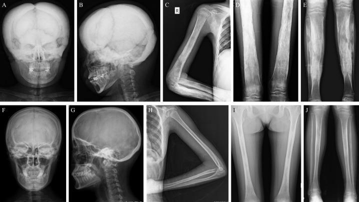
A composite image of 10 X-ray radiographs illustrating varying phenotypic expressions of Camurati-Engelmann Disease (CED), also known as progressive diaphyseal dysplasia. The image compares severe (A-E) and mild (F-J) radiological manifestations in adolescent and adult female patients. Panels A and B (severe) demonstrate significant hyperostosis and sclerosis of the skull and skull base, appearing radiopaque and thickened compared to the relatively normal skull morphology in F and G (mild). Panels C-E (severe) show marked cortical thickening of the diaphyses in long bones—including the humerus, radius, ulna, femur, and tibia—resulting in significant narrowing of the medullary canals. In contrast, panels H-J (mild) show only slight or focal cortical thickening, particularly in the ulna and tibia, with more preserved medullary spaces. The series serves as an educational reference for identifying characteristic symmetric, skeletal changes associated with CED, emphasizing the progression of bone density and marrow space obliteration in long bone shafts.
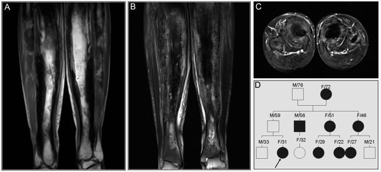
This composite educational graphic presents radiological and genetic data for a patient with Progressive Diaphyseal Dysplasia (PDD), also known as Camurati-Engelmann disease. Panels A, B, and C provide MRI views of the bilateral femora. Panel A (coronal T1-weighted image) demonstrates significantly thickened cortices with characteristic low signal intensity (SI). Panels B (coronal) and C (axial) utilize T2 fat suppression sequences to reveal heterogeneous signal intensity within the affected bone, indicating non-uniform bone marrow involvement and cortical changes. Key diagnostic features include irregular endosteal surfaces and a narrowed medullary cavity. Panel D contains a three-generation family pedigree illustrating an autosomal dominant inheritance pattern. The pedigree identifies the index case (proband) with an arrow, showing multiple affected individuals (black circles/squares) and an asymptomatic carrier (gray square). This visual set correlates clinical imaging findings of symmetric diaphyseal sclerosis and cortical thickening with genetic transmission, typical of hereditary sclerosing bone dysplasias.
osteoblastic metastasis prostate diffuse sclerosis axial skeleton CT scan

This composite figure presents clinical imaging of a patient with prostate cancer, focusing on bone metastasis evaluation. Image (A) is a whole-body radionuclide bone scan (PET/CT) in the anterior view, showing tracer uptake in the axial skeleton, kidneys, and bladder. A red box and black arrow highlight an area of interest in the right hip region. Image (B) provides a magnified CT scan of the right femoral head, revealing subchondral sclerosis, mixed dense foci, and internal cystic changes consistent with a subchondral bone cyst. Image (C) is a corresponding PET scan showing a focal area of increased radioactive tracer uptake at the same anatomical site, indicating high metabolic activity or bone turnover. Image (D) is a fused PET/CT image, co-registering the metabolic ‘hot spot’ with the structural lesions identified on CT. These findings are characteristic of osteoblastic and osteolytic changes associated with metastatic prostate cancer. The images demonstrate the clinical utility of multi-modal imaging in identifying the location, morphology, and metabolic significance of skeletal lesions.
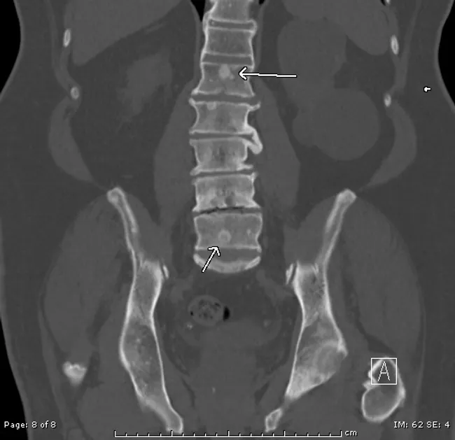
Computed tomography (CT) scan of the pelvis and lower spine performed in a coronal plane with bone-window technique demonstrates multiple osteoblastic, sclerotic lesions involving the sacrum and contiguous lumbar vertebrae. In this patient with a history of prostatic adenocarcinoma (Gleason score 5+4=9), the image highlights densely radiopaque foci along the sacral alae and across the L1–L5 pedicles and endplates, with subtle vertical striations and preserved cortical margins. The pattern is classic for hematogenous, prostatic metastasis to bone, producing reactive sclerosis and meshwork coalescence within the vertebral bodies. No obvious large lytic destruction or acute fracture is seen on the current study, though mild endplate irregularity at the sacral junction is present. The arrows bracket the most conspicuous metastases, illustrating pelvic ring involvement and axial skeleton dissemination. This radiologic finding has significant clinical implications: confirms metastatic disease, supports staging in patients with prostate cancer, influences therapeutic planning (androgen deprivation therapy, hormonal therapy, chemotherapy, radiopharmaceuticals), and serves as a baseline to monitor response or progression during systemic treatment. Clinically, correlation with PSA levels, prior imaging, and treatment history is essential to distinguish progression from treatment response and to guide follow-up imaging strategies. Correlation with symptoms and systemic biomarkers improves patient-specific management plans.
myelofibrosis mastocytosis skeletal imaging diagnosis
mkdir -p /tmp/workspace/diffuse-sclerosis-seminar && ls /tmp/workspace/diffuse-sclerosis-seminar/
Can you combine all sessions and give it in a single ready to go slides in pdf format for seminar
pyknodysostosis dense bones acro-osteolysis mandible wormian bones
This composite diagnostic image features three radiographs illustrating skeletal manifestations consistent with a systemic bone disorder such as pyknodysostosis. Panel A is an anteroposterior (AP) X-ray of both hands, showing short, stubby phalanges with notable acro-osteolysis (resorption of the distal tufts) and associated soft tissue changes indicating nail dysplasia. Panel B presents a lateral skull radiograph demonstrating a large, prominent cranial vault with delayed closure of the fontanelles and sutures, multiple intra-sutural Wormian bones, hypoplasia of the facial bones, and a small, underdeveloped mandible with an increased (obtuse) mandibular angle. Panel C includes AP and lateral views of the lower leg, identifying a transverse fracture of the mid-tibial shaft. Collectively, these images illustrate key diagnostic features of osteosclerosis and skeletal dysplasia, highlighting both cranial and peripheral bone abnormalities along with a predisposition to pathological fractures.
**Imaging Modality:** Posteroanterior (PA) conventional radiograph of the skull. **Anatomical Region:** Craniofacial skeleton, including the calvarium, orbits, nasal cavity, maxilla, and mandible. **Observed Pathology and Visual Features:** * **Cranial Vault:** The skull demonstrates a brachycephalic configuration with a noticeably widened transverse diameter. There is evidence of a persistent, open anterior fontanel and widened cranial sutures. The calvarium appears diffusely thickened with increased bone density. * **Facial Skeleton:** Marked generalized osteosclerosis is present throughout the facial bones. There is significant hypoplasia of the maxilla and mandible, contributing to a diminished midface and lower face height. * **Sinuses:** There is a lack of visible pneumatization of the paranasal sinuses, consistent with sinus aplasia or high-grade hypoplasia. * **Dental/Mandibular:** The mandible appears small and dense, with crowded or unerupted permanent dentition visible. **Diagnostic Context:** These radiographic findings—specifically the combination of delayed suture closure, osteosclerosis, and midface hypoplasia—are highly characteristic of pyknodysostosis. **Key Features for Retrieval:** Brachycephaly, open fontanelle, osteosclerosis, maxillary hypoplasia, nonpneumatized sinuses, pyknodysostosis, skeletal dysplasia.
mastocytosis diffuse bone sclerosis mixed lytic sclerotic spine pelvis
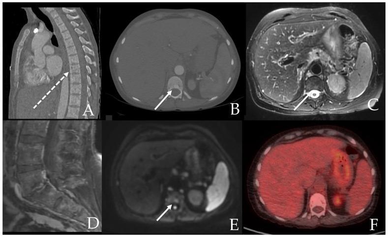
A multi-panel diagnostic imaging series demonstrating skeletal manifestations of aggressive systemic mastocytosis in a 71-year-old female. Panel A shows a sagittal CT of the spine revealing diffuse, patchy sclerosis interspersed with focal lytic lesions (dashed arrow). Panel B (axial CT) highlights a specific hyperdense sclerotic lesion in a vertebral body (arrow). Corresponding axial MRIs show this lesion is mildly hyperintense on T2-weighted imaging (C) and Diffusion-Weighted Imaging (E), suggesting increased cellularity or marrow edema. Panel D depicts a sagittal post-contrast T1-weighted MRI with mild, variable enhancement across the vertebral segments. Panel F shows a fused PET/CT scan demonstrating that the sclerotic bone lesions are non-PET hypermetabolic. The collection serves as a clinical comparison of CT, MRI (T1, T2, DWI), and PET modalities in evaluating heterogeneous (mixed lytic and sclerotic) bone marrow involvement associated with mast cell disorders.
This dual-panel figure displays computed tomography (CT) scans of the skeletal system in a patient with systemic mastocytosis. Image A is a sagittal CT of the lumbar and sacral spine, demonstrating diffuse osteosclerosis of the axial skeleton. White arrows indicate multiple vertebral bodies showing pathologically increased bone density with a patchy, sclerotic appearance. Image B is a coronal CT of the pelvis and proximal femurs, where black arrows highlight a diffuse loss of normal trabecular architecture. The proximal femurs exhibit a heterogeneous, osteolytic, and somewhat 'mottled' internal structure, indicative of bone marrow infiltration. The images collectively illustrate the varied osseous manifestations of mast cell infiltration, ranging from lytic-appearing trabecular loss to profound focal or diffuse sclerosis. These findings are clinically significant for the radiological evaluation of hematologic malignancies and metabolic bone diseases.
osteoblastic prostate cancer bone metastasis diffuse sclerosis pelvis CT radiograph
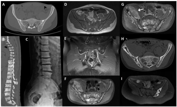
This composite figure presents various radiological modalities illustrating primary and secondary bone tumors affecting the spine and pelvis. (A) Axial CT shows an iliac bone enostosis (bone island). (B, C) Sagittal CT reconstruction and lateral radiograph demonstrate diffuse osteoblastic metastases in the lumbar spine, characterized by multifocal vertebral sclerosis typical of metastatic prostate cancer. (D) Axial T1-weighted MRI and (E) coronal post-contrast fat-saturated T1-weighted MRI reveal leukemic infiltration of the sacrum and iliac bones, evidenced by diffuse T1 hypointensity and patchy contrast enhancement indicating bone marrow replacement. (F-H) Axial PD fat-saturated MRI, post-contrast T1-weighted MRI, and axial CT depict a large Ewing sarcoma of the right iliac bone with a significant soft tissue component (arrow) and aggressive cortical destruction. (I) Axial fat-saturated PD MRI shows an aneurysmal bone cyst of the left iliac bone, characterized by multi-loculated fluid-fluid levels. The collection serves to educate on distinguishing benign variants like enostosis from aggressive primary malignancies and systemic marrow infiltrative processes.
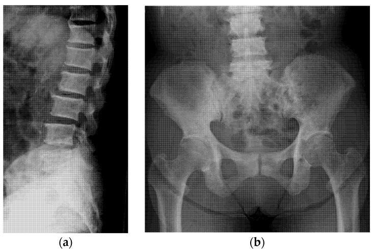
Two-panel plain film radiograph of the axial skeleton illustrating diffuse osteoblastic activity. Image (a) is a lateral projection of the thoracolumbar spine showing generalized, marked increase in radiodensity (sclerosis) within the vertebral bodies, resulting in a 'chalky' or 'ivory' appearance of the vertebrae. The intervertebral disc spaces appear relatively preserved. Image (b) is an anteroposterior (AP) view of the pelvis and lumbar spine, demonstrating widespread sclerotic changes throughout the sacrum and bilateral iliac bones. Notably, the proximal femurs maintain normal cortical and medullary radiodensity, highlighting the axial distribution of the pathology. These radiographic findings are characteristic of osteoblastic metastases, commonly seen in secondary malignancies such as prostate or gastric cancer. The educational focus is on the recognition of diffuse bone sclerosis as a manifestation of systemic disease and the clinical utility of plain radiographs in identifying metastatic bone lesions.
Camurati Engelmann diaphyseal dysplasia femur tibia cortical thickening
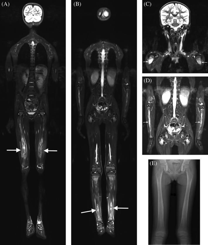
This composite diagnostic image showcases imaging findings of Camurati-Engelmann disease in a pediatric patient. Panels A-D present Whole-Body Short Tau Inversion Recovery (STIR) Magnetic Resonance Imaging (MRI) in the coronal plane. These scans demonstrate bilateral, symmetrical hyperintense signal intensity (edema) within the bone marrow of the diaphyses and metadiaphyseal regions of the femur (A), tibia (B), humerus (C), and radius/ulna (D). The pathology is characterized by concentric cortical thickening, surface irregularities, and fusiform diaphyseal enlargement. Notably, the epiphyseal regions are consistently spared across all long bones, and there is an absence of restricted diffusion. Panel E provides a corresponding clinical radiograph (X-ray) of the femurs, illustrating the radiographic correlate of slight fusiform cortical thickening along the femoral shafts. The findings are pathognomonic for progressive diaphyseal dysplasia, highlighting the characteristic symmetrical long bone involvement and sparing of the joints.
A composite image of 10 X-ray radiographs illustrating varying phenotypic expressions of Camurati-Engelmann Disease (CED), also known as progressive diaphyseal dysplasia. The image compares severe (A-E) and mild (F-J) radiological manifestations in adolescent and adult female patients. Panels A and B (severe) demonstrate significant hyperostosis and sclerosis of the skull and skull base, appearing radiopaque and thickened compared to the relatively normal skull morphology in F and G (mild). Panels C-E (severe) show marked cortical thickening of the diaphyses in long bones—including the humerus, radius, ulna, femur, and tibia—resulting in significant narrowing of the medullary canals. In contrast, panels H-J (mild) show only slight or focal cortical thickening, particularly in the ulna and tibia, with more preserved medullary spaces. The series serves as an educational reference for identifying characteristic symmetric, skeletal changes associated with CED, emphasizing the progression of bone density and marrow space obliteration in long bone shafts.