
Adrenaline it's mechanism how to give how munch to dilute
adrenaline epinephrine receptor mechanism adrenergic diagram
![This pathophysiology diagram illustrates the role of the Beta-2 Adrenergic Receptor (ADRB2) in modulating inflammatory responses during Rhinovirus (RV) infection. The visual is divided into three primary sections: [1] depicts RV particles binding to Intercellular Adhesion Molecule 1 (ICAM-1) on the surface of airway epithelial cells (ECs). [2] demonstrates that ADRB2 activation inhibits the expression of ICAM-1 and the subsequent release of cytokines and chemokines from these epithelial cells. [3] focuses on systemic immune cell modulation, showing that ADRB2 signaling exerts inhibitory effects (indicated by red T-bars) on multiple cell types: in macrophages (Mφ), it suppresses TNF-α, IL-6, IL-1β, and various CCL chemokines; in mast cells (MC), it inhibits TNF-α, histamine, and leukotriene release; in CD4+ T cells, it suppresses IFN-γ production; and in CD8+ T cells, it blocks TNF-α, IFN-γ, and cytolytic activity. The diagram serves as an educational resource for understanding neuro-immune interactions and the therapeutic mechanism of beta-agonists in dampening virus-induced airway inflammation.](https://cdn.orris.care/cdss_images/pmc_clinical_VQA_bdc00632428101dee8d73e300a2b0ae2a72521d5b25266eddbef38d212ed1f84.jpg)
This pathophysiology diagram illustrates the role of the Beta-2 Adrenergic Receptor (ADRB2) in modulating inflammatory responses during Rhinovirus (RV) infection. The visual is divided into three primary sections: [1] depicts RV particles binding to Intercellular Adhesion Molecule 1 (ICAM-1) on the surface of airway epithelial cells (ECs). [2] demonstrates that ADRB2 activation inhibits the expression of ICAM-1 and the subsequent release of cytokines and chemokines from these epithelial cells. [3] focuses on systemic immune cell modulation, showing that ADRB2 signaling exerts inhibitory effects (indicated by red T-bars) on multiple cell types: in macrophages (Mφ), it suppresses TNF-α, IL-6, IL-1β, and various CCL chemokines; in mast cells (MC), it inhibits TNF-α, histamine, and leukotriene release; in CD4+ T cells, it suppresses IFN-γ production; and in CD8+ T cells, it blocks TNF-α, IFN-γ, and cytolytic activity. The diagram serves as an educational resource for understanding neuro-immune interactions and the therapeutic mechanism of beta-agonists in dampening virus-induced airway inflammation.
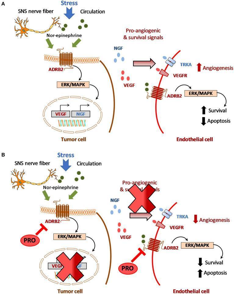
This pathophysiology diagram illustrates the crosstalk between adrenergic signaling and angiogenesis in the tumor microenvironment, specifically in Head and Neck Squamous Cell Carcinoma (HNSCC). Panel A depicts a stimulatory pathway where stress triggers Nor-epinephrine release from SNS nerve fibers or circulation. This activates beta-2 adrenergic receptors (ADRB2) on tumor cells, signaling via ERK/MAPK to upregulate VEGF and NGF. These pro-angiogenic factors bind to TRKA and VEGFR on endothelial cells, stimulating angiogenesis and survival while inhibiting apoptosis. Panel B presents a hypothetical therapeutic model using Propranolol (PRO), a non-selective beta-blocker. PRO binds to ADRB2 on both tumor and endothelial cells, effectively blocking the adrenergic signaling cascade. This inhibition results in suppressed expression of pro-angiogenic signals, leading to reduced angiogenesis, decreased cell survival, and increased apoptosis of endothelial cells. The diagram highlights the potential for targeting the adrenergic-angiogenic axis to disrupt tumor neovascularization and disease progression.
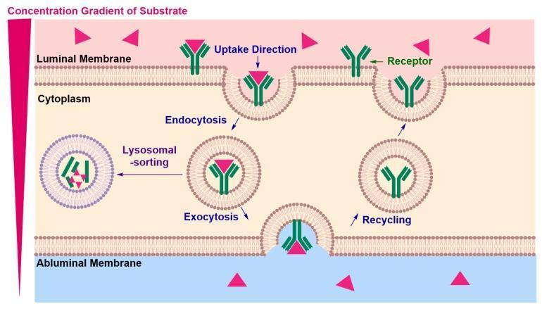
A medical pathophysiology diagram illustrating the mechanism of receptor-mediated transcytosis (RMT) across a vascular endothelial cell, such as the blood-brain barrier. The process is initiated at the luminal membrane where a substrate (red triangles) binds to a specific Y-shaped receptor (green). This triggers endocytosis, involving membrane invagination and the formation of an intracellular endosome containing the receptor-cargo complex. The diagram shows two potential pathways for the endosome: lysosomal sorting, leading to cargo degradation, or exocytosis at the abluminal membrane to release the cargo into the interstitial space. Following release, the receptor is shown undergoing a recycling process back to the luminal membrane for subsequent use. A visual concentration gradient is depicted on the left, indicating that transport typically occurs down the concentration gradient from the luminal to the abluminal side. This visual is relevant for understanding targeted drug delivery and nutrient transport mechanisms into the central nervous system.
Adrenaline (Epinephrine) - Complete Reference
Mechanism of Action
| Receptor | Effect |
|---|---|
| α-1 | Vasoconstriction (raises SVR and blood pressure), nasal mucosal shrinkage |
| α-2 | Inhibits further neurotransmitter release (presynaptic) |
| β-1 | Positive inotropy (↑ contractility), chronotropy (↑ heart rate), dromotropy (↑ conduction speed), lipolysis |
| β-2 | Bronchodilation, coronary vasodilation, smooth muscle relaxation, glycogenolysis, uterine relaxation |
- Guyton and Hall Textbook of Medical Physiology, p. 758
- K.J. Lee's Essential Otolaryngology, p. 223
Indications
- Anaphylaxis (drug of choice - first line)
- Cardiac arrest (ACLS/resuscitation)
- Severe bronchospasm / asthma
- Adjunct to local anaesthesia (prolongs duration, reduces bleeding)
- Nasal surgery (topical vasoconstriction)
- Severe contrast reactions
Routes of Administration
1. Intramuscular (IM) - PREFERRED for Anaphylaxis
- Site: Anterolateral thigh (mid-outer thigh) - fastest absorption
- Concentration used: 1:1000 (= 1 mg/mL)
- Adult dose: 0.3-0.5 mg IM (= 0.3-0.5 mL of 1:1000)
- Paediatric dose: 0.01 mg/kg IM (max 0.5 mg per dose)
- Repeat: Every 5-15 minutes as needed (repeat doses needed in up to 35% of anaphylaxis cases)
- Autoinjectors available: 0.1 mg (7.5-14 kg), 0.15 mg (15-29 kg), 0.3 mg (≥30 kg)
Epinephrine should be injected within <4 minutes of recognising anaphylaxis - delayed administration is the major contributor to fatalities.
2. Intravenous (IV) - For Cardiac Arrest / Severe Shock
- Use with extreme caution - can cause lethal arrhythmia; cardiac monitoring mandatory
- Concentration: 1:10,000 (= 0.1 mg/mL)
- Dose: 0.01 mg/kg (= 0.1 mL/kg of the 1:10,000 dilution)
- Slow, continuous, low-dose infusion preferred if repeated doses are needed
3. Subcutaneous (SC)
- Less preferred than IM (slower, less predictable absorption)
- 0.01 mg/kg; max 0.3 mg
4. Topical
- Nasal mucosa / mucous membranes: vasoconstriction for surgery or haemostasis
- Aerosolized (inhaled)
- Onset: within 5 minutes; Duration: <1 hour
Dilutions - Summary Table
| Dilution | Concentration | Common Use |
|---|---|---|
| 1:1000 | 1 mg/mL (= 1 g per 1,000 mL) | IM anaphylaxis, topical |
| 1:10,000 | 0.1 mg/mL (= 1 g per 10,000 mL) | IV/IO in arrest, IV bolus |
| 1:100,000 | 0.01 mg/mL | Mixed with local anaesthetics |
| 1:200,000 | 0.005 mg/mL | Local anaesthetic + vasoconstrictor |
Key Dosing Reference (Anaphylaxis)
| Patient | Route | Dose |
|---|---|---|
| Adult | IM (anterolateral thigh) | 0.5 mg of 1:1000 (= 0.5 mL) |
| Child | IM (anterolateral thigh) | 0.01 mg/kg of 1:1000 (max 0.5 mg) |
| Infant/small child | IM | Autoinjector 0.1 mg (if 7.5-14 kg) |
| Cardiac arrest (adult) | IV | 1 mg of 1:10,000 every 3-5 min |
With Local Anaesthetics
- Prolong duration - prevents interstitial drug washout
- Reduce bleeding - vasoconstriction at injection site
- Reduce systemic toxicity - slows absorption
- K.J. Lee's Essential Otolaryngology, p. 223
- Red Book 2021, p. 177-178
- Never give 1:1000 IV directly - it must be diluted to at least 1:10,000 for IV use
- Always use cardiac monitoring with IV administration
- IM into the anterolateral thigh is faster and safer than the deltoid or SC for anaphylaxis
- No absolute contraindications in anaphylaxis - the risk of not giving it always outweighs risk of giving it
How munch to dilute in normal saline
Adrenaline Dilution in Normal Saline - Practical Guide
1. Making 1:10,000 (for IV bolus - cardiac arrest / severe anaphylaxis)
| Step | Action |
|---|---|
| Take | 1 mL of 1:1000 adrenaline (1 mg) |
| Add | 9 mL of normal saline |
| Result | 10 mL of 1:10,000 (0.1 mg/mL) |
- Give 1 mg (10 mL) IV every 3-5 min in cardiac arrest
- Give 0.1 mL/kg IV slowly in anaphylaxis with cardiovascular collapse
2. Continuous IV Infusion (ICU / severe shock / refractory anaphylaxis)
| Preparation | Concentration |
|---|---|
| 1 mg (1 mL of 1:1000) in 250 mL NS | 4 mcg/mL |
| 1 mg (1 mL of 1:1000) in 500 mL NS | 2 mcg/mL |
| 4 mg (4 mL of 1:1000) in 250 mL NS | 16 mcg/mL |
3. Topical / Surgical Use (e.g. nasal surgery, wound haemostasis)
| Dilution | How to Make | Use |
|---|---|---|
| 1:1000 | Ready to use (no dilution) | Direct topical on mucosa |
| 1:2000 | 1 mL adrenaline + 1 mL NS | Nasal mucosa packing |
| 1:10,000 | 1 mL + 9 mL NS | Wound / surgical field |
4. With Local Anaesthetic (e.g. Lidocaine)
| Dilution Needed | How to Make |
|---|---|
| 1:100,000 (10 mcg/mL) | Add 0.1 mL of 1:1000 to 10 mL local anaesthetic |
| 1:200,000 (5 mcg/mL) | Add 0.1 mL of 1:1000 to 20 mL local anaesthetic |
Quick Dilution Formula
To find volume needed: Volume (mL) = Desired dose (mg) ÷ Final concentration (mg/mL)
Critical Safety Rules
- Never give 1:1000 (1 mg/mL) undiluted IV - causes fatal arrhythmia
- For IV use, always dilute to at least 1:10,000 (0.1 mg/mL) minimum
- Give IV bolus slowly with cardiac monitoring
- Label the syringe clearly after diluting - concentration errors are a leading cause of adrenaline fatalities
- For IM anaphylaxis - no dilution needed, use 1:1000 directly into the thigh
1 mL adrenaline + 9 mL normal saline = 10 mL of 1:10,000 - safe for IV use
In easy wording