
management of acute otitis media
"acute otitis media"[MeSH Terms] AND management
acute otitis media tympanic membrane otoscopy
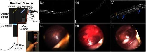
This composite educational graphic details a first-generation handheld optical coherence tomography (OCT) probe and its clinical application in otology. On the left, a schematic diagram labels the internal optics of the handheld scanner, including an LED fiber bundle, collimator, CCD camera, MEMS cold mirror, lens, and an integrated 5-inch display screen. Below the schematic is a photograph of the physical device being held. The right side presents comparative clinical data from pediatric subjects. Panels (a–c) show cross-sectional OCT images, while (d–f) provide corresponding digital otoscopy views. Column 1 (a, d) depicts a normal ear with a healthy tympanic membrane. Column 2 (b, e) illustrates acute otitis media, with otoscopy showing significant erythema and inflammation. Column 3 (c, f) demonstrates chronic otitis media, where the OCT image reveals a thickened tympanic membrane with complex multi-layered structures (indicated by arrows), and otoscopy shows visible white biofilms or middle ear effusion. This visual summarizes the integration of surface video and subsurface cross-sectional imaging for diagnosing middle ear pathologies in primary care settings.
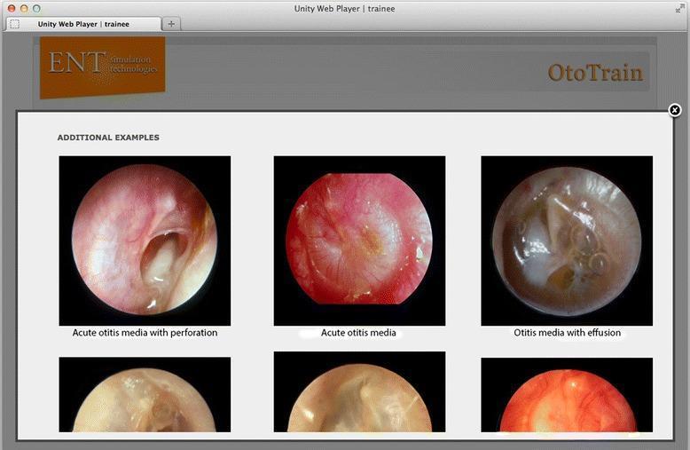
A comparison chart of otoscopic diagnostic images from a medical simulator, focusing on pathologies of the tympanic membrane (TM). The visual contains three labeled primary circular otoscopic views: 1) 'Acute otitis media with perforation,' showing an inflamed, erythematous TM with a large, irregular central perforation through which the middle ear space is visible; 2) 'Acute otitis media,' depicting an intensely hyperemic, opaque, and bulging TM with loss of normal landmarks and light reflex; and 3) 'Otitis media with effusion,' illustrating a translucent, amber-colored TM with visible air-fluid levels and bubbles in the middle ear space. These clinical photographs serve as educational examples for ENT trainees to differentiate between infectious stages (AOM) and non-purulent fluid accumulation (OME). The interface includes secondary supportive images below each category to demonstrate variability in presentation. This content is designed for medical students and residents to master diagnostic otoscopy and middle ear pathology recognition.
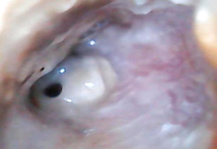
This clinical photograph displays an otoscopic view of the right ear, highlighting pathological findings of the external auditory canal (EAC) and tympanic membrane. The EAC shows significant inflammatory changes, characterized by diffuse erythema and edema of the canal walls. Visible purulent secretions are present within the lumen, suggesting an active infectious process such as chronic suppurative otitis media or acute exacerbation. The tympanic membrane is clearly visualized and exhibits a single, well-defined perforation located in the anterior-inferior quadrant. The perforation appears as a dark, circular opening amidst the remaining translucent, light-reflective portions of the membrane. Normal landmarks like the handle of the malleus are obscured by the surrounding inflammatory tissue and exudate. This image serves as a clinical example of suppurative middle ear disease with associated tympanic membrane rupture and secondary external canal inflammation, relevant for otolaryngology training in diagnostic otoscopy.
Management of Acute Otitis Media (AOM)
Definition and Diagnosis
| Scenario | Findings Required |
|---|---|
| 1 | Moderate to severe bulging of the tympanic membrane (TM) |
| 2 | Mild bulging of the TM + acute onset ear pain (<48 h) OR intense TM erythema |
| 3 | Acute otorrhoea not due to otitis externa or foreign body |
Erythema alone is insufficient - it can result from crying or fever.
Common Pathogens
- Streptococcus pneumoniae - ~23-30% of cases
- Non-typeable Haemophilus influenzae - ~29% (most common in some series)
- Moraxella catarrhalis (Branhamella) - ~10%
- Viral co-infection is common (rhinovirus, RSV, parainfluenza)
Management Algorithm
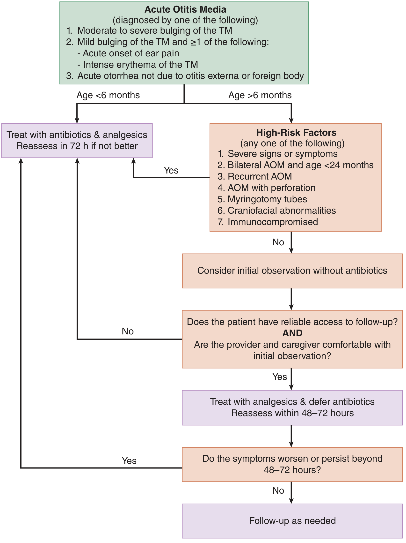
Step 1: Pain Control (All Patients)
- Ibuprofen 10 mg/kg PO every 6 hours PRN
- Acetaminophen 15 mg/kg PO/PR every 4 hours PRN
- Oxycodone 0.1 mg/kg PO every 4 hours PRN
- Hydrocodone 0.2 mg/kg PO every 6 hours PRN
- Antipyrine/benzocaine 2-3 drops every 1-2 hours PRN
- Lidocaine 2% aqueous 2-3 drops every 1-2 hours PRN
- Contraindicated if TM perforation or tympanostomy tubes present
Step 2: Antibiotics vs. Observation
Immediate Antibiotics Required
- Age < 6 months (always treat)
- Severe signs/symptoms (temperature ≥39°C, severe otalgia)
- Age < 24 months with bilateral AOM
- Recurrent AOM
- AOM with TM perforation
- Patient has myringotomy tubes
- Craniofacial abnormalities (cleft palate, Down syndrome)
- Immunodeficiency
Initial Observation Acceptable (no immediate antibiotics)
- Age 6-23 months with unilateral AOM, mild symptoms (ear pain <48 h, temp <39°C)
- Age ≥24 months with unilateral OR bilateral AOM, mild/moderate symptoms
- Reliable access to follow-up within 48-72 hours
- Provider and caregiver both comfortable with observation
A "safety-net/wait-and-see prescription" can be given at the initial visit with instructions to fill only if symptoms worsen or do not improve within 48-72 hours. - Tintinalli's Emergency Medicine, p. 799
Step 3: Antibiotic Therapy
First-Line
- Higher dose achieves middle ear concentrations exceeding the MIC for highly resistant S. pneumoniae
- Duration: 10 days for children < 2 years or severe disease; 5-7 days for mild/moderate disease in children ≥2 years
Alternatives / Penicillin Allergy
| Situation | Drug |
|---|---|
| Non-severe penicillin allergy | Cefdinir, cefuroxime, or cefpodoxime |
| Severe penicillin allergy (anaphylaxis) | Azithromycin or clarithromycin (less effective against S. pneumoniae) |
| Unable to take oral | Ceftriaxone IM/IV (single dose or 3-day course) |
| Concurrent purulent conjunctivitis ("conjunctivitis-otitis syndrome") | Amoxicillin-clavulanate (covers H. influenzae which causes both) |
| Received amoxicillin in past 30 days | Amoxicillin-clavulanate |
Step 4: Treatment Failure
| Failure Step | Management |
|---|---|
| 1st failure (amoxicillin fails) | Switch to amoxicillin-clavulanate OR ceftriaxone IM (covers beta-lactamase-producing M. catarrhalis and H. influenzae) |
| 2nd failure | Clindamycin + 3rd-generation cephalosporin; OR refer to ENT for tympanocentesis and culture |
Complications and Their Management
- TM perforation - most common; typically heals spontaneously after resolution
- Mastoiditis - requires IV antibiotics; CT scan; cortical mastoidectomy if needed
- Facial nerve paralysis - wide myringotomy + systemic antibiotics; mastoidectomy if mastoiditis suspected; usually full recovery
- Intracranial spread - meningitis, epidural/subdural abscess, lateral sinus thrombosis, otitic hydrocephalus - require IV antibiotics + surgical drainage + CT/MRI
- Appropriate antibiotics + myringotomy and ventilating tube placement
- Tympanocentesis for culture and sensitivity
- CT/MRI if intracranial complication suspected
Special Populations
- Adults: Usually treated with antibiotics. Amoxicillin remains first-line.
- Neonates/young infants: Always treat with antibiotics; facial nerve paralysis is a recognized complication (usually incomplete, associated with intact bulging TM).
- Immunocompromised: Always treat; broader coverage may be needed.
Recurrent AOM (≥3 episodes in 6 months or ≥4 in 12 months)
- Tympanostomy tube placement is the primary surgical intervention
- Reduces recurrence rate and restores hearing during periods of effusion
- Prophylactic antibiotics are not routinely recommended
What NOT to Do
- Decongestants and antihistamines - a 2025 Cochrane meta-analysis (PMID 41307300) found no benefit and potential harm in children; do not use
- Topical otic drops in the presence of TM perforation or tympanostomy tubes
- Intranasal steroids, systemic steroids - not indicated for AOM
Otoscopic Reference
- Tintinalli's Emergency Medicine: A Comprehensive Study Guide, pp. 798-802
- Cummings Otolaryngology Head and Neck Surgery, p. 2703
- Scott-Brown's Otorhinolaryngology Head & Neck Surgery Vol 2
- 2025 Cochrane review: Decongestants and antihistamines for AOM in children (PMID 41307300)
- 2025 systematic mapping of AOM CPGs: PMID 40717508