
Iga nephropathy from Harrison
IgA nephropathy mesangial deposits immunofluorescence histology
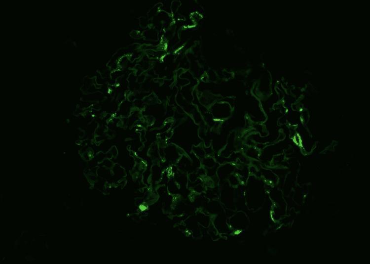
This diagnostic immunofluorescence image demonstrates a renal glomerulus exhibiting a characteristic staining pattern for IgA nephropathy. Against a dark background, a bright green fluorescent signal is localized primarily within the mesangial regions. The signal presents as granular, irregular deposits concentrated in the mesangial matrix, reflecting the entrapment of immunoglobulin A (IgA) immune complexes. The overall structure of the glomerulus shows a complex, lobulated architecture with visible capillary loops. While some areas demonstrate intense, discrete granular fluorescence, others show a more diffuse or attenuated signal, which may correlate with underlying morphological changes such as mesangial hypercellularity or segmental sclerosis. This imaging modality is essential in nephropathology for differentiating between various forms of glomerulonephritis based on the specific distribution (mesangial vs. capillary wall) and nature (granular vs. linear) of immune deposits.
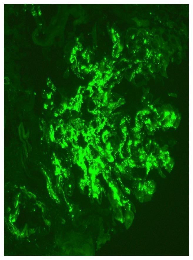
This diagnostic image is a high-magnification (x400) immunofluorescence microscopy of a renal biopsy specimen. The visual field demonstrates a single glomerulus exhibiting a classic pathological pattern of intense, apple-green fluorescent staining. The primary finding is the global, 3+ intensity mesangial deposition of immunoglobulin A (IgA). The deposits are characterized by a granular and confluent distribution primarily restricted to the mesangial matrix and stalk regions of the glomerular tuft, highlighting the branched architecture of the mesangium. There is a notable absence of significant capillary loop or basement membrane staining, which is highly characteristic of IgA nephropathy (Berger disease). The surrounding renal parenchyma is dark, providing high contrast to the localized immunocomplex accumulation within the glomerular architecture. This diagnostic finding is a hallmark educational example used to confirm the diagnosis of primary IgA nephropathy or Henoch-Schönlein purpura nephritis in clinical pathology.
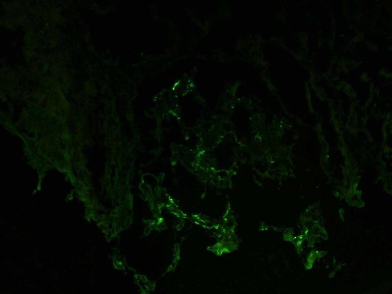
This diagnostic image is a direct immunofluorescence (DIF) micrograph of a renal biopsy specimen. It shows positive staining for IgA antibodies within a glomerulus. The fluorescence pattern is characterized by granular, segmental deposits primarily localized in the mesangium, creating a scattered apple-green signal against a dark, non-reactive background. The distribution is discontinuous and varies in intensity, which is a hallmark finding in IgA nephropathy (Berger's disease). The image illustrates the specific immunological deposition that leads to glomerular injury, highlighting the importance of immunofluorescence in differentiating various types of glomerulonephritis. Key educational concepts include the recognition of mesangial immune complex deposition and the diagnostic utility of antisera specific for immunoglobulin subclasses in nephropathology.
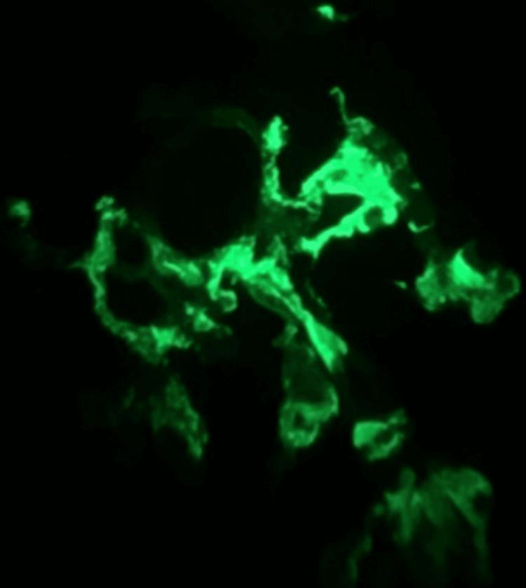
This diagnostic image shows immunofluorescence microscopy of a renal biopsy specimen, specifically demonstrating glomerular IgA staining. Set against a dark background, the image reveals intense apple-green fluorescence within the mesangium and along some capillary loops of a glomerulus. The staining pattern is granular and predominantly mesangial, characterized by irregular clumps and lobulated deposits of IgA immune complexes. This visual finding is a hallmark diagnostic feature of IgA nephropathy (Berger disease) or IgA vasculitis (Henoch-Schönlein purpura) in a clinical context of palpable purpura and subnephrotic proteinuria. The distribution illustrates the localization of immunoglobulin A deposits within the glomerular tuft, which is essential for differentiating immune-mediated glomerulonephritides.
IgA Nephropathy (Berger's Disease)
Definition & Epidemiology
- Sex: Male preponderance
- Age: Peak in the 2nd and 3rd decades of life
- Familial clustering: Rare
- Geographic variation: 30-45% prevalence along the Asian and Pacific Rim, 20% in Southern Europe, 10% in Northern Europe and North America (may reflect true ethnic variation or detection differences)
Associations / Secondary IgA Deposits
- Chronic liver disease, Crohn's disease, celiac disease
- Chronic bronchiectasis, idiopathic interstitial pneumonia
- Dermatitis herpetiformis, mycosis fungoides
- Ankylosing spondylitis, HIV infection, Sjogren's syndrome
Pathogenesis
- Genetic component: Susceptibility shown (uncommonly) to have genetic basis depending on geography/"founder effects." No single causal gene identified.
- Central mechanism: Abnormalities in O-glycosylation of the hinge region of polymeric IgA1
- Poorly galactosylated IgA1 exposes N-acetyl-galactosamine in truncated hinge regions
- This is recognized by IgG or IgA1 antibodies → immune complex formation in circulation or in situ after glomerular deposition
- Galactose-deficient IgA1 activates complement via the alternative and lectin pathways
- Viral/antigen exposure or hereditary defects in alternative complement pathway proteins may influence disease manifestation
- Elevated serum IgA levels in 20-50% of patients; IgA in skin biopsies in 15-55%
Biopsy / Pathology
- IgA deposits are typically J-chain containing polymeric IgA1
- Light microscopy may show: DPGN, segmental sclerosis, or rarely segmental necrosis with cellular crescent formation (presents as RPGN)
- A kidney biopsy is necessary to confirm the diagnosis despite elevated serum IgA or skin deposits
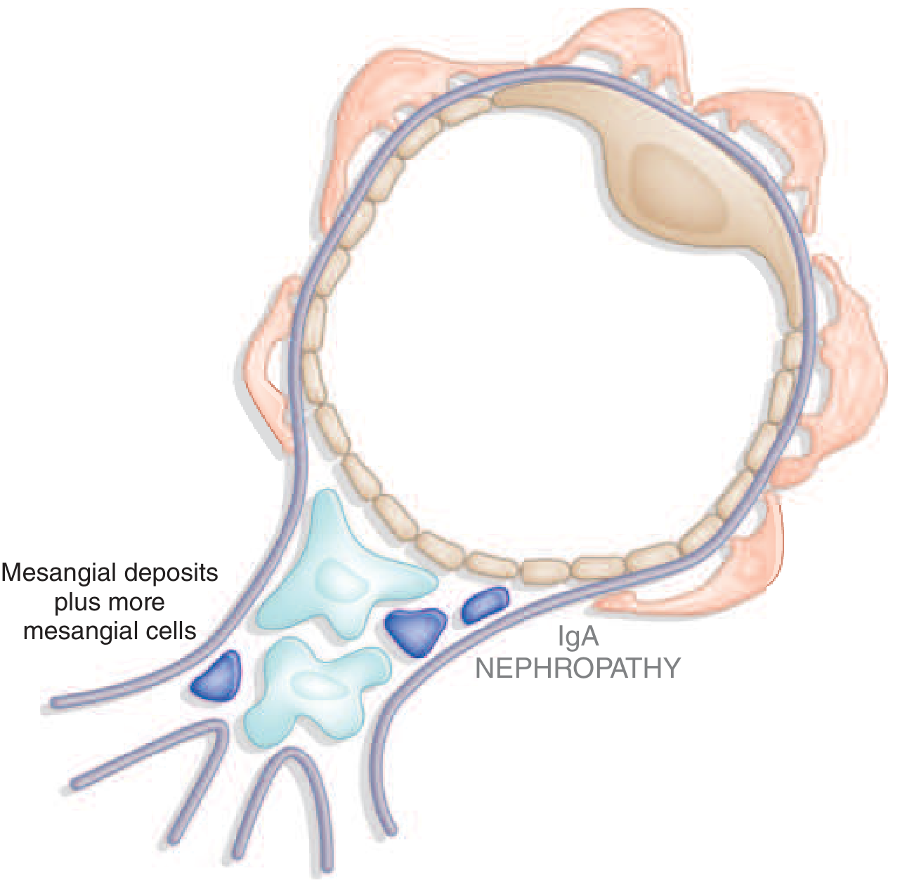
Oxford Classification / MEST-C Score
| Letter | Feature |
|---|---|
| M | Mesangial hypercellularity |
| E | Endocapillary hypercellularity |
| S | Segmental glomerulosclerosis |
| T | Tubular interstitial fibrosis |
| C | Crescents |
Clinical Presentations
- Persistent asymptomatic microscopic hematuria
- Recurrent episodes of macroscopic hematuria during or immediately following an upper respiratory infection, often with proteinuria ("synpharyngitic hematuria")
- Nephrotic syndrome: uncommon
- Proteinuria may appear late in disease course
- Rarely: acute kidney failure with rapidly progressive picture (RPGN)
Prognosis
- Can be a benign disease - some patients achieve complete remission
- Others have ongoing hematuria with well-preserved kidney function
- Slow progression to ESKD in only 25-30% of patients over 20-25 years
- Risk varies considerably among populations
- Hypertension or proteinuria
- Absence of episodes of macroscopic hematuria
- Male sex
- Older age of onset
Treatment
| Intervention | Evidence/Comment |
|---|---|
| ACE inhibitors | Supported by both large multi-disease studies and small IgA-specific studies; used for proteinuria or declining kidney function |
| Steroids / immunosuppressives | Conflicting results |
| Budesonide (oral targeted-release) | Shown to reduce proteinuria and improve preserved kidney function in patients at risk of rapid progression |
| SGLT2i (dapagliflozin) | Preliminary studies show reduced adverse kidney outcomes |
| Sparsentan (endothelin antagonist) | Preliminary studies show reduced adverse kidney outcomes |
| Tonsillectomy | Suggested benefit in select patients in small studies |
| Fish oil | Suggested benefit in select patients in small studies |
| RPGN presentation | Steroids + cytotoxic agents + plasmapheresis |