
Macular Rashes over hand face and trunk 16 yr no fever what are the diagnosis
macular rash hands face trunk secondary syphilis pityriasis rosea
pityriasis rosea herald patch trunk rash
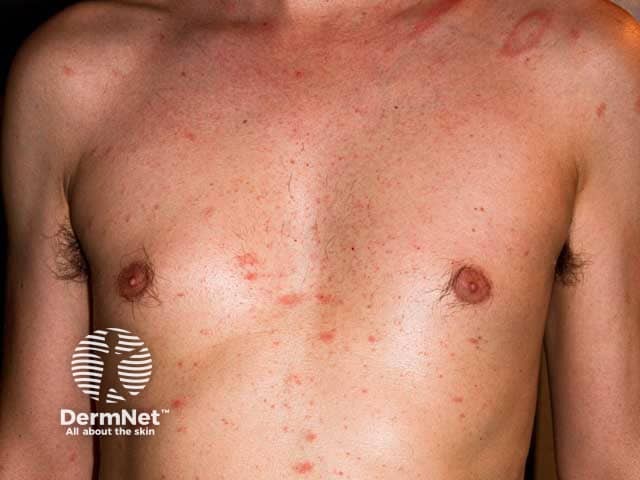
Clinical photography of the skin in an adult male demonstrating pityriasis rosea with a herald patch on the left shoulder and a secondary eruption across the anterior trunk. Modality: Clinical color photograph of cutaneous lesions. Anatomical location: trunk and left shoulder; body region: anterior chest, shoulders; left shoulder herald patch. Visual features: a focal, oval, erythematous patch with a fine, peripheral, collarette scale characteristic of a herald patch, measuring several centimeters in diameter; surrounding smaller, discrete, pink-to-reddish papules and plaques arranged along the trunk in a roughly perpendicular or 'Christmas tree' distribution. The skin surface shows mild surface scaling, slight erythema, and no purulence or crusting. The overall appearance is consistent with pityriasis rosea in the classic stage, with the herald patch preceding or accompanying generalized lesions. Diagnostic significance: This pattern supports a self-limited, benign inflammatory dermatosis with uncertain etiology, possibly viral (HHV-6/7) association; prognosis is favorable, treatment typically supportive (emollients, anti-pruritics). Differential diagnoses include tinea corporis, pityriasis versicolor, secondary syphilis, and eczema. Clinical correlation is important: young adults, acute onset, trunk-dominant rash, PR often resolves in 6-8 weeks. This image is useful for education, pattern recognition, and dermatology teaching materials regarding herald patch and classic pityriasis rosea presentation criteria.
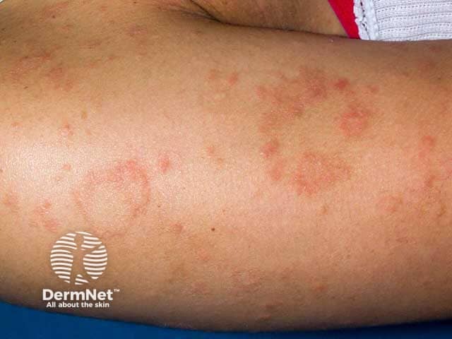
This is a clinical dermatology photograph of in vivo human skin on the forearm, illustrating a herald patch with accompanying pityriasis rosea–type lesions in a patient with skin of color. Imaging modality: clinical photography; technique: standard digital single-shot close-up under diffuse studio lighting or natural daylight. Primary subject: an oval to circular erythematous-to-pink patch with fine peripheral scaling (collarette scale) and slight central clearing, consistent with a herald patch of pityriasis rosea. Surrounding skin shows multiple smaller patches or papules at various stages of eruption, creating a subtle, tree-like distribution that may become more pronounced with trunk involvement in subsequent weeks. In darker phototypes, erythema can be muted, and residual postinflammatory hyperpigmentation may dominate as brown-pigmented patches after resolution. The lesion margins are well defined, the surface may show delicate scale, and there is no vesiculation or crusting observed. The image highlights characteristic pigmentary presentation in skin of color and emphasizes diagnostic cues: herald patch with collarette scaling, annular geometry, and the expected eruption sequence preceding a generalized rash. Clinically, this supports pityriasis rosea over mimics such as tinea corporis, eczema, or psoriasis, and informs patient counseling, prognosis, and follow-up assessment.
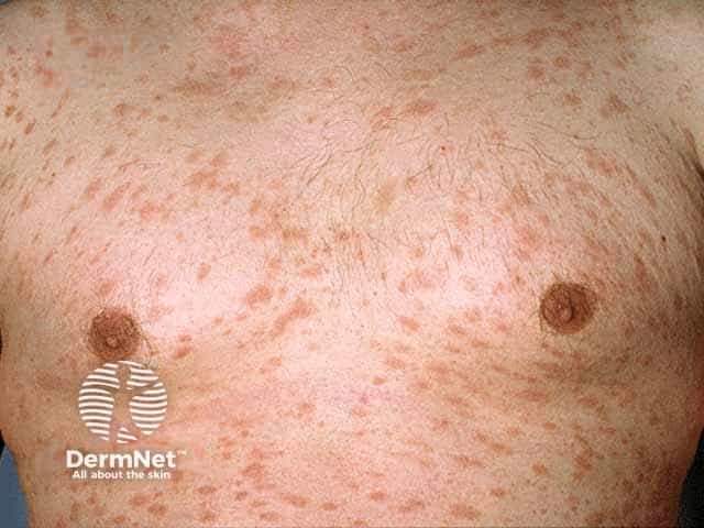
Imaging modality: Clinical photography of the anterior chest using standard color digital photography. Perspective: frontal view of the chest and upper trunk. Anatomical location: anterior thorax/pectoral region of the trunk, bilateral chest wall. Skin layer: epidermis and dermis involved with superficial scaling. Visual features: numerous erythematous to pink-brown oval and round macules and plaques distributed across the chest, some coalescing into larger patches; fine, peripheral scale is visible at lesion margins; lesions have slightly raised but flat morphology with a matte surface; no vesicles or crusts noted; mild surrounding erythema; skin texture appears normal elsewhere. Distribution: trunk-dominant eruption with symmetrical involvement of the chest, consistent with pityriasis rosea; clinical pattern may reflect a herald patch history in some individuals. Pathology: histology not available in this image; clinical likelihood favored by characteristic trunk involvement and collarette scaling. Diagnostic significance: supports diagnosis of pityriasis rosea when integrated with patient history and exam; helps distinguish from differential diagnoses such as tinea versicolor, eczema, seborrheic dermatitis, psoriasis, and secondary syphilis. Clinical relevance: useful for dermatology education, telemedicine documentation, and monitoring treatment response to topical steroids, calcineurin inhibitors, or antihistamines for pruritus. Suggested coding terms: pityriasis rosea, trunk eruption, chest rash, collarette scaling, herald patch (if present).
secondary syphilis maculopapular rash palms
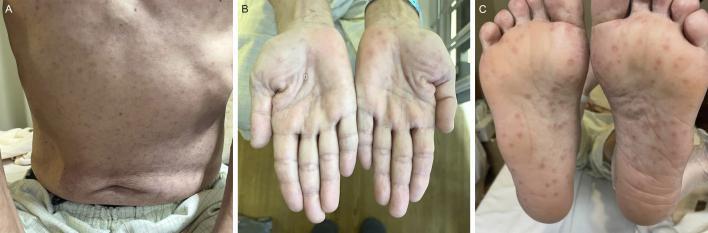
A clinical photograph series illustrating dermatological manifestations of secondary syphilis across three anatomical regions. Panel A shows the posterior trunk displaying a diffuse, symmetric, erythematous maculopapular rash. Panel B and C depict the bilateral palms and soles, respectively, featuring characteristic well-demarcated, rounded, erythematous to reddish-brown macules and papules. Some acral lesions exhibit subtle peripheral scaling, consistent with Biett's collarette. This classic 'copper-colored' rash distribution, specifically involving the palms and soles, is a pathognomonic clinical sign for secondary syphilis. The presentation highlights the generalized nature of the Treponema pallidum infection during the disseminated stage. This visual resource is intended for medical education regarding sexually transmitted infections and differential diagnosis of palmoplantar eruptions.
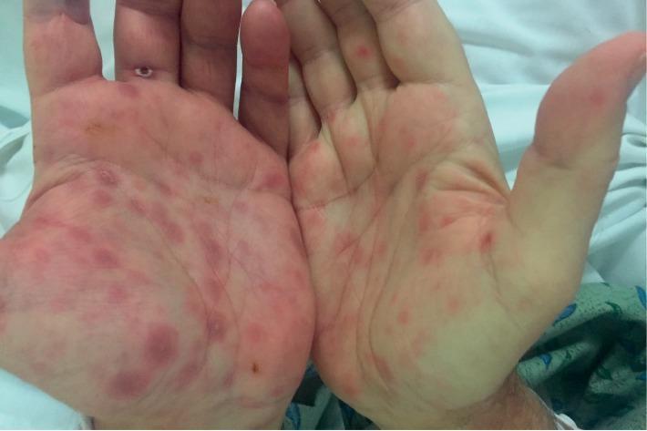
Clinical photograph of the palmar surfaces of both hands, demonstrating a bilateral, symmetric maculopapular eruption. The lesions are well-demarcated, varying in color from erythematous to dusky reddish-purple. The morphology consists of multiple round-to-oval macules and slightly raised papules, approximately 5-10 mm in diameter, distributed across the palms and extending onto the ventral surfaces of the fingers. Some lesions appear deeper in color with a copper-colored or ham-colored hue, characteristic of secondary syphilis. The surrounding skin is largely unremarkable, without significant edema or diffuse desquamation. In the clinical context of a genital chancre and high RPR titers, these findings are highly suggestive of the palmoplantar rash associated with secondary syphilis (syphilis stage II). This image serves as a classic diagnostic example of infectious dermatology for medical education, emphasizing the importance of examining the palms and soles in suspected systemic infections.
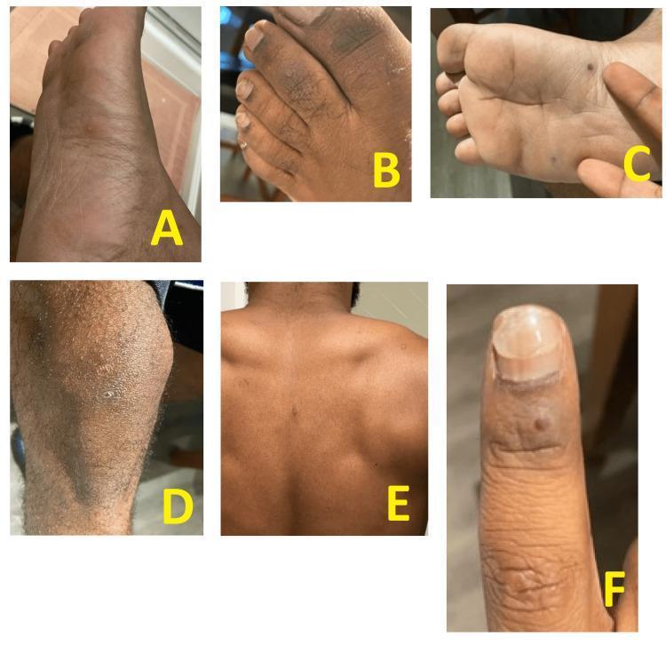
Six clinical photographs (labeled A-F) demonstrate a disseminated maculopapular exanthem and isolated pustules on a patient with dark skin. Images A, B, and C focus on the feet, showing scattered erythematous papules and distinct small, dark-colored pustules on the plantar surface and toes. Image D shows a diffuse erythematous maculopapular rash on the lower leg with interspersed small pustules. Image E displays sparse, individual papules across the back, and image F highlights a solitary papule on the dorsal aspect of a finger near the nail. The lesions are non-grouped and vary from small, raised red bumps (papules) to darker, fluid-filled elevations (pustules). The distribution is widespread, involving the palms, soles, extremities, and trunk. This clinical presentation is characteristic of systemic infectious processes, such as secondary syphilis or viral exanthems (e.g., mpox), and is useful for teaching dermatologic manifestations of systemic disease in diverse skin phototypes.
Differential Diagnosis: Macular Rash - Hands, Face & Trunk (Afebrile, 16 yr)
1. Secondary Syphilis (Must Rule Out First)
- The rash is classically non-pruritic, macular or maculopapular, beginning on the trunk and proximal extremities
- Palm and sole involvement is the hallmark - highly specific for syphilis
- May also show mucous membrane patches, condyloma lata, and lymphadenopathy
- Fever, myalgias, sore throat can be present but are often absent or mild
- RPR / VDRL (screening) + TPHA/FTA-ABS (confirmatory) are diagnostic
- Symptom to Diagnosis (4th Ed): "Secondary syphilis should always be considered in the differential diagnosis of rash in sexually active patients"
2. Pityriasis Rosea (Most Common in this Age Group)
- Most common in 15-40 year olds, often spring/fall onset
- Often starts with a single "herald patch" (oval, 2-5 cm, salmon-colored, fine scaling) 1-2 weeks before the generalized eruption
- Generalized eruption on trunk and proximal arms in a "Christmas tree" pattern along skin tension lines
- Face, palms, soles are rarely involved (atypical pityriasis rosea)
- Lesions show a collarette of scale with inner open edge - diagnostic finding
- Usually self-limiting (4-16 weeks); associated with HHV-6/7 reactivation
- Tintinalli's Emergency Medicine: "Syphilis is another key item on the differential, particularly with palm and sole involvement, and a rapid plasma reagin should be considered"
3. Systemic Lupus Erythematosus (SLE)
- Young adolescent females are a classic demographic for SLE
- Malar (butterfly) rash across cheeks/nose sparing nasolabial folds
- Additional findings: discoid rash, photosensitivity rash, oral ulcers, arthritis, serositis
- Fever may be absent early in disease
- ANA, anti-dsDNA, anti-Smith antibodies; CBC (cytopenias), urinalysis (proteinuria)
- SLICC 2012 / ACR-EULAR 2019 criteria for diagnosis
4. Drug Eruption (Morbilliform / Drug Rash)
- Most common drug reaction pattern - diffuse macular or maculopapular eruption
- Classically starts centrally on trunk, spreads to face and extremities
- Onset typically 1-2 weeks after starting a new drug (antibiotics - beta-lactams, sulfa drugs; NSAIDs, anticonvulsants)
- Usually non-pruritic to mildly pruritic, symmetric, often no fever in simple morbilliform eruptions
- Key question: any new medication in the past 2-3 weeks?
5. Viral Exanthem
| Virus | Key Features |
|---|---|
| Parvovirus B19 (Fifth disease) | "Slapped cheek" face + lacy reticular rash trunk/extremities; adolescents can present atypically |
| EBV (Infectious Mono) | Maculopapular rash especially if given ampicillin/amoxicillin; with pharyngitis, LAP |
| Rubella | Macular rash face → trunk → extremities; post-auricular lymphadenopathy; mild/absent fever |
| HHV-6/7 | Usually younger children but can cause pityriasis rosea in adolescents |
6. Guttate Psoriasis
- Triggered by streptococcal pharyngitis (throat swab helpful)
- Raindrop-like, small (< 1 cm) scaly red papules on trunk, especially in children and young adults
- Distinct from pityriasis rosea: no herald patch, no collarette scale
- Can involve face
7. Tinea Versicolor (Pityriasis Versicolor)
- Hypopigmented or hyperpigmented macules on trunk, neck, proximal arms
- Fine "furfuraceous" scale when scratched; KOH scraping shows "spaghetti and meatballs" pattern
- Adolescents and young adults; associated with Malassezia overgrowth
8. Subacute Cutaneous Lupus Erythematosus (SCLE)
- Photosensitive, annular or papulosquamous macules/plaques on sun-exposed areas (face, arms, trunk)
- Anti-Ro/SSA antibodies; may occur without systemic SLE
Diagnostic Approach Summary
| Test | Rules Out / Rules In |
|---|---|
| RPR/VDRL + TPHA | Secondary syphilis |
| ANA, anti-dsDNA, CBC, UA | SLE |
| KOH scraping | Tinea versicolor |
| Throat swab / ASO titer | Guttate psoriasis (post-strep) |
| Medication history | Drug eruption |
| Parvovirus B19 IgM | Fifth disease |
| EBV monospot / serology | EBV exanthem |
Key clinical pearl: If palm/sole involvement is present, syphilis moves to the top of the list. If there is a preceding single oval "herald patch," pityriasis rosea is most likely. If it is a young adolescent female with facial rash and arthralgia, always screen for SLE.
- Tintinalli's Emergency Medicine, p. 1679 - Pityriasis Rosea section
- Symptom to Diagnosis (4th Ed), p. 103 - Secondary Syphilis highlights