Here is a complete, diagrammatic breakdown of both pathways - sourced from Lippincott Biochemistry 8e and Harper's Illustrated Biochemistry 32e.
Glycogenesis & Glycogenolysis
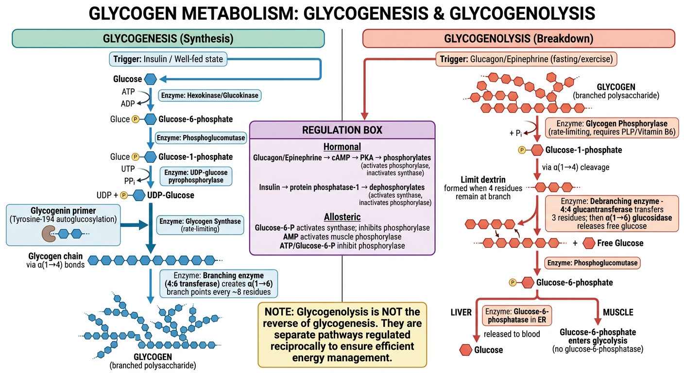
Key principle: Glycogenolysis is NOT the reverse of glycogenesis. They are entirely separate pathways using different enzymes, different intermediates, and different regulation. - Harper's Illustrated Biochemistry, 32e
AI-Generated Master Diagram
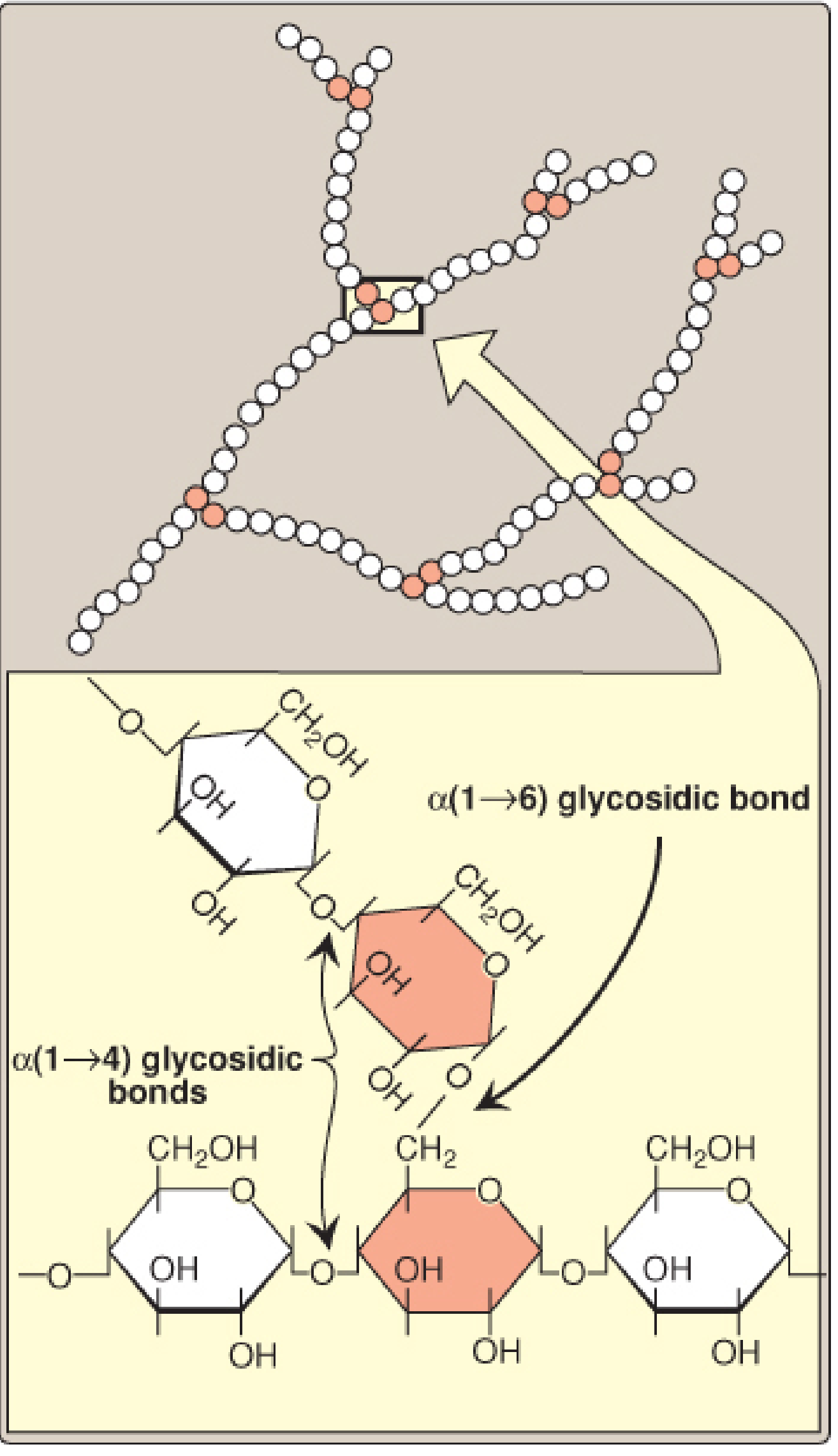
Glycogen Structure
Glycogen is a highly branched polysaccharide of α-D-glucose:
- α(1→4) glycosidic bonds form the linear backbone chains
- α(1→6) glycosidic bonds form branch points, located every ~8 glucosyl residues apart
➡️ GLYCOGENESIS (Synthesis)
Trigger: Well-fed state, insulin, high glucose
Location: Cytosol of liver and muscle
Energy cost: ATP + UTP consumed
GLUCOSE
   │  Hexokinase / Glucokinase (ATP → ADP)
   ▼
Glucose-6-phosphate (G6P)
   │  Phosphoglucomutase (G1,6-bisphosphate intermediate)
   ▼
Glucose-1-phosphate (G1P)
   │  UDP-glucose pyrophosphorylase (+ UTP → PPi)
   │  [PPi → 2Pi by pyrophosphatase — drives reaction forward]
   ▼
UDP-Glucose  ←── Activated glucose donor
   │
   │  [Glycogenin — autoglucosylation at Tyrosine-194]
   │  [Builds a short α(1→4) primer of ≥4 glucose residues]
   ▼
Primer (short glucosyl chain)
   │  Glycogen Synthase (RATE-LIMITING)
   │  [Transfers glucose from UDP-glucose → nonreducing end]
   │  [Forms α(1→4) bonds]
   ▼
Linear chain (amylose-like)
   │  Branching enzyme (Amylo-4,6-glucan transferase / 4:6 transferase)
   │  [Transfers ≥6 residue block to C6-OH, forming α(1→6) branch]
   ▼
GLYCOGEN ✓
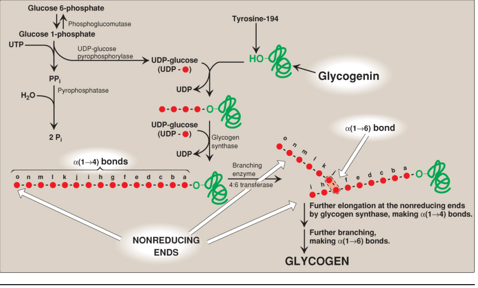
Full pathway diagram from Lippincott Biochemistry 8e:
⬅️ GLYCOGENOLYSIS (Breakdown)
Trigger: Fasting (glucagon), exercise/stress (epinephrine), low blood glucose
Location: Cytosol
Key product: Glucose-1-phosphate (enters directly into metabolism, no ATP wasted)
GLYCOGEN
   │  Glycogen Phosphorylase (RATE-LIMITING)
   │  [Phosphorolysis of α(1→4) bonds at nonreducing ends]
   │  [Requires pyridoxal phosphate / Vitamin B6 as coenzyme]
   │  [Continues until 4 glucose residues remain at each branch → "limit dextrin"]
   ▼
Glucose-1-phosphate (most residues) + Limit Dextrin
   │  Debranching Enzyme (bifunctional):
   │    1. Glucantransferase (4:4 transferase): moves 3 of 4 residues to another chain
   │    2. α(1→6)-glucosidase: cleaves last residue → FREE glucose (nonphosphorylated)
   ▼
Glucose-1-phosphate (bulk) + free Glucose (branch residues)
   │  Phosphoglucomutase
   ▼
Glucose-6-phosphate
   │
   ├── LIVER: Glucose-6-phosphatase (in ER lumen)
   │         → Free Glucose → released to blood ✓
   │         (maintains blood glucose homeostasis)
   │
   └── MUSCLE: No glucose-6-phosphatase
              → Enters glycolysis directly
              (fuels muscle contraction only)
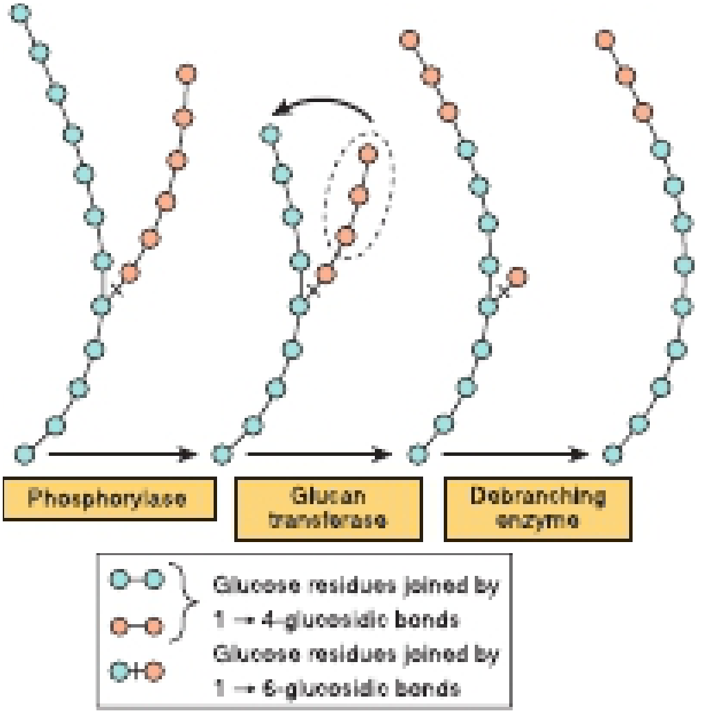
Steps of glycogenolysis - phosphorylase, glucan transferase, debranching enzyme:
⚙️ REGULATION
Hormonal (Covalent) Regulation
| Hormone | Signal | Effect on Glycogen Synthase | Effect on Glycogen Phosphorylase |
|---|
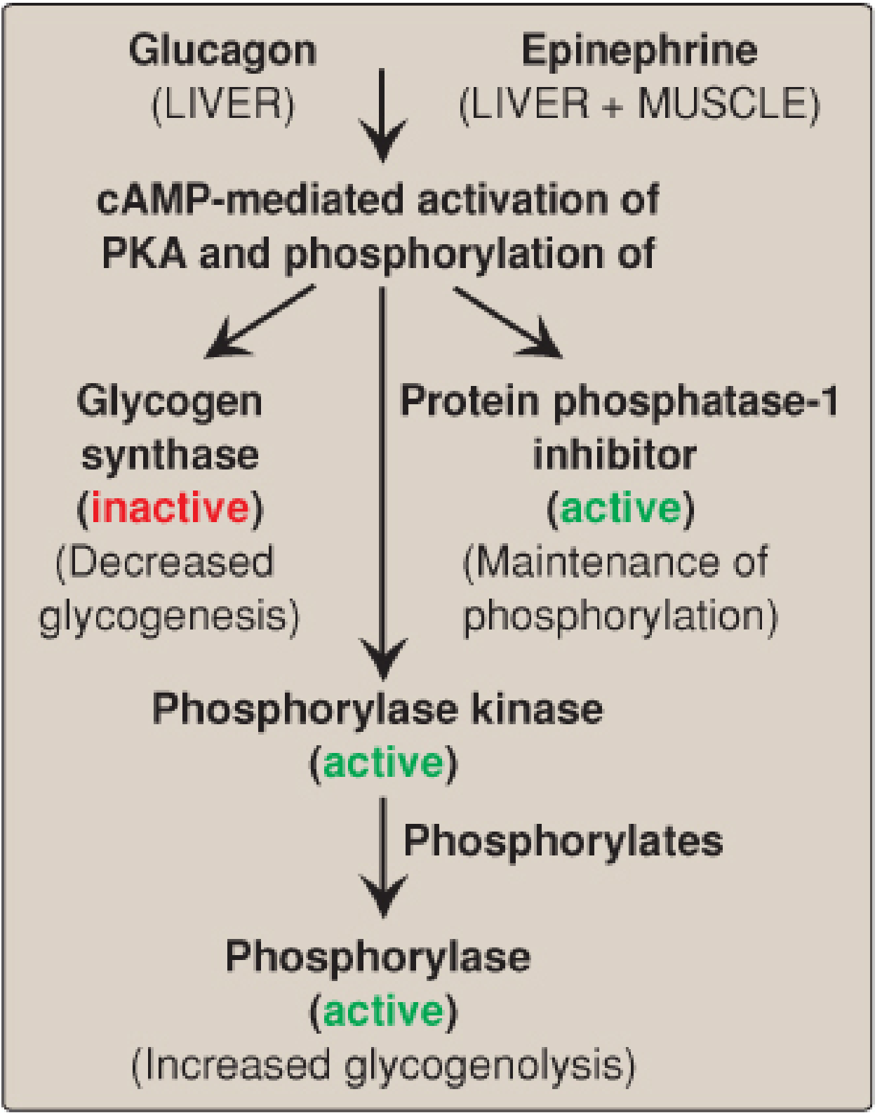
| Glucagon (liver) | cAMP ↑ → PKA activated | Phosphorylated → INACTIVE | Phosphorylated → ACTIVE |
| Epinephrine (liver + muscle) | cAMP ↑ → PKA activated | Phosphorylated → INACTIVE | Phosphorylated → ACTIVE |
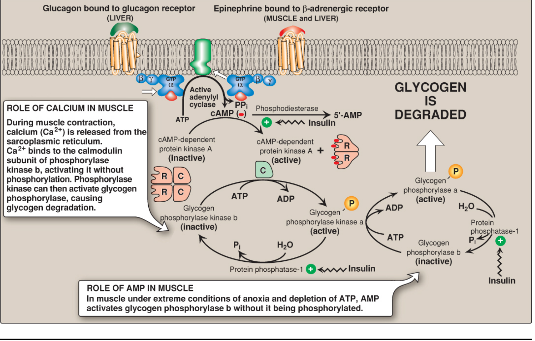
| Insulin | Activates Protein Phosphatase-1 | Dephosphorylated → ACTIVE | Dephosphorylated → INACTIVE |
Cascade for glycogenolysis activation (Glucagon/Epinephrine):
Full signaling cascade (PKA, phosphorylase kinase, role of Ca²⁺ and AMP in muscle):
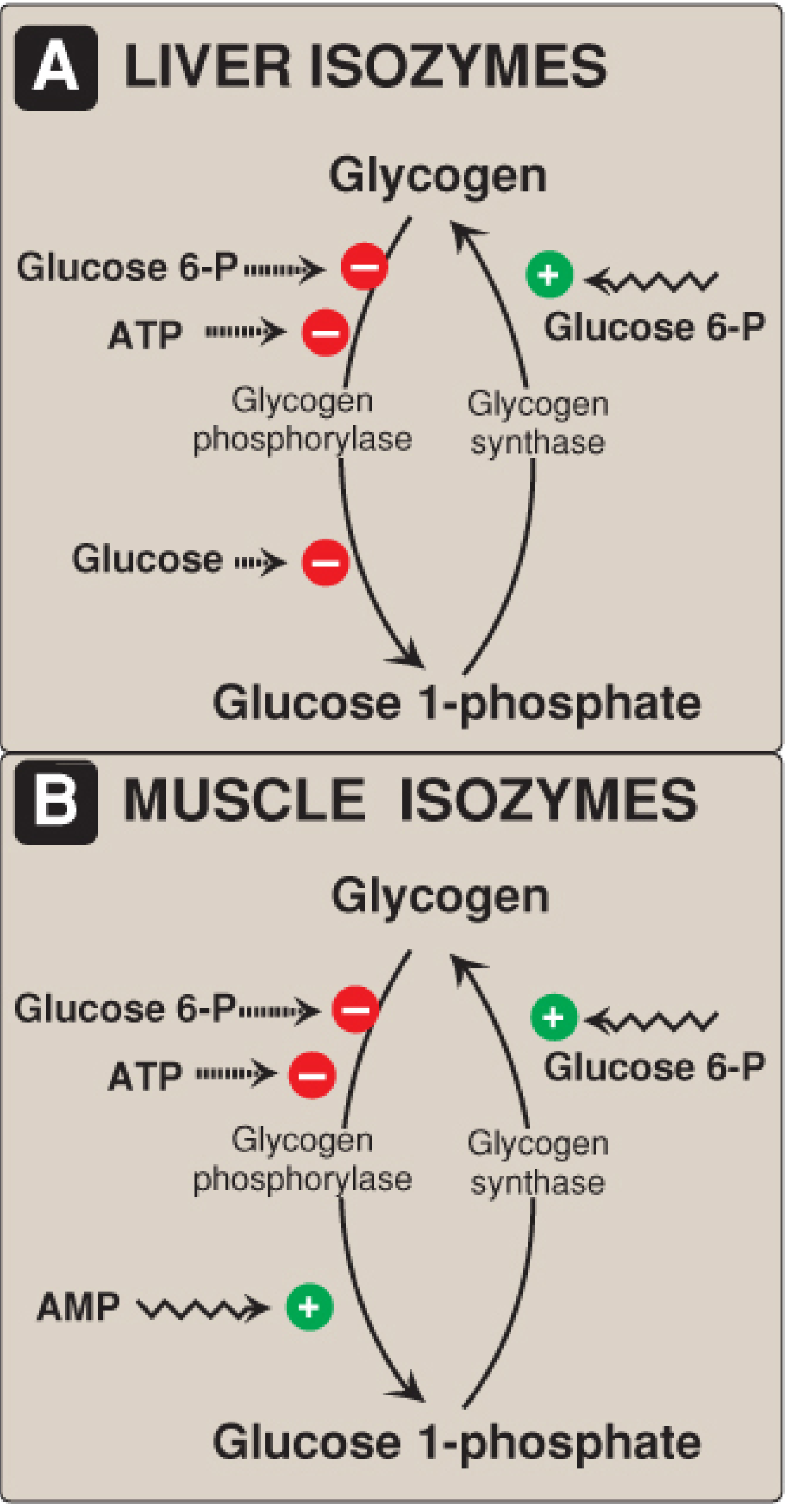
Allosteric Regulation
| Effector | Effect on Synthase | Effect on Phosphorylase | Tissue |
|---|
| Glucose-6-phosphate (high) | ✅ Activates ("b" → active) | ❌ Inhibits | Liver + Muscle |
| ATP (high) | - | ❌ Inhibits | Liver + Muscle |
| Free Glucose (high) | - | ❌ Inhibits | Liver only |
| AMP (high/anoxia) | - | ✅ Activates "b" without phosphorylation | Muscle only |
| Ca²⁺ (muscle contraction) | - | Activates via CaM → phosphorylase kinase | Muscle only |
📍 Tissue-Specific Differences
| Feature | LIVER | MUSCLE |
|---|
| Purpose | Blood glucose homeostasis | Local energy for contraction |
| Glucagon response | Yes (glucagon receptor present) | No |
| Glucose-6-phosphatase | Present → free glucose exported | Absent → G6P enters glycolysis |
| Allosteric phosphorylase activator | Free glucose (inhibits, not activates) | AMP (activates) |
| Glycogen content | ~100g (depleted after ~24h fast) | ~400g (not depleted by short fast) |
⚠️ Glycogen Storage Diseases (GSDs) - Quick Reference
| Type | Disease | Deficient Enzyme | Pathway Affected |
|---|
| 0 | Aglycogenosis | Glycogen Synthase | Glycogenesis ↓ |
| I (Ia) | Von Gierke | Glucose-6-phosphatase | Glycogenolysis (final step) |
| II | Pompe | α(1→4)-glucosidase (lysosomal) | Lysosomal glycogen |
| III | Cori/Forbes | Debranching enzyme | Glycogenolysis |
| IV | Andersen | Branching enzyme | Glycogenesis |
| V | McArdle | Muscle phosphorylase | Muscle glycogenolysis |
| VI | Hers | Liver phosphorylase | Liver glycogenolysis |
Sources: Biochemistry, 8th ed - Lippincott Illustrated Reviews, pp. 376-395; Harper's Illustrated Biochemistry, 32e, pp. 184-186