
Write in detail about optic neuritis for pg ophthalmology examination .
Reading File
Finding Sources
Finding Sources
Searching PubMed
"optic neuritis"
Searching the Web
optic neuritis diagnosis management guideline 2024 MRI MOGAD NMOSD
Reading File
Reading File
Finding Sources
Searching PubMed
"optic neuritis" AND treatment
Reading File
Reading File
Searching Images
typical acute demyelinating optic neuritis mild optic disc edema fundus photograph
Reading Web Page
https://www.aao.org/young-ophthalmologists/yo-info/article/5…
Reading File
Finding Sources
Reading Article
PMID: 41391387
query_images_db
Optic Neuritis
Definition
Optic neuritis (ON) is an acute inflammatory optic neuropathy, most commonly caused by demyelination of the optic nerve. In routine ophthalmic usage, it usually refers to typical demyelinating optic neuritis, often associated with multiple sclerosis (MS). It produces acute or subacute monocular visual loss, dyschromatopsia, visual-field loss, and frequently pain on eye movement.
It is a clinical syndrome, not a single disease. Modern evaluation must distinguish typical MS-associated ON from MOG antibody-associated disease (MOGAD), aquaporin-4 antibody-positive neuromyelitis optica spectrum disorder (AQP4-NMOSD), infectious optic neuritis, neuroretinitis, inflammatory/infiltrative disease, and ischemic optic neuropathy.
Kanski's Clinical Ophthalmology, 10th ed., p. 766; Bradley and Daroff's Neurology in Clinical Practice, p. 259.
Classification
1. By site of optic nerve involvement
-
Papillitis (anterior optic neuritis)
- Inflammation involving the optic nerve head.
- Disc edema is visible on fundus examination.
- May show mild hyperemia and blurred disc margins.
- Usually no marked hemorrhages or extensive hard exudates in typical demyelinating ON.
-
Retrobulbar neuritis
- Inflammation posterior to the optic disc.
- Fundus is initially normal.
- Classic teaching: “The patient sees nothing and the doctor sees nothing.”
- Common form of typical ON in adults.
-
Neuroretinitis
- Optic-disc edema with a delayed macular star of hard exudates.
- Consider infections, especially Bartonella henselae (cat-scratch disease), syphilis, Lyme disease, tuberculosis, and other inflammatory causes.
- It should not be casually labelled as typical demyelinating ON.
2. By etiology
| Group | Examples |
|---|---|
| Demyelinating | Isolated ON, MS-associated ON, MOGAD, AQP4-NMOSD |
| Parainfectious/postinfectious | After viral exanthem, vaccination, or systemic infection, more frequent in children |
| Infectious | Syphilis, tuberculosis, Lyme disease, cat-scratch disease, herpes zoster, viral infections, sinus-related infection, cryptococcosis |
| Noninfectious inflammatory/systemic | Sarcoidosis, SLE, Sjögren syndrome, vasculitis, Behçet disease |
| Infiltrative/neoplastic | Leukemia, lymphoma, optic nerve glioma, metastasis, meningeal infiltration |
| Drug/toxic/nutritional mimics | Ethambutol, methanol, nutritional optic neuropathy |
| Hereditary mimics | Leber hereditary optic neuropathy (LHON) |
Kanski's Clinical Ophthalmology, 10th ed., pp. 766, 769-770.
Typical Demyelinating Optic Neuritis
Epidemiology
Typical ON commonly affects:
- Young adults, usually 20-45 years
- Females more often than males
- Usually unilateral at presentation
- Often associated with MS, either as a first demyelinating event or during established disease
It is less often bilateral simultaneously in typical MS-related ON. Marked bilateral or sequential ON should raise suspicion for MOGAD or AQP4-NMOSD.
Pathogenesis
In typical demyelinating ON:
- Immune-mediated inflammation targets myelin in the optic nerve.
- T lymphocytes, macrophages, and microglial activation cause inflammation and demyelination.
- Demyelination produces conduction block, causing acute visual dysfunction.
- Axonal injury may occur, resulting in retinal nerve fibre layer and ganglion cell loss.
- Repeated inflammation or severe attacks can lead to optic atrophy and permanent visual disability.
Optical coherence tomography (OCT) after the acute episode often demonstrates thinning of the peripapillary retinal nerve fibre layer (RNFL) and macular ganglion-cell-inner plexiform layer (GCIPL), reflecting axonal loss.
Bradley and Daroff's Neurology in Clinical Practice, p. 259.
Clinical Features
Symptoms
The classic triad is:
- Subacute unilateral visual loss
- Pain on eye movement
- Dyschromatopsia
Visual loss
- Vision declines over hours to several days.
- Severity is variable, ranging from mild blur to profound loss.
- Visual loss generally worsens for a few days and then stabilizes.
- Spontaneous visual recovery usually begins within 2-4 weeks.
Pain
- Periocular or retro-orbital pain is present in about 90% of typical cases.
- Pain is characteristically aggravated by eye movement.
- It often precedes visual loss.
- Pain generally lasts 3-5 days.
- Persistent pain beyond about a week is atypical and warrants reassessment.
Dyschromatopsia
- Colour desaturation is often disproportionate to the reduction in Snellen acuity.
- Red desaturation is useful clinically: red objects appear faded, darker, or washed out in the affected eye.
Contrast sensitivity
- Reduced contrast sensitivity is common and can persist despite recovery of high-contrast visual acuity.
Photopsia
- Patients may complain of flashes or flickering lights, particularly with eye movements.
Uhthoff phenomenon
- Visual blurring worsens transiently with raised body temperature, exercise, fever, or hot baths.
- It reflects impaired conduction in demyelinated axons.
Pulfrich phenomenon
- Due to delayed conduction in one optic nerve, a moving object may appear to follow an abnormal elliptical path.
- It may produce difficulty judging motion, especially while driving.
Signs
| Finding | Typical feature |
|---|---|
| Visual acuity | Variable reduction, often unilateral |
| Colour vision | Disproportionately reduced |
| Contrast sensitivity | Reduced |
| Pupils | Relative afferent pupillary defect (RAPD) in unilateral/asymmetric disease |
| Visual field | Central, centrocecal, arcuate, altitudinal, diffuse depression, or other scotoma |
| Fundus | Normal in most cases initially, or mild disc edema in about one-third |
| Optic disc edema | Usually mild, without extensive hemorrhages or macular exudates |
| Later stage | Temporal pallor or generalized optic atrophy |
Typical fundus appearance when papillitis is present:
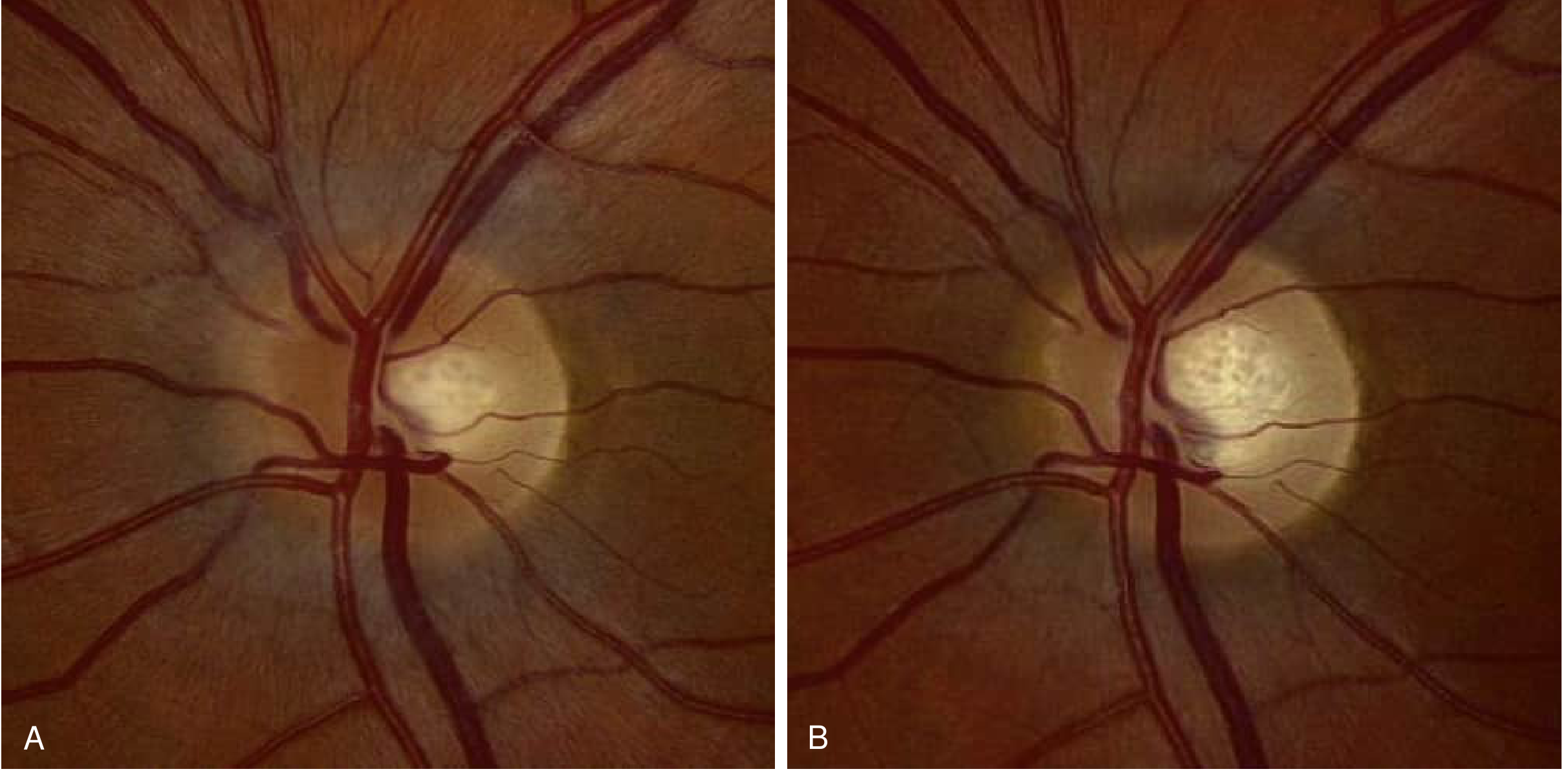
In typical ON, mild disc edema may be seen, but severe disc swelling, marked peripapillary hemorrhages, vitreous inflammation, retinal exudation, or a macular star should prompt a search for another diagnosis. Bradley and Daroff's Neurology in Clinical Practice, p. 259.
Typical versus Atypical Optic Neuritis
This distinction is highly important in examinations and clinical practice.
| Feature | Typical MS-associated ON | Atypical ON |
|---|---|---|
| Age | Young adult | Child, older adult, or atypical age |
| Laterality | Usually unilateral | Bilateral simultaneous or rapidly sequential |
| Pain | Common, especially on eye movement | Absent, prolonged, severe, or unusual pain |
| Visual loss | Mild to moderate in many cases | Very severe, often worse than 6/60 or 20/200 |
| Fundus | Normal or mild disc edema | Severe edema, hemorrhages, exudates, macular star, vitreous cells |
| Recovery | Usually good | Poor, absent, or steroid-dependent recovery |
| Relapse | Possible but not frequent | Frequent relapses suggest MOGAD, CRION, or systemic disease |
| MRI | Short-segment optic nerve lesion, MS-type brain lesions may occur | Long optic nerve lesion, chiasmal involvement, perineural enhancement, orbital/infiltrative lesions |
| Important associations | MS | MOGAD, AQP4-NMOSD, sarcoidosis, infection, CRION, malignancy |
Red flags for atypical ON
- Age under 12 years or over 50 years
- Painless visual loss
- Severe loss at nadir
- Bilateral simultaneous involvement
- Severe optic-disc edema
- Disc/peripapillary hemorrhages
- Macular star or prominent retinal exudates
- Vitritis, uveitis, retinitis, or vasculitis
- No improvement within 2-4 weeks
- Progressive worsening beyond 2 weeks
- Steroid-dependent or recurrent disease
- Systemic symptoms such as fever, weight loss, meningism, rash, pulmonary disease, oral ulcers, arthritis, or myelopathy
The AAO red-flag review emphasizes severe or bilateral disease and marked disc edema as reasons to urgently consider MOGAD and NMOSD.
Important Demyelinating Disorders
1. Multiple sclerosis-associated optic neuritis
Typical MS-associated ON is usually:
- Unilateral
- Painful
- Associated with a normal disc or mild disc edema
- Followed by good visual recovery
MRI brain is important because the presence of typical white matter demyelinating lesions substantially increases the future risk of MS.
In the Optic Neuritis Treatment Trial follow-up, the 15-year risk of MS was approximately 72% in patients with one or more characteristic brain MRI lesions, compared with approximately 25% in those with a normal MRI at presentation.
Bradley and Daroff's Neurology in Clinical Practice, p. 259.
2. AQP4-NMOSD
AQP4-NMOSD is an antibody-mediated astrocytopathy. Optic neuritis may be:
- Severe
- Bilateral or recurrent
- Associated with poor visual recovery
- Involving a long segment of optic nerve
- Extending posteriorly to the optic chiasm or optic tract
- Associated with longitudinally extensive transverse myelitis, typically spanning 3 or more vertebral segments
Test serum AQP4-IgG, preferably by a cell-based assay.
Acute attacks require urgent aggressive therapy because permanent visual loss can occur. IV corticosteroids are used, with early plasma exchange for severe disease or inadequate response. Kanski's Clinical Ophthalmology, 10th ed., pp. 781-782.
3. MOG antibody-associated disease
MOGAD may present with:
- Bilateral simultaneous or sequential ON
- Marked optic-disc edema
- Severe visual loss
- Prominent orbital pain
- Longitudinal optic nerve involvement
- Perineural optic nerve sheath enhancement on MRI
- Relapsing, steroid-responsive or steroid-dependent disease
Visual recovery may be better than in AQP4-NMOSD, but relapse is important and may cause cumulative disability.
Test serum MOG-IgG by a live cell-based assay, ideally before major immunotherapy when feasible. Kanski's Clinical Ophthalmology, 10th ed., p. 781.
4. Chronic relapsing inflammatory optic neuropathy
CRION is characterized by recurrent, painful optic neuritis with marked corticosteroid responsiveness and relapse on steroid withdrawal. It is a diagnosis of exclusion after evaluating for MOGAD, AQP4-NMOSD, sarcoidosis, infection, and infiltrative disease.
Investigations
1. Basic ophthalmic assessment
Perform and document:
- Best-corrected visual acuity in each eye
- Colour vision, such as Ishihara plates or red desaturation
- Contrast sensitivity
- Pupillary examination for RAPD
- Slit-lamp examination for anterior chamber or vitreous inflammation
- Dilated fundus examination
- Intraocular pressure
- Fundus photography
- Automated perimetry
- OCT of RNFL and macular GCIPL
Visual-field defects
There is no pathognomonic field defect. Common defects include:
- Central scotoma
- Centrocecal scotoma
- Diffuse depression
- Arcuate defects
- Altitudinal defects
- Peripheral constriction
An altitudinal defect in an older person, especially with vascular risk factors and a swollen optic disc, should prompt consideration of anterior ischemic optic neuropathy.
2. MRI brain and orbit with gadolinium
MRI of the brain and orbits with contrast and fat-suppressed sequences is the key imaging investigation.
It helps to:
- Confirm optic nerve inflammation and enhancement
- Determine lesion length and location
- Identify optic nerve sheath/perineural involvement
- Detect brain lesions typical of MS
- Assess chiasmal or optic tract involvement
- Exclude compressive, infiltrative, or orbital pathology
- Estimate risk of conversion to MS
A 2026 systematic review and meta-analysis found pooled MRI sensitivity of 84.0% and specificity of 86.4% for acute ON; contrast-enhanced T1 imaging had the highest sensitivity among evaluated sequences. See the MRI meta-analysis. Thus, a normal MRI does not by itself exclude ON.
3. Serology and targeted tests
Order based on presentation, especially in atypical ON:
- Serum AQP4-IgG
- Serum MOG-IgG
- Syphilis serology
- Tuberculosis testing where epidemiologically appropriate
- Bartonella serology when neuroretinitis is suspected
- HIV testing when indicated
- ACE, chest imaging, and other sarcoidosis evaluation when appropriate
- ANA, ANCA, ESR, CRP, and systemic autoimmune work-up when indicated
- Vitamin B12, folate, and toxic/nutritional evaluation in bilateral chronic optic neuropathy
4. Cerebrospinal fluid examination
CSF is not mandatory in straightforward typical ON but is useful when diagnosis is uncertain, atypical disease is suspected, or MS/NMOSD/infection is being assessed.
Possible tests:
- Cells, protein, glucose
- Oligoclonal bands and IgG index
- Infectious studies, including VDRL and PCR where indicated
- Cytology or flow cytometry if malignancy/infiltration is suspected
5. Visual evoked potentials
VEP typically shows:
- Increased P100 latency due to demyelination
- Reduced amplitude in significant axonal loss
It is useful for detecting previous or subclinical optic nerve demyelination, but MRI and OCT are more commonly central to modern work-up.
Differential Diagnosis
| Condition | Features favoring it over typical ON |
|---|---|
| Non-arteritic anterior ischemic optic neuropathy | Usually >50 years, often painless, altitudinal defect, vascular risk factors, crowded disc in fellow eye |
| Arteritic anterior ischemic optic neuropathy due to giant cell arteritis | Age >50 years, severe sudden visual loss, pallid disc edema, scalp tenderness, jaw claudication, high ESR/CRP |
| Compressive optic neuropathy | Insidious progressive loss, optic atrophy, proptosis, dysmotility, persistent progression |
| Leber hereditary optic neuropathy | Young man, painless sequential bilateral central loss, family history, telangiectatic microangiopathy |
| Neuroretinitis | Disc edema followed by macular star, frequently infectious |
| Papilledema | Usually bilateral, visual acuity initially relatively preserved, headache/raised intracranial pressure, enlarged blind spot |
| Toxic/nutritional optic neuropathy | Gradual bilateral symmetrical centrocecal scotomas, often painless |
| Functional visual loss | Inconsistent findings, no RAPD in claimed profound unilateral loss, normal objective tests |
| Optic perineuritis | Often more severe/prolonged pain, disc edema, MRI sheath enhancement rather than intrinsic nerve enhancement, steroid-responsive but relapsing |
Management
Principles
- Confirm that this is optic neuritis and identify atypical features.
- Urgently exclude arteritic ischemic optic neuropathy, infection, compression, and infiltrative disease where relevant.
- Obtain MRI brain and orbit with contrast.
- Test AQP4-IgG and MOG-IgG in severe, bilateral, recurrent, or atypical cases.
- Treat the acute attack appropriately.
- Refer jointly to neuro-ophthalmology and neurology when demyelinating disease is suspected.
Treatment of typical demyelinating optic neuritis
Observation
Observation is reasonable in mild, typical ON because spontaneous visual recovery is common.
High-dose corticosteroids
High-dose corticosteroids:
- Accelerate visual recovery
- Do not significantly improve final long-term visual outcome in typical ON
- May transiently reduce short-term risk of a further demyelinating event, but do not eliminate long-term MS risk
A conventional ONTT-derived regimen is:
- IV methylprednisolone 1 g/day for 3 days
or equivalent high-dose IV corticosteroid treatment - Followed in selected patients by an oral corticosteroid taper
Exact regimen and suitability should be decided with the treating neuro-ophthalmology/neurology team.
Important ONTT point
Oral prednisone alone in conventional doses should not be used for typical ON, because it was associated with an increased risk of ON recurrence. High-dose IV steroids may hasten recovery, but do not materially improve final visual acuity in typical disease. Bradley and Daroff's Neurology in Clinical Practice, p. 259.
Adverse effects of steroids
Counsel and monitor for:
- Hyperglycemia
- Hypertension
- Gastritis
- Mood changes, insomnia, psychosis
- Infection risk
- Fluid retention
- Rare avascular necrosis with repeated courses
Treatment of NMOSD- or MOGAD-associated ON
These disorders should not be managed as routine MS-associated ON.
Acute therapy
- Prompt high-dose IV corticosteroids
- Early escalation to plasma exchange (PLEX) for severe visual loss, AQP4-NMOSD, or insufficient steroid response
- IV immunoglobulin may be considered in selected steroid-refractory cases, particularly where PLEX is not feasible or in specific clinical contexts
For NMOSD, delay in treatment can lead to irreversible visual loss. The AAO discussion of NMOSD/MOGAD acute management stresses the importance of rapid initiation of therapy and early PLEX in NMOSD.
Relapse prevention
Long-term immunotherapy is determined by the underlying disease:
- AQP4-NMOSD: therapies may include rituximab, eculizumab, inebilizumab, satralizumab, or other specialist-directed immunosuppression.
- MOGAD: relapse prevention is individualized and may include prolonged steroid taper, IVIG, rituximab, mycophenolate, azathioprine, or other specialist-directed therapy.
Do not start MS disease-modifying therapy until the disorder has been accurately classified, because treatment strategies differ between MS, AQP4-NMOSD, and MOGAD.
Prognosis
Visual prognosis
Typical demyelinating ON generally has a favorable visual prognosis:
- Recovery often starts within 2-4 weeks.
- Vision continues improving over weeks to months.
- High-contrast Snellen visual acuity may return to normal or near-normal.
- However, subtle deficits can persist:
- Reduced contrast sensitivity
- Colour vision impairment
- Visual-field defects
- Glare sensitivity
- Exercise/heat-induced visual symptoms
- OCT evidence of RNFL and GCIPL thinning
Visual prognosis is worse with:
- Very severe initial visual loss
- Recurrent attacks
- AQP4-NMOSD
- Delayed treatment in severe inflammatory ON
- Significant axonal loss on OCT
- Marked optic atrophy
Risk of multiple sclerosis
Risk depends mainly on brain MRI findings at the initial attack.
- Typical ON with MS-pattern lesions on MRI: high future MS risk.
- Typical ON with normal MRI: lower but not zero risk.
- Atypical clinical features reduce the likelihood that the episode represents MS-associated ON.
Bradley and Daroff's Neurology in Clinical Practice, p. 259.
Examination Summary: Short Answer Format
Optic neuritis is an acute inflammatory optic neuropathy, most commonly demyelinating and associated with MS. Typical ON occurs in a young adult, usually as unilateral painful subacute visual loss with dyschromatopsia, RAPD, central scotoma, and a normal fundus or mild disc edema. MRI brain and orbit with gadolinium confirms optic nerve inflammation, evaluates MS risk, and detects features suggestive of MOGAD or AQP4-NMOSD. High-dose IV corticosteroids hasten visual recovery but do not significantly alter final visual outcome in typical ON; oral prednisone alone should be avoided. Severe, bilateral, recurrent, steroid-dependent, or atypical ON requires urgent work-up for MOGAD, AQP4-NMOSD, infection, sarcoidosis, and infiltrative disease, with early plasma exchange considered for severe NMOSD-associated attacks.
High-yield viva points
- Pain on eye movement is characteristic of typical ON.
- RAPD occurs in unilateral or asymmetric optic neuropathy.
- Fundus is normal in most cases of retrobulbar neuritis.
- Disc edema occurs in about one-third of typical ON cases.
- Severe disc edema, hemorrhages, macular star, painless loss, bilateral disease, and poor recovery are atypical.
- MRI brain lesions are the strongest predictor of later MS.
- OCT documents RNFL and GCIPL loss after the acute episode.
- IV corticosteroids speed recovery but do not improve final vision in typical ON.
- Avoid conventional-dose oral prednisone alone.
- Think of MOGAD in bilateral ON with marked disc edema.
- Think of AQP4-NMOSD in severe ON, chiasmal/posterior involvement, and longitudinally extensive transverse myelitis.
Recent-evidence note: MRI performance data have been updated by a 2026 systematic review, PMID 41391387. This supports MRI as a useful adjunct but reinforces that ON remains primarily a clinical diagnosis and a negative MRI cannot fully exclude it.
How to differentiate between each type of optic neuritis, make a table
Reading File
Finding Sources
Searching PubMed
"optic neuritis"
Searching the Web
AAO differentiate typical optic neuritis MOGAD NMOSD neuroretinitis optic perineuritis
Differentiation of Types of Optic Neuritis
A. Anatomical clinical types
| Feature | Papillitis (anterior ON) | Retrobulbar neuritis | Neuroretinitis | Optic perineuritis |
|---|---|---|---|---|
| Primary site | Optic nerve head | Optic nerve behind globe | Optic nerve head plus outer retina/macula | Optic nerve sheath, rather than nerve primarily |
| Fundus at presentation | Swollen, hyperemic disc with blurred margins | Usually normal initially | Disc edema initially, followed days later by macular star | Disc edema may occur; retinal signs variable |
| Classic description | Visible optic-disc inflammation | “Patient sees nothing, doctor sees nothing” | Disc edema + macular star | Painful optic neuropathy with sheath inflammation |
| Pain on eye movement | Common | Common | Variable | Often prominent and prolonged |
| Visual loss | Acute/subacute | Acute/subacute | Variable, often less severe initially | Subacute, may be progressive |
| Macular star | Absent | Absent | Characteristic, appears after 1-2 weeks | Absent |
| Common causes | Typical demyelinating ON, MOGAD, infection | MS-associated ON | Bartonella infection, idiopathic, syphilis, Lyme, TB | Idiopathic, MOGAD, sarcoidosis, syphilis, TB, IgG4-related disease |
| MRI | Anterior optic nerve enhancement | Intrinsic optic nerve enhancement | May show optic nerve inflammation | Sheath enhancement: “tram-track” on axial and “doughnut” on coronal images |
| Important differential | Papilledema, AION | Functional loss, compressive neuropathy | Hypertensive retinopathy, diabetic papillopathy | MOGAD, orbital inflammation, sarcoidosis, optic nerve meningioma |
| Treatment principle | Treat cause; steroids for demyelinating disease when indicated | Same | Treat underlying infection if present; do not give steroids alone until infection considered | Steroid-responsive but often needs slow taper and etiological work-up |
Exam point: Typical demyelinating ON is retrobulbar in most adults; approximately one-third may have mild optic-disc edema, constituting papillitis. Marked disc edema, hemorrhages, vitritis, or a macular star are atypical for routine MS-associated ON. Bradley and Daroff's Neurology in Clinical Practice, p. 259.
B. Etiological differentiation: most useful PG table
| Feature | Typical MS-associated ON | MOGAD-associated ON | AQP4-NMOSD-associated ON | CRION | Parainfectious ON | Infectious ON / neuroretinitis |
|---|---|---|---|---|---|---|
| Typical age | 18-45 years | Children and young adults, but any age | Adults, often middle-aged | Any age, usually adults | Common in children | Any age, depends on cause |
| Sex | Female predominance | No strong fixed pattern | Marked female predominance | Variable | No consistent pattern | Variable |
| Laterality | Usually unilateral | Frequently bilateral or rapidly sequential | Often bilateral or sequential | Usually unilateral at each attack, may alternate | Often bilateral | Variable, may be unilateral |
| Pain on eye movement | Very common | Common, often severe | May occur | Marked pain is typical | Variable | Variable |
| Degree of visual loss | Mild to moderate in many cases | Often severe at nadir, but recovery often good | Often severe, with poorer recovery | Moderate to severe, progressively disabling with relapses | Variable | Variable |
| Optic disc appearance | Normal in most, or mild edema | Prominent disc edema, sometimes with hemorrhages | May be normal or swollen; severe edema possible | Disc edema may occur | Papillitis often present | Disc edema, vitritis, retinitis, macular star, or hemorrhages may occur |
| Visual recovery | Generally good | Usually good after attack, but repeated relapses cause damage | Often incomplete, poorer prognosis | Improves dramatically with steroids but relapses on tapering/withdrawal | Usually good | Depends on infection and timeliness of treatment |
| Relapse tendency | Possible but not defining | Common in relapsing MOGAD | Common unless preventive therapy used | Defining feature: recurrent, steroid-dependent ON | Usually monophasic | Depends on persistent/untreated infection |
| Brain MRI | Typical MS white matter lesions may be present | Brain MRI may be normal or show ADEM-like/cortical lesions | May show characteristic periependymal, hypothalamic, brainstem lesions | Often no typical MS lesions | May be normal or show ADEM-like lesions | May show meningeal, orbital, retinal, or CNS disease depending on cause |
| Orbital MRI | Usually unilateral, short anterior segment optic nerve lesion | Long-segment, often bilateral optic nerve enhancement; anterior involvement and sheath/perineural enhancement | Long-segment lesion, often posterior optic nerve, chiasm, or optic tract | Optic nerve enhancement; exclude MOGAD, NMOSD, sarcoid | Optic nerve enhancement, often bilateral | Variable; may show nerve, sheath, retinal, meningeal, or sinus involvement |
| Serum antibody | No specific antibody | MOG-IgG positive by cell-based assay | AQP4-IgG positive by cell-based assay | MOG-IgG and AQP4-IgG should be negative before diagnosis | Usually negative | Targeted infectious serology/PCR positive |
| Associated systemic/CNS feature | Other MS symptoms: sensory symptoms, myelitis, internuclear ophthalmoplegia | ADEM in children, myelitis, cortical encephalitis | Longitudinally extensive transverse myelitis, area postrema syndrome | No systemic disorder after exclusion | Viral illness, fever, exanthem, recent infection/vaccination | Fever, lymphadenopathy, rash, uveitis, retinitis, meningitis, sinus/orbital disease |
| Acute treatment | Observation or high-dose IV methylprednisolone | IV methylprednisolone; prolonged taper often considered; PLEX if severe/refractory | IV methylprednisolone plus early PLEX in severe disease | Steroids with slow taper; consider long-term immunosuppression | Usually steroids after excluding active infection | Specific antimicrobial therapy; corticosteroids only when appropriate and usually with antimicrobial cover |
| Long-term treatment | MS disease-modifying therapy if MS/high-risk clinically isolated syndrome | Relapse prevention if relapsing | Long-term immunotherapy is usually indicated | Steroid-sparing immunosuppression often needed | Usually none | Treat underlying infection |
Kanski's Clinical Ophthalmology, 10th ed., pp. 766, 769-770, 781-782.
MRI differentiation in one glance
| Disorder | Characteristic orbital MRI pattern |
|---|---|
| MS-associated ON | Usually unilateral, focal short-segment enhancement, commonly anterior intraorbital optic nerve |
| MOGAD-ON | Longitudinally extensive optic nerve enhancement, often bilateral; anterior optic nerve involvement; optic-disc edema; optic nerve sheath/perineural enhancement can occur |
| AQP4-NMOSD-ON | Long lesion, often bilateral; posterior optic nerve involvement with extension to optic chiasm and optic tract is suggestive |
| Optic perineuritis | Circumferential optic nerve sheath enhancement, “tram-track” on axial MRI and “doughnut” on coronal MRI |
| Compressive optic neuropathy | Mass lesion, optic canal/orbital apex lesion, gradual course rather than acute inflammatory enhancement |
The AAO MRI red-flag summary describes short unilateral lesions as more typical of MS-ON, perineural involvement as suggestive of MOGAD, and posterior/chiasmal disease as more suggestive of NMOSD.
Important mimics to differentiate from optic neuritis
| Condition | Differentiating clues |
|---|---|
| Non-arteritic anterior ischemic optic neuropathy (NAION) | Usually age >50 years, painless sudden loss, vascular risk factors, altitudinal field defect, sectoral disc edema, crowded fellow disc |
| Arteritic AION due to giant cell arteritis | Age >50 years, profound visual loss, pallid disc edema, jaw claudication, headache, scalp tenderness, raised ESR/CRP |
| Papilledema | Usually bilateral disc edema due to raised intracranial pressure; visual acuity often initially preserved; enlarged blind spot, headache, transient visual obscurations |
| Leber hereditary optic neuropathy | Young male, painless sequential bilateral central visual loss, peripapillary telangiectasia, family history through maternal line |
| Compressive optic neuropathy | Slow progressive visual loss, optic atrophy, proptosis/diplopia, optic disc may be normal early, MRI reveals lesion |
| Toxic/nutritional optic neuropathy | Painless, bilateral symmetrical progressive centrocecal scotomas, dyschromatopsia, exposure history or nutritional deficiency |
| Functional visual loss | Inconsistent visual findings, normal pupillary reactions despite reported profound unilateral visual loss, normal OCT and imaging |
Rapid PG examination algorithm
-
Young adult + unilateral painful visual loss + RAPD + dyschromatopsia + normal/mildly swollen disc
→ Typical MS-associated ON until proven otherwise. -
Bilateral or sequential ON + severe disc edema + long optic nerve lesion/perineural enhancement
→ Suspect MOGAD. Send serum MOG-IgG. -
Severe visual loss + bilateral involvement + posterior/chiasmal lesion + myelitis
→ Suspect AQP4-NMOSD. Send serum AQP4-IgG and initiate urgent treatment. -
Recurrent painful ON that improves on steroids but returns during taper
→ Suspect CRION, but first exclude MOGAD, NMOSD, sarcoidosis, infection, and infiltrative disease. -
Disc edema followed by macular star, especially with cat exposure or fever
→ Neuroretinitis, commonly Bartonella-associated. -
Painless acute visual loss in a patient older than 50 years
→ Assume ischemic optic neuropathy until excluded. Urgently rule out giant cell arteritis.
Recent-evidence note: MRI is helpful but not definitive. A 2026 systematic review found pooled MRI sensitivity of 84.0% and specificity of 86.4% for acute ON, so diagnosis must remain clinicoradiological rather than MRI-only (PMID 41391387).
Write short note on scotoma and difference between positive and negative scotoma
Reading File
Finding Sources
Searching PubMed
"scotoma" AND visual field
Searching the Web
scotoma positive negative scotoma ophthalmology definition
Scotoma
A scotoma is a localized area of partial or complete loss of vision within the visual field, surrounded by relatively normal vision. It may be detected by confrontation testing, Amsler grid, or formal perimetry.
A central or centrocecal scotoma is commonly seen in optic neuritis, while arcuate scotoma and nasal step are characteristic patterns in glaucoma. A central visual-field defect may be readily noticed by the patient, whereas peripheral defects can remain asymptomatic.
Classification of scotomas
| Type | Description | Common examples/causes |
|---|---|---|
| Central scotoma | Defect involving fixation | Optic neuritis, macular disease, toxic/nutritional optic neuropathy |
| Centrocecal scotoma | Defect extending from fixation to blind spot | Optic neuritis, tobacco-alcohol amblyopia, LHON |
| Paracentral scotoma | Defect adjacent to fixation | Glaucoma, macular disease |
| Arcuate (Bjerrum) scotoma | Arcuate defect following retinal nerve-fibre bundles from blind spot to nasal field | Glaucoma |
| Seidel scotoma | Sickle-shaped extension from blind spot, often an early arcuate defect | Early glaucoma |
| Ring scotoma | Annular field defect around central field | Retinitis pigmentosa, advanced glaucoma |
| Altitudinal scotoma | Loss of upper or lower half of the field, respecting horizontal meridian | Anterior ischemic optic neuropathy |
| Junctional scotoma | Ipsilateral central scotoma with contralateral superotemporal defect | Optic nerve-chiasm junction lesion |
| Physiological scotoma | Normal blind spot corresponding to optic disc, where there are no photoreceptors | Normal finding |
Positive versus Negative Scotoma
| Feature | Positive scotoma | Negative scotoma |
|---|---|---|
| Definition | A field defect that is subjectively perceived by the patient as an abnormal visual sensation or visible spot | A field defect that is not perceived by the patient and is found only on visual-field testing |
| Patient description | “Black spot,” “dark patch,” “grey area,” “blurred spot,” “flashing lights,” “zig-zag lines,” or “shimmering area” | Usually no subjective complaint of a spot or missing area |
| Nature | May be a dark scotoma or a positive visual phenomenon such as scintillation | Represents absence or reduction of visual sensitivity without a perceived visual phenomenon |
| Detection | Patient may identify it spontaneously; confirmed by perimetry or Amsler grid | Usually detected by confrontation testing or automated/manual perimetry |
| Examples | Retinal hemorrhage may produce a perceived dark spot; scintillating scotoma in migraine aura | Glaucomatous arcuate scotoma, early optic nerve defects, physiological blind spot |
| Clinical significance | Suggests the patient is aware of the defect or associated visual phenomenon | Can be clinically important despite being asymptomatic, especially in glaucoma |
Key examination point
- Positive scotoma: The patient sees something abnormal in that area of the field.
- Negative scotoma: The patient does not see the missing area and may be unaware of the defect until perimetry is done.
Relative versus absolute scotoma
This is a separate classification and should not be confused with positive/negative scotoma.
| Type | Meaning |
|---|---|
| Relative scotoma | A stronger or brighter stimulus can be seen in the affected area, but a usual test stimulus is missed |
| Absolute scotoma | No test stimulus is perceived even at maximum brightness |
One-line viva answer: A positive scotoma is perceived by the patient as a dark, coloured, flashing, or shimmering spot, whereas a negative scotoma is an unperceived area of visual-field loss detected only by examination or perimetry.
This is a shared conversation. Sign in to Orris to start your own chat.