
Draw a diagram of arch support heel pad and heel cup
A clear labeled anatomical diagram showing three orthotic/footwear support devices side by side: 1. Arch Support - a foot insole with a raised contoured arch in the middle, with labels pointing to the arch support area, metatarsal region, and heel area 2. Heel Pad - a small oval cushioned pad that fits under the heel, with labels showing the cushioning layer and placement under the calcaneus 3. Heel Cup - a deep cupped rigid or semi-rigid orthotic device that cradles the heel, with labels showing the side walls, base, and how it cups the calcaneus Each device shown from a side profile and top-down view. Clean medical illustration style on white background with clear annotation lines and text labels.
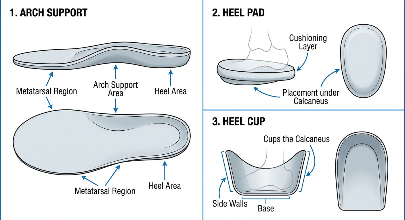
- A full-length or 3/4 insole with a raised contoured section under the medial longitudinal arch
- Distributes pressure evenly across the foot and corrects overpronation
- A small, flat, oval-shaped cushion placed directly under the heel
- Absorbs shock at the calcaneus - commonly used for plantar fasciitis, heel spurs, and Achilles tendinopathy
- Usually made of silicone, foam, or felt
- A rigid or semi-rigid cupped device with raised side walls that cradle and centralize the heel's fat pad
- Prevents the fat pad from spreading laterally, improving cushioning efficiency
- Used for heel pain, calcaneal stress fractures, and Sever's disease in children
Draw the diagram of metatarsal pad ucbl foot orthosis medial wedge and lateral wedge
A clear labeled medical diagram showing four foot orthotic devices arranged on a white background: 1. METATARSAL PAD - small teardrop or dome-shaped pad, shown placed just proximal (behind) the metatarsal heads on the plantar surface of the foot, side view and top view, with labels pointing to the dome/raised area, placement proximal to metatarsal heads, and the spreading effect on metatarsals 2. UCBL FOOT ORTHOSIS (University of California Biomechanics Laboratory) - a deep-seated, total contact rigid shell orthosis that encompasses the heel and midfoot, with high medial and lateral walls and a deep heel cup, shown in side profile and posterior view, labels pointing to medial wall, lateral wall, deep heel cup, and forefoot platform 3. MEDIAL WEDGE - a triangular cross-section insole wedge that is thicker on the medial (inner) side, shown in coronal cross-section view and from behind, with arrows showing how it tilts the foot into varus (supination), labels: medial side thicker, lateral side thinner, corrects valgus/pronation 4. LATERAL WEDGE - a triangular cross-section insole wedge that is thicker on the lateral (outer) side, shown in coronal cross-section view and from behind, with arrows showing how it tilts the foot into valgus, labels: lateral side thicker, medial side thinner, corrects varus/supination, used in knee OA Clean anatomical illustration style, clear annotation lines, each device clearly separated with a title label, white background
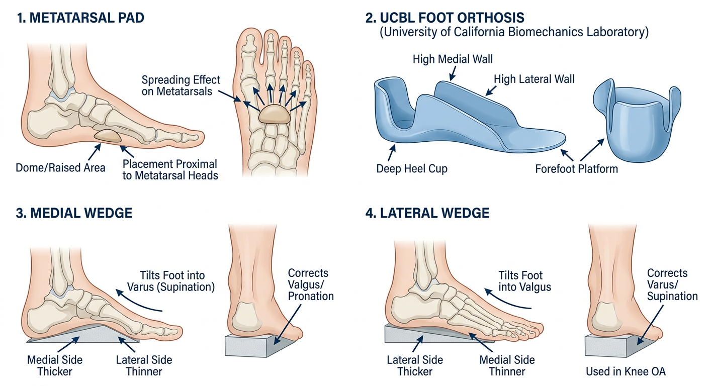
- A small dome-shaped pad placed just proximal to the metatarsal heads (not under them)
- Offloads pressure from the metatarsal heads by redistributing weight proximally
- Used for metatarsalgia, Morton's neuroma, and plantar plate tears
- A rigid, total-contact shell with deep heel cup and high medial/lateral walls
- Controls rearfoot and midfoot motion - especially hyperpronation (planovalgus)
- Indicated for flexible flatfoot, posterior tibialis tendon dysfunction, and pediatric foot deformities
- Thicker on the medial (inner) side, tapering toward the lateral side
- Tilts the foot into varus (supination), correcting excessive valgus/pronation
- Used for planovalgus deformity and overpronation
- Thicker on the lateral (outer) side, tapering toward the medial side
- Tilts the foot into valgus, offloading the medial compartment of the knee
- Commonly used for medial compartment knee osteoarthritis and varus foot deformity
Locked kafo and scott r craig kafo
A detailed labeled medical diagram showing two types of Knee-Ankle-Foot Orthoses (KAFO) side by side on white background: 1. LOCKED KAFO (left side): - Full leg orthosis from thigh to foot shown in frontal and lateral view - Knee joint with drop-lock ring mechanism clearly shown - a metal ring that drops down to lock the knee in full extension - Labeled components: thigh cuff (proximal), knee joint with drop-lock/ring lock mechanism, calf band, ankle joint (with limited motion), foot plate - Upright metal bars on medial and lateral sides - Shows locked knee in full extension position - Indication label: used for quadriceps weakness, polio, paraplegia 2. SCOTT-CRAIG KAFO (right side): - Specialized KAFO designed for paraplegic ambulation - Features: offset knee joint (posterior to knee axis), bail lock/bail-activated knee lock mechanism (a U-shaped wire bail) - Double stopped ankle joint set in slight dorsiflexion (5-7 degrees) - Crossbar connecting the two uprights at the ankle - Foot plate with toe piece - High thoracic band optional - Labeled components: bail lock knee joint, offset knee joint, double-stopped ankle joint, foot plate, crossbar, thigh band, calf band - Side view showing the slight forward lean/stable posture it creates Clean anatomical orthotic illustration style, clear annotation lines and text, each KAFO labeled with its name as title, white background, professional medical diagram quality
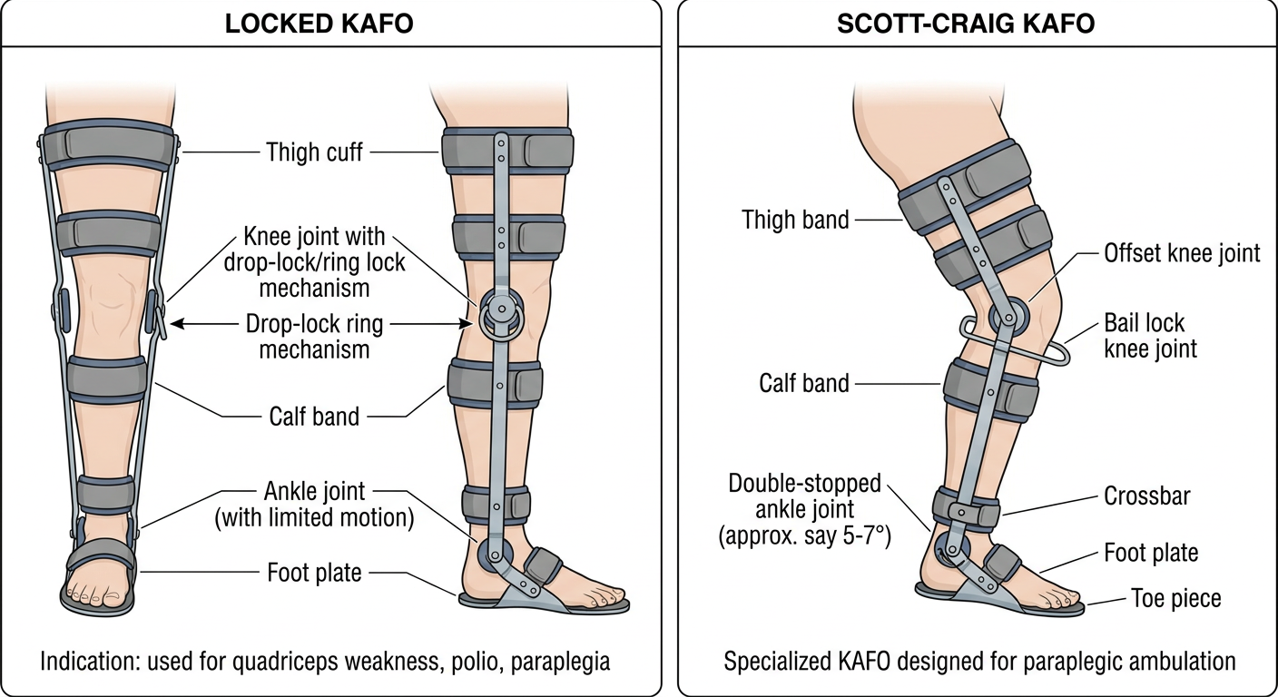
- A conventional KAFO with a drop-lock (ring lock) at the knee joint - a metal ring slides down to lock the knee in full extension
- Medial and lateral metal uprights run from thigh to foot plate
- Components: thigh cuff, drop-lock knee joint, calf band, ankle joint (usually limited motion), foot plate
- Indications: quadriceps weakness or paralysis, polio sequelae, spastic paraparesis, genu recurvatum
- Lock must be manually released to sit down, which limits functional ease
- Offset knee joint - positioned posterior to the knee's mechanical axis, creating passive knee extension stability through body weight
- Bail lock (bail-activated lock) - a U-shaped wire bail behind the knee that automatically unlocks when the user sits back and re-locks on standing
- Double-stopped ankle joint - set at 5-7° dorsiflexion to promote forward momentum during swing phase
- Crossbar connecting bilateral uprights for structural rigidity
- Allows hands-free locking/unlocking via the bail, making it far more functional than a simple ring lock KAFO
- Indications: complete or incomplete thoracic/lumbar paraplegia for therapeutic or functional ambulation
Stance control kafo plastic kafo conventional kafon
A detailed labeled medical diagram showing three types of Knee-Ankle-Foot Orthoses (KAFO) side by side on a white background: 1. STANCE CONTROL KAFO (left): - Full leg orthosis shown in lateral and frontal view - Knee joint with automatic stance-control mechanism (polycentric or ratchet mechanism) that locks during stance phase and unlocks during swing phase - Shows knee in locked position during stance (foot flat on ground, body weight through leg) - Shows knee flexing freely during swing phase (foot off ground) - Labeled components: thigh shell, stance-control knee joint mechanism, swing-phase free knee flexion arrow, calf shell, ankle-foot section - Small mechanical diagram showing the locking/unlocking mechanism triggered by ground reaction force - Indication: quadriceps weakness with preserved hip flexion 2. PLASTIC KAFO (center): - Modern thermoplastic/polypropylene KAFO shown in frontal and lateral view - Solid plastic thigh shell (proximal) and calf-foot shell (distal) - Plastic knee joint hinges on medial and lateral sides - Smooth contoured plastic shells that conform to limb shape - Labeled components: thermoplastic thigh shell, plastic knee joint (medial and lateral hinges), plastic calf shell, plastic ankle-foot orthosis (AFO) integrated section, trim lines, foot plate - Lightweight and cosmetically appealing design - Indication: post-stroke, spastic conditions, moderate weakness 3. CONVENTIONAL KAFO (right): - Traditional metal and leather KAFO in frontal and lateral view - Metal uprights (medial and lateral) running full length of leg - Leather or padded thigh band, knee pad/strap, calf band - Metal knee joint (drop-lock ring or dial lock shown) - Metal ankle joint (Klenzak spring or fixed) - Leather-covered stirrup attached to shoe - Labeled components: thigh band (leather), medial upright, lateral upright, knee joint with ring lock, calf band, ankle joint (Klenzak), stirrup, shoe attachment - Heavy and bulky appearance compared to plastic version - Indication: severe weakness, heavy-duty use, edematous limbs All three shown at same scale for easy comparison, clean medical illustration style, clear annotation lines, white background, titles above each KAFO
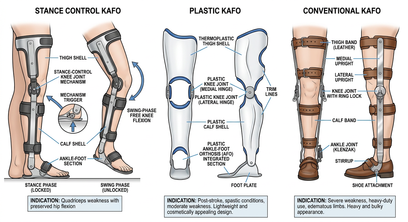
- Features a microprocessor or mechanical knee joint that locks automatically during stance phase (weight bearing) and unlocks freely during swing phase (foot off ground)
- Triggered by ground reaction forces - no manual locking required
- Allows a more natural gait pattern with knee flexion during swing
- Components: thigh shell, stance-control knee mechanism, calf shell, integrated AFO section
- Indications: quadriceps weakness with intact hip flexors (post-polio, incomplete SCI, MS), where a locked KAFO would cause gait deviation
- Made from thermoplastic (polypropylene) shells custom-molded to the limb
- Plastic knee hinges (medial and lateral) allow controlled motion or can be set to limit range
- Lighter, more cosmetically acceptable, and easier to don/doff than conventional metal
- Integrates seamlessly with regular footwear
- Limitations: not suitable for fluctuating edema or very heavy patients
- Indications: stroke, post-polio, spastic paresis, moderate quadriceps weakness
- Traditional design with metal uprights (medial and lateral), leather bands (thigh, calf, knee), and metal joints
- Ankle joint options: Klenzak spring assist, fixed, or limited motion
- Knee joint: drop-lock (ring lock), dial lock, or bail lock
- Attached to the shoe via a stirrup (metal plate welded to shoe shank)
- Advantages: durable, adjustable, tolerates edema and volume changes
- Indications: severe paralysis, heavy-duty agricultural/work use, patients with fluctuating limb volume, pediatric growing patients
| Feature | Conventional | Plastic | Stance Control |
|---|---|---|---|
| Material | Metal + leather | Thermoplastic | Mixed/metal |
| Weight | Heavy | Light | Moderate |
| Gait quality | Poor (locked knee) | Moderate | Best |
| Cost | Low | Moderate | High |
| Edema tolerance | Good | Poor | Moderate |