
Explain pneumonia in detail according to Microbiology 2nd mbbs
pneumonia lobar consolidation chest radiograph
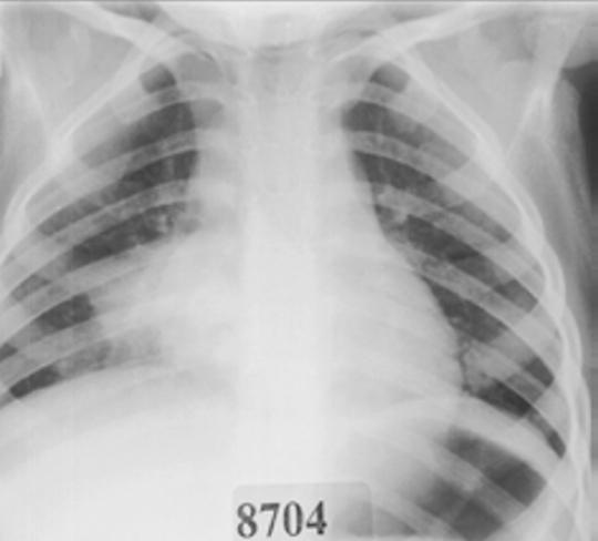
This frontal pediatric chest radiograph displays a diagnostic image of the thorax demonstrating classic features of lobar pneumonia. The primary finding is a large, dense, and relatively homogenous area of increased radiopacity located within the right lower lung field. This consolidation extends from the mid-lung zone down to the right hemidiaphragm, which it partially obscures, indicating an alveolar filling process. Key radiographic features include visible air bronchograms—linear and branching lucencies representing air-filled bronchi outlined by the surrounding consolidated lung parenchyma. The opacity appears non-segmental and occupies a substantial portion of the right lower lobe. The mediastinal structures, including the heart and trachea, appear centrally located without significant deviation. This image serves as a clinical example of bacterial pneumonia, typically associated with pathogens such as Streptococcus pneumoniae, highlighting the consolidation and air bronchogram signs essential for medical student and radiologic technologist education.
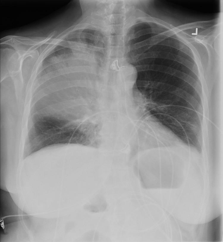
This anterior-posterior (AP) chest radiograph displays a prominent area of heterogeneous opacity and consolidation localized to the right upper lobe. The consolidation is characterized by increased radiodensity with ill-defined margins, indicative of lobar pneumonia, specifically associated here with Legionella pneumophila infection. In contrast, the left lung field remains relatively clear with normal bronchovascular markings. Visible medical interventions include a central venous catheter projecting over the superior mediastinum and multiple external monitor leads traversing the thoracic and upper abdominal regions. The cardiac silhouette and diaphragmatic contours are visible, though partially obscured on the right side by the overlying consolidation. This imaging provides a classic example of acute bacterial pneumonia in an adult patient, demonstrating clear lobar involvement and the presence of clinical support apparatus common in acute care settings.
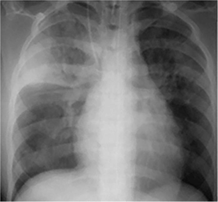
Frontal chest radiograph demonstrating significant bilateral pulmonary opacities. A prominent, dense area of lobar consolidation is visible in the right upper lobe, sharply demarcated inferiorly by the horizontal (minor) fissure. Within this consolidated region, branching lucencies representing air bronchograms are evident, indicating alveolar space filling with patent airways. Additionally, there is patchy, multifocal consolidation involving the right lower lung zone and the left upper and mid-lung zones, particularly in the perihilar distributions. An endotracheal tube is visualized with its tip in the mid-trachea. These radiographic findings are characteristic of severe inflammatory or infectious processes such as acute pneumonia or pulmonary tuberculosis. The image serves as an educational example of lobar vs. patchy consolidation and the utility of the silhouette sign and minor fissure in anatomical localization of lung pathology.
Streptococcus pneumoniae gram stain diplococci sputum
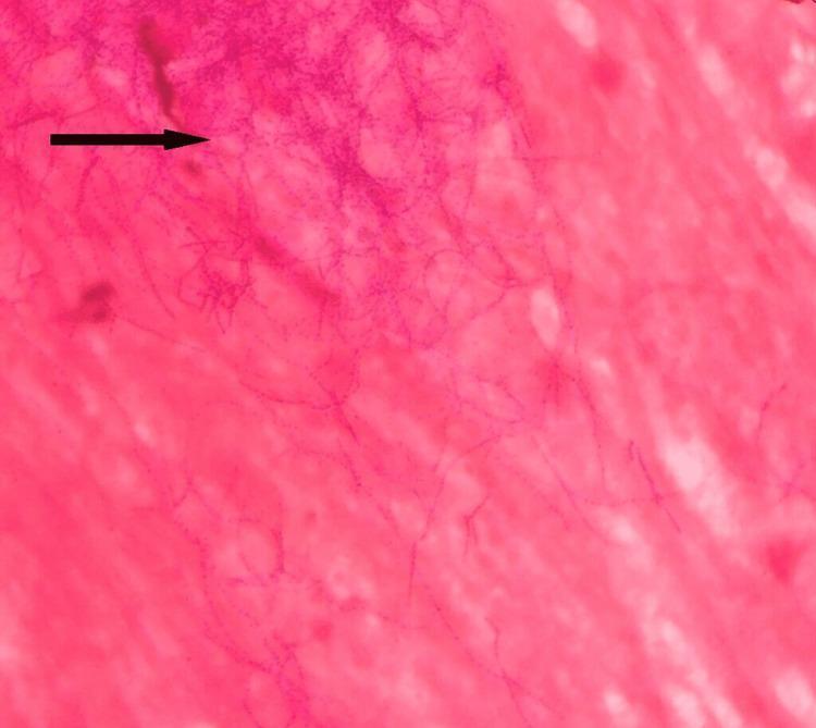
Diagnostic light microscopy image featuring a Gram stain of a clinical sputum sample at high magnification. The visual field demonstrates multiple Gram-positive (dark purple) microorganisms set against a pink-stained proteinaceous and cellular background. The pathogens exhibit a distinctive morphology characterized by long, slender, filamentous rods with frequent branching patterns. A characteristic 'beaded' appearance is evident along the length of the filaments, where the crystal violet stain appears irregularly distributed in alternating dark and light segments. These morphological features—Gram-positive staining, branching filaments, and beaded appearance—are classic diagnostic indicators for Nocardia species. This image serves as an educational resource for medical microbiology, illustrating the differentiation between typical pyogenic bacteria and branching actinomycetes in respiratory infections.
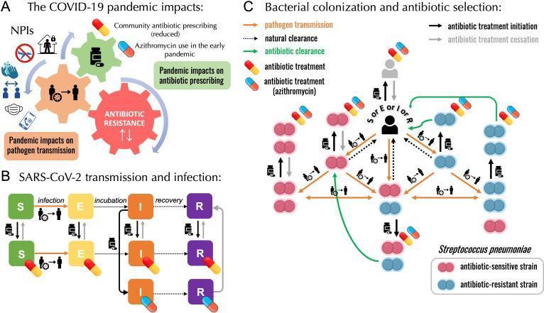
This medical infographic illustrates a mathematical modeling framework examining the impacts of the COVID-19 pandemic on the transmission and antibiotic resistance of Streptococcus pneumoniae. The diagram is divided into three sections: (A) Pandemic impacts on transmission and prescribing, showing how Non-Pharmaceutical Interventions (NPIs) like masking and social distancing influence pathogen transmission and healthcare-seeking behavior, impacting 'Community antibiotic prescribing' and 'ANTIBIOTIC RESISTANCE' through an interlocking gear visual. (B) A compartmental SEIR (Susceptible-Exposed-Infected-Recovered) model for SARS-CoV-2 transmission, incorporating antibiotic treatment pathways for general community prescribing and specific azithromycin use. (C) A detailed bacterial colonization and selection diagram. It uses diplococci icons to differentiate between antibiotic-sensitive (pink) and antibiotic-resistant (blue) S. pneumoniae strains. The flow uses color-coded arrows to represent pathogen transmission (orange dashed), natural clearance (black dashed), and antibiotic clearance (solid green). Clinical dynamics such as antibiotic treatment initiation (black solid arrows) and cessation (gray solid arrows) are mapped against human host icons to illustrate bystander selection and the competitive coexistence of bacterial strains during drug exposure.
Pneumonia - 2nd MBBS Microbiology
Definition
Classification
1. Based on Anatomical Pattern
| Type | Description | Common Cause |
|---|---|---|
| Lobar pneumonia | Consolidation of an entire lobe | S. pneumoniae (most common) |
| Bronchopneumonia | Patchy consolidation around bronchioles | S. aureus, H. influenzae, in children/elderly |
| Interstitial pneumonia | Inflammation of alveolar walls and interstitium | Viruses, Mycoplasma |
2. Based on Epidemiology (Clinically Important)
- Community-Acquired Pneumonia (CAP): Acquired outside hospital
- Hospital-Acquired (Nosocomial) Pneumonia (HAP): Onset >48 hours after admission
- Ventilator-Associated Pneumonia (VAP): In mechanically ventilated patients
- Aspiration Pneumonia: Due to inhalation of oropharyngeal/gastric contents
3. Based on Clinical Presentation
- Typical pneumonia - abrupt onset, productive cough, lobar consolidation (bacterial)
- Atypical pneumonia - gradual onset, dry cough, diffuse interstitial infiltrates (Mycoplasma, Chlamydia, Legionella, viruses)
Etiology (Causative Organisms)
Bacterial Pneumonias
A. Streptococcus pneumoniae (Pneumococcus) - Most Common
- Morphology: Gram-positive, lancet-shaped diplococci, encapsulated
- Virulence factor: Polysaccharide capsule (antiphagocytic) - most important
- Pathogenesis: Organisms colonize the nasopharynx -> aspiration into lungs -> rapid multiplication in alveolar fluid -> RBCs leak into alveoli (congestion) -> neutrophil infiltration (red hepatization) -> macrophage infiltration (grey hepatization) -> resolution when anticapsular antibodies develop
- Predisposing conditions: Preceding viral respiratory infection (especially influenza), chronic pulmonary disease, alcoholism, congestive heart failure, diabetes mellitus, chronic renal disease, splenic dysfunction/splenectomy
- Clinical features:
- Abrupt onset with severe shaking chill
- Sustained high fever (39-41°C)
- Productive cough with blood-tinged (rusty) sputum
- Pleuritic chest pain (pleurisy)
- Lobar consolidation - usually lower lobes
- Dullness to percussion, bronchial breath sounds, crepitations
- Resolution in 2-3 weeks with antibiotics
- Lab diagnosis:
- Sputum Gram stain: Gram-positive lancet-shaped diplococci
- Culture of blood, sputum, pleural fluid
- Urinary antigen test (useful when cultures negative)
- Optochin sensitivity, bile solubility tests
- Treatment: Penicillin G (first-line); fluoroquinolones or vancomycin for penicillin-resistant strains
- Complications: Bacteremia, meningitis, endocarditis, pericarditis, empyema
B. Staphylococcus aureus
- Setting: Common after influenza epidemics; nosocomial pneumonia
- Gram stain: Gram-positive cocci in clusters
- CXR: Patchy infiltrates; may show cavitation, empyema
- Complications: Empyema, lung abscess, pneumatoceles (especially in children)
- Treatment: Nafcillin (MSSA); Vancomycin (MRSA)
C. Klebsiella pneumoniae
- Setting: Alcoholics, diabetics, elderly, nosocomial
- Gram stain: Gram-negative encapsulated rods (mucoid colonies)
- Clinical: Abrupt onset; upper lobe involvement common; currant jelly sputum (thick, blood-stained mucoid)
- CXR: Lobar consolidation, bulging fissure sign, cavitation
- Complications: Cavitation, empyema
- Treatment: Third- or fourth-generation cephalosporins; aminoglycosides for severe infections
D. Haemophilus influenzae
- Setting: Chronic cardiopulmonary disease, follows viral URTI
- Gram stain: Tiny Gram-negative coccobacilli
- CXR: Lobar consolidation
- Diagnosis: Culture on chocolate agar; requires X (hematin) and V (NAD) factors
- Treatment: Ampicillin if beta-lactamase negative; ceftriaxone if resistant
E. Pseudomonas aeruginosa
- Setting: Nosocomial, cystic fibrosis, immunocompromised
- Gram stain: Gram-negative rods
- CXR: Patchy infiltrates with cavitation
- Treatment: Antipseudomonal cephalosporin or carbapenem or piperacillin/tazobactam PLUS an aminoglycoside
F. Anaerobic Bacteria
- Setting: Aspiration pneumonia, periodontitis
- Gram stain: Mixed flora
- CXR: Patchy infiltrates in dependent lung zones
- Complications: Necrotizing pneumonia, lung abscess, empyema
- Treatment: Clindamycin
Atypical Pneumonias
G. Mycoplasma pneumoniae - "Walking Pneumonia"
- Unique features: No cell wall (not seen on Gram stain); smallest free-living organism
- Epidemiology: Most common in teenagers (5-15 years); accounts for >one-third of pneumonia in adolescents; worldwide; epidemics every 4-6 years; spread by droplets; incubation 2-3 weeks; attack rate in families ~60%
- Pathogenesis:
- Has a terminal organelle with proteins P1 and P30 that attach to sialic acid receptors on bronchial epithelial cilia
- ADP-ribosylating toxin interferes with ciliary action -> desquamation and inflammation
- Affects trachea, bronchi, bronchioles, peribronchial tissue
- Clinical features: Gradual onset; headache, malaise, low-grade fever; non-productive (dry) cough; tracheobronchitis or atypical (interstitial) pneumonia
- Gram stain of sputum: PMNs and monocytes; no bacterial pathogens visible
- CXR: Extensive patchy infiltrates (worse on X-ray than clinical appearance)
- Serology:
- Cold agglutinins (IgM antibodies against altered RBC antigen) - positive in ~two-thirds of symptomatic patients; may cause hemolysis and Raynaud phenomenon
- Complement fixation titer
- Treatment: Macrolides (azithromycin), tetracyclines (doxycycline), or fluoroquinolones (levofloxacin) - NOT beta-lactams (no cell wall)
- Complications: Skin rashes, hemolytic anemia, encephalitis, pericarditis, Stevens-Johnson syndrome
H. Legionella pneumophila - "Legionnaires' Disease"
- Setting: Middle-aged, elderly, immunocompromised; air-conditioning cooling towers, water systems (hospital outbreaks)
- Gram stain: Gram-negative rod (poorly stained by Gram; use silver stain or DFA)
- CXR: Lobar consolidation progressing to bilateral
- Diagnosis:
- Urinary antigen test (best rapid test)
- DFA staining of sputum
- Culture on BCYE agar
- Treatment: Azithromycin, levofloxacin, or doxycycline (preferred)
I. Chlamydophila pneumoniae
- Setting: Community-acquired; mimics Mycoplasma pneumonia
- Clinical: Gradual onset; dry cough; pharyngitis often present
- CXR: Patchy infiltrates
- Diagnosis: Serology (complement fixation, microimmunofluorescence)
- Treatment: Azithromycin, levofloxacin, or doxycycline
Viral Pneumonia
- Causative agents: Influenza viruses (most important), RSV (in infants), Parainfluenza (croup in children), Adenovirus, Metapneumovirus
- Pathology: Interstitial inflammation, hyaline membrane formation in alveoli, bronchiolitis, sloughing of ciliated cells
- CXR: Diffuse bilateral interstitial infiltrates
- Sputum culture: Normal oral flora; no bacterial pathogens
- Treatment: Supportive; oseltamivir for influenza; palivizumab prophylaxis for RSV in high-risk infants
Fungal Pneumonias
- Pneumocystis jirovecii (PCP):
- Caused by an atypical fungus; formerly called P. carinii
- Affects severely immunocompromised patients (AIDS, CD4 <200 cells/mm³)
- CXR: Bilateral perihilar "butterfly" infiltrates
- Diagnosis: BAL with methenamine silver stain, DFA, PCR
- Treatment: Trimethoprim-sulfamethoxazole (TMP-SMX); pentamidine as alternative
- Prophylaxis: TMP-SMX when CD4 <200
Summary Table: Key Characteristics of Selected Pneumonias
| Organism | Clinical Setting | Sputum Gram Stain | CXR | Key Complications | Treatment |
|---|---|---|---|---|---|
| S. pneumoniae | Chronic cardiopulmonary disease; post-viral URTI | Gram+ lancet diplococci | Lobar consolidation | Bacteremia, meningitis, empyema | Penicillin G; fluoroquinolones for resistant strains |
| H. influenzae | Chronic lung disease; post-viral URTI | Gram- coccobacilli | Lobar consolidation | Empyema, endocarditis | Ampicillin (if beta-lactamase -); ceftriaxone |
| S. aureus | Post-influenza; nosocomial | Gram+ cocci in clusters | Patchy infiltrates | Empyema, cavitation | Nafcillin/Vancomycin |
| K. pneumoniae | Alcoholism, DM; nosocomial | Gram- encapsulated rods | Lobar consolidation, upper lobe | Cavitation, empyema | 3rd/4th gen cephalosporin ± aminoglycoside |
| E. coli | Nosocomial | Gram- rods | Patchy infiltrates, pleural effusion | Empyema | 3rd gen cephalosporin |
| Pseudomonas | Nosocomial, CF | Gram- rods | Patchy + cavitation | Cavitation | Anti-pseudomonal beta-lactam + aminoglycoside |
| Anaerobes | Aspiration, periodontitis | Mixed flora | Dependent zone infiltrates | Abscess, empyema | Clindamycin |
| M. pneumoniae | Young adults; summer/fall | PMNs, no bacteria | Extensive patchy infiltrates | Hemolysis, rashes | Azithromycin/doxycycline (NO beta-lactams) |
| Legionella | Immunocompromised; water sources | Gram- (poorly stained) | Lobar -> bilateral | Respiratory failure | Azithromycin/levofloxacin |
| Influenza virus | Epidemic; immunosuppressed | Normal flora | Bilateral interstitial | Secondary bacterial pneumonia | Oseltamivir; supportive |
Pathological Stages of Lobar Pneumonia (Pneumococcal)
- Congestion (Days 1-2): Vascular engorgement, bacteria multiply in edema fluid, few neutrophils
- Red Hepatization (Days 3-4): Alveoli filled with RBCs, fibrin, and neutrophils; lung firm and liver-like appearance
- Grey Hepatization (Days 5-7): RBCs disintegrate; alveoli filled with neutrophils and macrophages; grey appearance
- Resolution (Day 8+): Macrophages clear debris; enzymatic digestion of exudate; normal architecture restored
Host Defense Mechanisms Against Pneumonia
- Epiglottal reflex - prevents aspiration
- Mucociliary escalator - cilia propel mucus upward
- Cough reflex - expels aspirated material
- Alveolar macrophages - phagocytose organisms
- Secretory IgA - in respiratory secretions
- Complement and opsonins - facilitate phagocytosis
Clinical Diagnosis Summary
| Feature | Typical Pneumonia | Atypical Pneumonia |
|---|---|---|
| Onset | Abrupt | Gradual |
| Fever | High (39-41°C) with rigors | Low-grade |
| Cough | Productive, purulent/rusty sputum | Dry, non-productive |
| CXR | Lobar consolidation | Bilateral interstitial/patchy |
| Consolidation signs | Yes (dullness, bronchial BS) | Minimal |
| Gram stain | Organisms visible | No organisms |
| Common agents | S. pneumoniae, Klebsiella, S. aureus | Mycoplasma, Legionella, Chlamydia, viruses |
| Response to beta-lactams | Good | Poor (Mycoplasma has no cell wall) |
Microbiological Diagnosis
- Sputum collection: Early morning, before antibiotics; must have <10 squamous epithelial cells and >25 PMNs per LPF for a valid sample
- Gram stain: Rapid presumptive identification
- Culture: Blood culture, sputum culture, BAL (bronchoscopy)
- Urinary antigen: For S. pneumoniae and Legionella (rapid, useful even after antibiotics)
- Serology: Cold agglutinins (Mycoplasma), complement fixation
- PCR: For viral and atypical pathogens
- Special stains: Silver stain (PCP, Legionella), DFA
Radiological Findings
Prevention (Vaccines)
- Pneumococcal vaccines:
- PCV13 (conjugate) - children <5 years and high-risk adults
- PPSV23 (polysaccharide) - adults >65 years and high-risk groups
- Influenza vaccine: Annual; reduces secondary bacterial pneumonia
- H. influenzae type b (Hib) vaccine: Part of routine childhood immunization
Key Exam Points for 2nd MBBS
- Most common cause of CAP: S. pneumoniae
- Most common nosocomial pneumonia organisms: Pseudomonas aeruginosa, S. aureus, Klebsiella
- Rusty sputum = S. pneumoniae | Currant jelly sputum = Klebsiella | Salmon-colored sputum = RSV
- Walking pneumonia = Mycoplasma pneumoniae (most common in teenagers)
- Cold agglutinins = Mycoplasma pneumoniae (IgM type)
- Legionella diagnosis = Urinary antigen test (best rapid test)
- No beta-lactam for Mycoplasma - it has no cell wall
- PCP = opportunistic infection in AIDS (CD4 <200)
- Alcoholic + upper lobe + currant jelly sputum = Klebsiella pneumoniae
- Post-influenza pneumonia = Think S. aureus (secondary bacterial pneumonia)
- Medical Microbiology 9e (Murray et al.), Chapters 19, 14
- Jawetz Melnick & Adelberg's Medical Microbiology 28e, Table 48-3, Cases 3 & 4
- Sherris & Ryan's Medical Microbiology 8e, Chapters 25, 31, 38