
Draw the schematic representation the functional areas of cerebral cortex
functional areas of cerebral cortex schematic diagram Brodmann areas
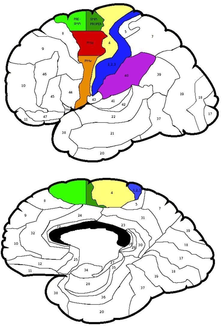
This medical anatomical diagram illustrates the functional subdivisions of the human cerebral cortex, focusing on Brodmann Areas (BA) relevant to motor and sensory processing. The visual contains two schematic views of the brain: a lateral surface view (top) and a medial surface view (bottom). Specific regions of interest are color-coded and labeled to show their spatial relationships. Highlighted areas include the Pre-SMA (light green) and SMA proper (dark green) within the medial frontal lobe. On the lateral surface, the dorsal premotor cortex (PMd, red) and ventral premotor cortex (PMv, orange) are depicted anterior to the primary motor cortex (BA 4, yellow). The primary somatosensory cortex (BA 1, 2, 3) is shown in blue, posterior to the central sulcus, while the supramarginal gyrus (BA 40) is highlighted in purple. Other Brodmann areas are numerically labeled but uncolored. This educational material is designed for neuroscience and neurology students to understand cortical topographical organization and regions involved in proprioception and limb movement control.
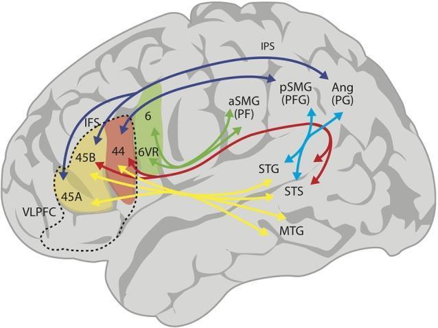
An anatomical diagram illustrating the structural and functional connectivity of language-related regions in the human brain. The schematic uses a lateral view of the cerebral cortex to map pathways between the frontal, parietal, and temporal lobes. Key regions include Broca's area (Brodmann areas 44 and 45A/B) within the ventrolateral prefrontal cortex (VLPFC), the inferior parietal lobe (areas PF/aSMG, PFG/pSMG, and PG/Ang), and the superior and middle temporal gyri (STG, MTG, STS). Color-coded arrows represent distinct neural fasciculi: red arrows denote the arcuate fasciculus (AF) connecting the posterior temporal lobe to areas 44 and 45B; dark blue arrows represent the superior longitudinal fasciculus (SLF) connecting the angular and supramarginal gyri to the VLPFC; and yellow arrows indicate the ventral pathway via the extreme capsule connecting the anterior temporal lobe to Broca's region. Light blue arrows show connections between the temporal and parietal lobes via the middle longitudinal fasciculus. This diagram demonstrates the dual-stream model of language processing, highlighting dorsal and ventral integration.
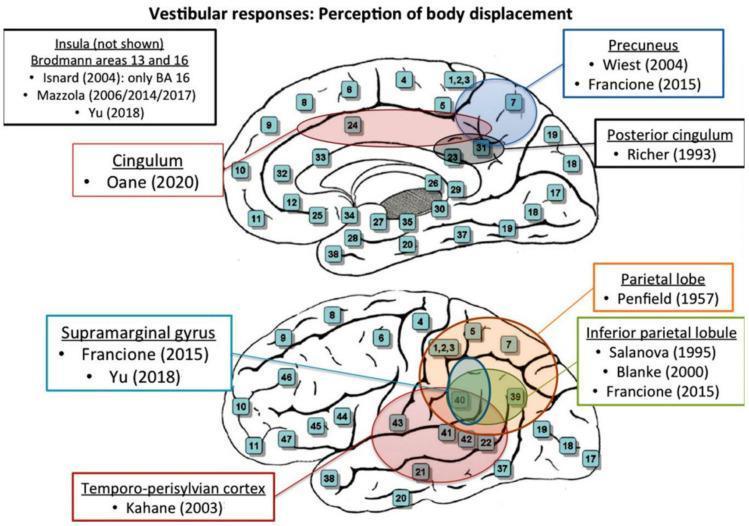
Two schematic topographical diagrams of the human brain illustrating cortical areas associated with vestibular responses, specifically the perception of body displacement. The top diagram shows a medial view of the cerebral hemisphere, highlighting the Precuneus (Brodmann area 7), Posterior cingulum (BA 31), and the Cingulum (BA 24, 32, 33). The bottom diagram depicts a lateral view, highlighting the Parietal lobe, Inferior parietal lobule (BA 39, 40), Supramarginal gyrus, and the Temporo-perisylvian cortex. Both diagrams include numbered stimulation sites corresponding to specific neuroanatomical locations. Text boxes identify key functional regions and cite primary literature (e.g., Penfield 1957, Blanke 2000, Oane 2020) that investigated these areas through electrical stimulation. A note specifies that the Insula (Brodmann areas 13 and 16) is also associated with these responses although not visually depicted in these specific planes. This educational graphic serves to map the extensive vestibular cortical network involved in spatial orientation and self-motion perception.
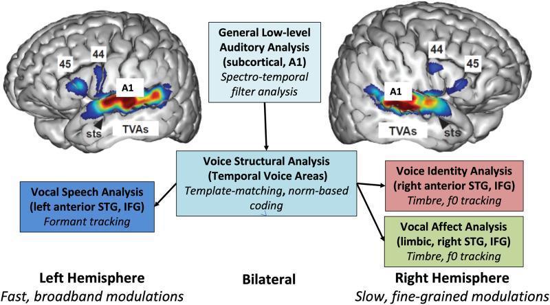
This medical schematic and neuroimaging diagram illustrates the functional processing hierarchy of auditory communicative signals in the human brain. The image features bilateral lateral views of the cerebral cortex with fMRI-style heatmaps highlighting the Temporal Voice Areas (TVAs) along the Superior Temporal Sulcus (STS) and Superior Temporal Gyrus (STG). Primary auditory cortex (A1) and Inferior Frontal Gyrus (IFG) regions, specifically Brodmann areas 44 and 45, are annotated. A central flowchart outlines the hierarchical processing sequence: 1) General Low-level Auditory Analysis (subcortical and A1) involving spectro-temporal filter analysis; 2) Bilateral Voice Structural Analysis (TVAs) utilizing template-matching and norm-based coding; and 3) Hemispherically lateralized downstream processing. The left hemisphere is associated with Vocal Speech Analysis (formant tracking) characterized by fast, broadband modulations. The right hemisphere is associated with Voice Identity and Vocal Affect Analysis (timbre and f0 tracking) involving the right anterior STG, IFG, and limbic structures, characterized by slow, fine-grained modulations. This infographic serves as a pedagogical tool for understanding the neuroanatomical basis of social signal processing and voice categorization.
Schematic diagram of the functional areas of the cerebral cortex, lateral view of the left hemisphere. Clearly labeled and color-coded regions: Primary Motor Cortex (precentral gyrus, BA 4) in red, Premotor Cortex (BA 6) in orange, Prefrontal Cortex (BA 9, 10, 11, 46) in yellow, Broca's Speech Area (BA 44, 45) in dark orange, Primary Somatosensory Cortex (postcentral gyrus, BA 1, 2, 3) in blue, Somatosensory Association Cortex (BA 5, 7) in light blue, Primary Visual Cortex (BA 17) in purple, Visual Association Cortex (BA 18, 19) in lavender, Primary Auditory Cortex (BA 41, 42) in green, Auditory Association Cortex / Wernicke's Area (BA 22) in light green, Angular Gyrus (BA 39) in teal, Supramarginal Gyrus (BA 40) in cyan. Major sulci labeled: central sulcus, lateral sulcus (Sylvian fissure), parieto-occipital sulcus. Lobes labeled: frontal, parietal, temporal, occipital. Clean educational medical illustration style with legend.
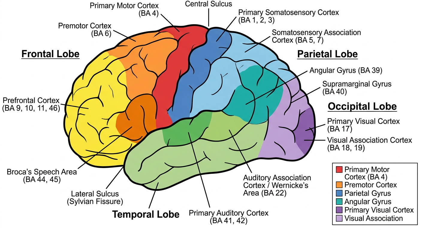
Functional Areas of the Cerebral Cortex
Frontal Lobe
| Area | Brodmann Area (BA) | Function |
|---|---|---|
| Primary Motor Cortex | BA 4 (precentral gyrus) | Voluntary movement; contralateral body control |
| Premotor Cortex | BA 6 | Planning and coordination of movement |
| Supplementary Motor Area | BA 6 (medial) | Bimanual coordination, motor sequencing |
| Prefrontal Cortex | BA 9, 10, 11, 46 | Executive function, personality, judgment, working memory |
| Broca's Area | BA 44, 45 | Motor speech production (dominant hemisphere) |
| Frontal Eye Field | BA 8 | Voluntary eye movements |
Parietal Lobe
| Area | Brodmann Area | Function |
|---|---|---|
| Primary Somatosensory Cortex | BA 1, 2, 3 (postcentral gyrus) | Touch, pain, temperature, proprioception |
| Somatosensory Association Cortex | BA 5, 7 | Integration of sensory information |
| Supramarginal Gyrus | BA 40 | Phonological processing, tactile attention |
| Angular Gyrus | BA 39 | Reading, writing, language comprehension, calculation |
Temporal Lobe
| Area | Brodmann Area | Function |
|---|---|---|
| Primary Auditory Cortex | BA 41, 42 | Hearing, frequency and pitch detection |
| Wernicke's Area | BA 22 | Language comprehension (dominant hemisphere) |
| Auditory Association Cortex | BA 21, 22 | Sound recognition, meaning |
Occipital Lobe
| Area | Brodmann Area | Function |
|---|---|---|
| Primary Visual Cortex (V1) | BA 17 (calcarine sulcus) | Basic visual processing |
| Visual Association Cortex (V2, V3, V4, V5) | BA 18, 19 | Form, color, motion, depth perception |
- Damage to BA 4 - contralateral spastic paralysis
- Damage to Broca's area (BA 44/45) - expressive (non-fluent) aphasia
- Damage to Wernicke's area (BA 22) - receptive (fluent) aphasia
- Damage to BA 17 - cortical blindness
- Damage to Angular gyrus (BA 39) - Gerstmann syndrome (acalculia, agraphia, finger agnosia, right-left disorientation)