
Define defets of blood and correlate with cbc and explain in detail
Reading File
Now let me search the library in parallel for all relevant topics.
Finding Sources
Finding Sources
Finding Sources
Finding Sources
Finding Sources
Finding Sources
I can see there's a Tietz Textbook of Laboratory Medicine which will be very relevant for CBC. Let me search specifically for hematology books and CBC parameters.
Finding Sources
Finding Sources
Finding Sources
Excellent! Now let me read the key textbook sections in parallel.
Reading File
Reading File
Reading File
Excellent data collected. Let me now fetch the blood smear images and read more CBC parameters from Tietz.
Reading File
Loading Image
Loading Image
I now have excellent, comprehensive textbook data to compose a detailed answer. Let me also fetch one more image showing abnormal RBC morphology.
Loading Image
I now have comprehensive textbook material and images. Let me compose the full, detailed answer.
Defects of Blood: Definitions, Classification, and CBC Correlation
Blood defects (hematological disorders) refer to abnormalities in the quantity, quality, or function of any blood component - red blood cells (RBCs), white blood cells (WBCs), or platelets. The Complete Blood Count (CBC) is the cornerstone investigation that quantifies and characterizes these abnormalities.
What the CBC Measures
A CBC of a whole blood sample consists of:
- RBC count (erythrocytes)
- WBC count (total and differential)
- Platelet count
- Hemoglobin (Hb) - direct measurement
- Red cell indices: MCV, MCH, MCHC, RDW - derived parameters
- Hematocrit (HCT)
- WBC differential (neutrophils, lymphocytes, monocytes, eosinophils, basophils)
- Tietz Textbook of Laboratory Medicine, 7th Edition
PART I: DEFECTS OF RED BLOOD CELLS (Erythrocyte Disorders)
A. ANEMIA (Decreased RBCs / Hemoglobin)
Anemia is defined as a reduction in the oxygen-carrying capacity of the blood. It is classified morphologically using CBC indices.
1. Microcytic Hypochromic Anemia
CBC Findings:
- Hb: Low
- MCV: <80 fL (microcytic)
- MCH: <27 pg
- MCHC: Low
- RDW: Elevated (especially in iron deficiency)
| CBC Parameter | Thalassemia Trait | Iron Deficiency (IDA) | Anemia of Chronic Disease (ACD) |
|---|---|---|---|
| RBC count | ↑/Normal | ↓ | ↓ |
| MCV | ↓ | ↓ | Normal/↓ |
| MCH | ↓ | ↓ | Normal/↓ |
| RDW | Normal | ↑ | Normal/↑ |
| Serum iron | Normal | ↓ | ↓ |
| TIBC/transferrin | Normal | ↑ | ↓ |
| Ferritin | Normal | ↓ | ↑/Normal |
- Tietz Textbook of Laboratory Medicine, 7th Ed
Clinical notes:
- In iron deficiency anemia, the RDW rises early (anisocytosis) before MCV falls - making RDW elevation the earliest CBC clue.
- In thalassemia trait, RBC count is often elevated or normal despite low MCV, which helps distinguish it from IDA where RBC is reduced.
- The MCV swells artifactually after 2-3 days of EDTA storage - samples should not be excessively delayed before analysis.
2. Macrocytic / Megaloblastic Anemia
CBC Findings:
- Hb: Low
- MCV: >100 fL (macrocytic)
- Hypersegmented neutrophils on smear (>5 lobes in >5% of neutrophils) - classic finding
- WBC and platelets may also be reduced (pancytopenia in severe cases)
- Reticulocytes: Low
Causes: Vitamin B12 deficiency, folate deficiency, alcohol, drugs (methotrexate, hydroxyurea)
Key correlation: Concurrent thalassemia and megaloblastic anemia can normalize the MCV, masking both conditions - the RDW and blood smear features are then the clues.
3. Normocytic Normochromic Anemia
CBC Findings:
- Hb: Low
- MCV: 80-100 fL (normal)
- MCH, MCHC: Normal
- RDW: May be elevated in mixed deficiency
Causes: Acute blood loss, hemolysis, aplastic anemia, anemia of chronic disease (early), renal failure (erythropoietin deficiency)
CBC clue for hemolysis: Reticulocytosis (elevated reticulocyte count) distinguishes hemolytic anemia (increased marrow output) from aplastic anemia (absent reticulocytes).
4. Key RBC Indices Explained
| Index | Formula | Normal | Clinical Significance |
|---|---|---|---|
| MCV | HCT / RBC × 10 | 80-100 fL | Cell size - microcytic vs macrocytic |
| MCH | Hb / RBC × 10 | 27-33 pg | Hb content per cell |
| MCHC | Hb / HCT × 100 | 32-36 g/dL | Hb concentration - low = hypochromic |
| RDW | CV of RBC volumes | 11-15% | Anisocytosis - mixed populations |
Important: The MCV reference range in infancy and childhood differs significantly from adults. MCV is high at birth (100-125 fL in term infants), falls to a nadir at ~1 year of age (~70-82 fL), and only reaches adult values after age 18. This must be remembered to avoid over-investigating low MCV in early childhood.
PART II: DEFECTS OF WHITE BLOOD CELLS (Leukocyte Disorders)
CBC WBC Component: Differential Count
| Cell | Normal Range | Function |
|---|---|---|
| Neutrophils | 40-70% (1.8-7.7 × 10⁹/L) | Bacterial/fungal defense |
| Lymphocytes | 20-40% | Viral defense, immunity |
| Monocytes | 2-8% | Phagocytosis, antigen presentation |
| Eosinophils | 1-4% | Parasitic/allergic response |
| Basophils | 0-1% | Inflammatory mediator release |
A. Neutrophilia (High Neutrophils on CBC)
- Bacterial infections, tissue damage, corticosteroids, pregnancy
- Toxic granulation (heavy staining of azurophilic granules) and vacuolation on smear suggest active infection or inflammation
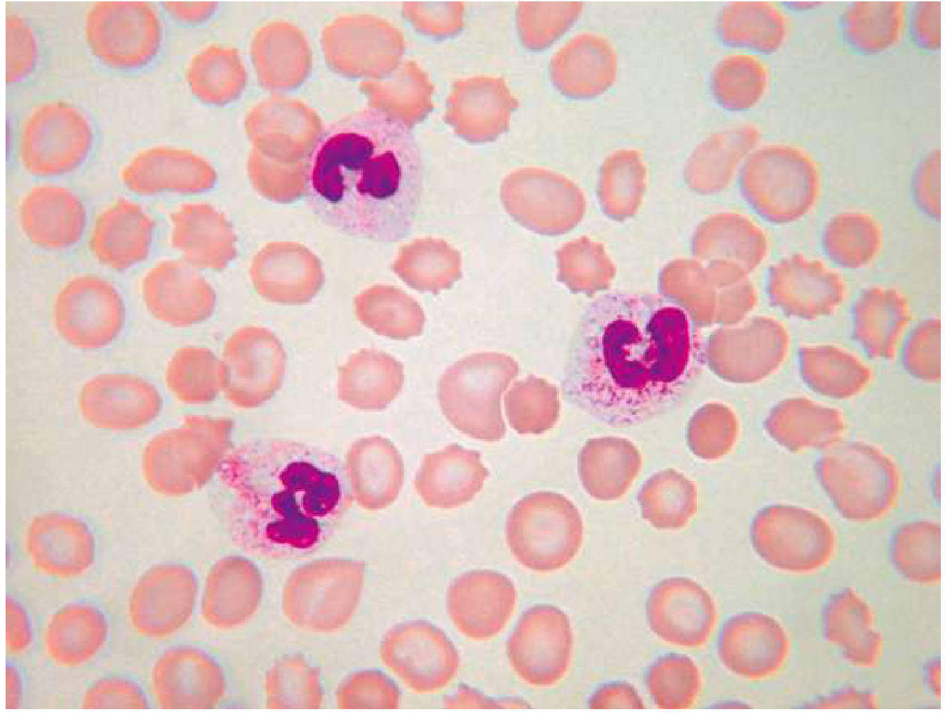
Toxic granulation: Heavy staining of neutrophil azurophilic granules, seen in infection, inflammation, tissue damage, and G-CSF administration. (Goldman-Cecil Medicine)
B. Neutropenia (Low Neutrophils)
- Absolute neutrophil count (ANC) <1.5 × 10⁹/L
- Causes: Viral infections, chemotherapy, autoimmune, aplastic anemia
- Risk of bacterial infection rises sharply when ANC <0.5 × 10⁹/L
C. Pelger-Huet Anomaly (Morphological Defect)
- Hypolobulation of neutrophil nuclei with increased chromatin clumping
- Inherited form: bilobed "pince-nez" shaped nuclei
- Acquired (pseudo-Pelger): seen in myelodysplastic syndromes (MDS), drugs (tacrolimus, mycophenolate)
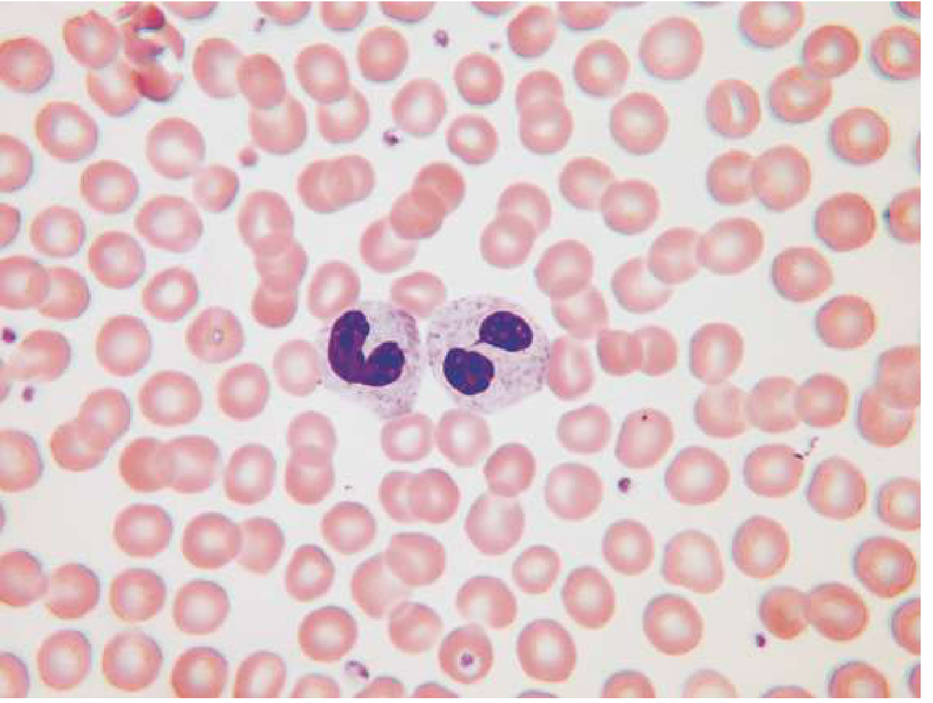
Pelger-Huet anomaly: Hypolobulated neutrophil nuclei with clumped chromatin. In MDS, the neutrophils may also be hypogranular. (Goldman-Cecil Medicine)
D. Dohle Bodies
- Pale blue-gray amorphous inclusions near the neutrophil cell membrane
- Seen in: infection, inflammation
- Similar but larger/more angular inclusions occur in May-Hegglin anomaly (MYH9-related disorder)
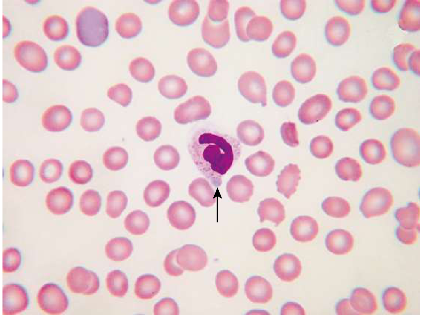
Döhle body (arrow): A pale blue-gray amorphous cytoplasmic inclusion in a neutrophil. Can result from infection and inflammation, or as part of May-Hegglin anomaly. (Goldman-Cecil Medicine)
E. Leukocytosis (Total WBC >11 × 10⁹/L)
- Bacterial infection (neutrophilic shift), viral infection (lymphocytosis), allergy/parasites (eosinophilia)
- Leukemoid reaction: WBC >50 × 10⁹/L with left shift (bands, metamyelocytes) - mimics leukemia
- Leukemia: Blast cells on differential; confirmed by bone marrow biopsy
F. Leukopenia (Total WBC <4 × 10⁹/L)
- Viral infections (HIV, EBV), chemotherapy, aplastic anemia, B12/folate deficiency
- CBC in aplastic anemia: Pancytopenia (low RBC + low WBC + low platelets) with very low reticulocyte count
PART III: DEFECTS OF PLATELETS (Thrombocyte Disorders)
A. Thrombocytopenia (Low Platelets)
Definition: Platelet count <150,000/μL (150 × 10⁹/L)
Clinical severity by CBC count:
- 50,000-150,000/μL: Mild - usually no spontaneous bleeding but aggravated post-trauma
- 20,000-50,000/μL: Can aggravate post-traumatic bleeding
- <20,000/μL: Risk of spontaneous, non-traumatic bleeding; intracranial hemorrhage is the most feared complication
- PT and PTT remain normal in isolated thrombocytopenia
Causes (from Robbins Pathology):
| Category | Examples |
|---|---|
| Decreased production | Aplastic anemia, leukemia, B12/folate deficiency, alcohol, HIV, myelodysplastic syndrome |
| Immunologic destruction | ITP (immune thrombocytopenic purpura), SLE, drug-induced (heparin, quinidine, sulfa), HIV, post-transfusion |
| Non-immunologic destruction | DIC, thrombotic microangiopathy (TTP, HUS), giant hemangiomas |
| Sequestration | Hypersplenism |
| Dilution | Massive transfusions |
- Robbins, Cotran & Kumar Pathologic Basis of Disease
Important CBC rules for thrombocytopenia:
- Always confirm an automated count <60 × 10⁹/L with a peripheral blood smear before altering clinical management (never transfuse platelets for unexpected thrombocytopenia without microscopic confirmation)
- EDTA-induced pseudothrombocytopenia: In vitro platelet clumping in EDTA-anticoagulated tubes causes falsely low automated counts - confirmed by redrawing in citrate or heparin, or viewing smear for platelet aggregates
- Platelet satellitism: Platelets surrounding neutrophils - another EDTA artifact
- Fibrin strands on smear suggest coagulation activation and erroneous counts
B. Thrombocytosis (High Platelets >400,000/μL)
- Reactive: Iron deficiency, infection, post-splenectomy, inflammation
- Primary (essential thrombocythemia): Myeloproliferative neoplasm; paradoxically can cause both thrombosis and bleeding
C. Qualitative Platelet Defects
CBC count may be normal, but bleeding time and platelet function assay (PFA-100) are abnormal.
| Disorder | Defect | Key CBC/Lab Finding |
|---|---|---|
| Glanzmann thrombasthenia | Absent GPIIb/IIIa (fibrinogen receptor) | Normal count, abnormal aggregation |
| Bernard-Soulier syndrome | Absent GPIb (vWF receptor) | Normal/low count + giant platelets on smear |
| von Willebrand disease | Deficient/abnormal vWF | Normal CBC; prolonged PFA; ↓ vWF antigen |
| Drug-induced (aspirin) | COX-1 inhibition | Normal CBC; abnormal aggregation |
PART IV: COMBINED / MULTI-LINEAGE DEFECTS
Pancytopenia (Low RBC + WBC + Platelets on CBC)
| Cause | Key Diagnostic Clue |
|---|---|
| Aplastic anemia | Very low reticulocytes, hypocellular bone marrow |
| B12/Folate deficiency | High MCV, hypersegmented neutrophils |
| Bone marrow infiltration (leukemia, myeloma, metastases) | Leukoerythroblastic picture (nucleated RBCs + immature WBCs on smear) |
| Hypersplenism | Splenomegaly; normal bone marrow |
| HIV | Cytopenias across all lineages |
Summary: CBC as a Diagnostic Tool
CBC Result                    → Think First
─────────────────────────────────────────────────────
Low Hb + Low MCV + High RDW  → Iron Deficiency Anemia
Low Hb + Low MCV + Normal RDW → Thalassemia Trait
Low Hb + High MCV             → B12/Folate deficiency
Low Hb + Normal MCV           → Acute loss, hemolysis, renal failure
Pancytopenia + Low Retics     → Aplastic anemia
Pancytopenia + High MCV       → Megaloblastic anemia
High WBC + Blasts             → Acute leukemia (urgent smear/BM biopsy)
Low Platelets + Normal PT/PTT → Primary thrombocytopenia (ITP)
Low Platelets + High PT/PTT   → DIC or TTP
Key Takeaway
The CBC is not merely a count - it is a functional diagnostic map of the bone marrow and peripheral blood. Each parameter (Hb, MCV, MCH, MCHC, RDW, WBC differential, platelet count) contributes a piece to diagnosing defects across the three major blood cell lineages. The peripheral blood smear remains the gold standard to validate automated counts and identify morphological defects (toxic granulation, hypersegmented neutrophils, Pelger-Huet anomaly, giant platelets, schistocytes) that numbers alone cannot reveal.
- Tietz Textbook of Laboratory Medicine, 7th Edition
- Robbins, Cotran & Kumar Pathologic Basis of Disease
- Goldman-Cecil Medicine, International Edition
This is a shared conversation. Sign in to Orris to start your own chat.