
Teach me rheumatic fever
rheumatic fever Jones criteria Aschoff nodules mitral stenosis
Patients with mitral stenosis may or may not have a history of rheumatic fever, but the classic pathology and echocardiographic morphology must be appreciated. After initial medical management, the criteria for timing of intervention may include pulmonary hypertension in addition to symptoms of dyspnea. More importantly, physicians should know the impact of valve morphology, various scoring systems, and their corresponding hemodynamic and echocardiographic features that influence the short- and long-term outcomes of the valvuloplasty or valve replacement and help in informed decision-making.
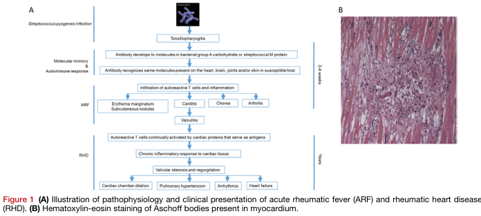
Summary : This figure illustrates the pathophysiology and clinical presentation of acute rheumatic fever (ARF) and rheumatic heart disease (RHD), and shows a histological image of Aschoff bodies in myocardium. flowchart and photo: # Panel A: Pathophysiology and Clinical Presentation Flowchart ## Nodes : • Streptococcus pyogenes infection (text node) • Tonsillopharyngitis (rectangle) • Antibody develops to molecules in bacterial group A carbohydrate or streptococcal M protein (rectangle) • Antibody recognizes same molecules present on the heart, brain, joints and/or skin in susceptible host (rectangle) • Infiltration of autoreactive T cells and inflammation (rectangle) • Erythema marginatum, Subcutaneous nodules (rectangle, branch) • Carditis (rectangle, branch) • Chorea (rectangle, branch) • Arthritis (rectangle, branch) • Valvulitis (rectangle) • Autoreactive T cells continually activated by cardiac proteins that serve as antigens (rectangle) • Chronic inflammatory response to cardiac tissue (rectangle) • Valvular stenosis and regurgitation (rectangle) • Cardiac chamber dilation (rectangle, branch) • Pulmonary hypertension (rectangle, branch) • Arrhythmia (rectangle, branch) • Heart failure (rectangle, branch) ## Connectors : • Linear downward arrows connect each main step. • Branching arrows from "Infiltration of autoreactive T cells and inflammation" to four clinical features: erythema marginatum/subcutaneous nodules, carditis, chorea, arthritis. • Branching arrows from "Valvular stenosis and regurgitation" to four complications: cardiac chamber dilation, pulmonary hypertension, arrhythmia, heart failure. ## Layout : • Vertical, left-to-right flow with two main phases: ARF (2–4 weeks) and RHD (years). • Branches at clinical presentation and complications. • Timeline markers on the right: "2–4 weeks" for ARF, "Years" for RHD. # Panel B: Histological Image ## Scene Overview : • Main subject: Myocardial tissue section stained with hematoxylin-eosin. • Central focus: Cluster of cells (Aschoff body) within pink-stained cardiac muscle fibers. • Colour palette: Pink (muscle), purple/blue (nuclei and inflammatory infiltrate). ## Technical Details : • Staining: Hematoxylin-eosin. • Magnification and scale bar: Not specified. • Tissue: Myocardium. ## Spatial Relationships : • Aschoff body is centrally located, surrounded by elongated muscle fibers. • Dense cellular infiltrate contrasts with the striated background. # Analysis : • The flowchart in Panel A details the progression from streptococcal infection to autoimmune-mediated heart damage, highlighting both acute and chronic clinical manifestations and complications. • The histological image in Panel B visually confirms the presence of Aschoff bodies, a hallmark of rheumatic carditis, within the myocardium.
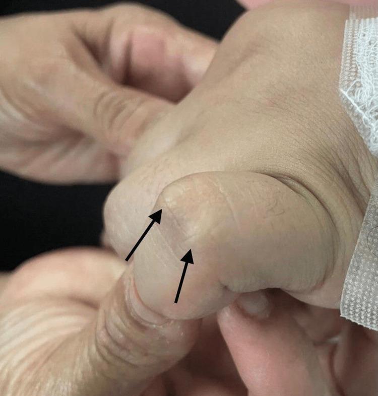
Clinical photograph showing the dorsal surface of a human hand, specifically focusing on the interphalangeal joints. Two prominent subcutaneous nodules are visible over the joint space, indicated by black arrows. These nodules appear as firm, well-circumscribed, painless elevations under the skin. They are flesh-colored to slightly pale, with smooth overlying skin and no signs of acute inflammation, erythema, or ulceration. This finding is a characteristic major Jones criterion for the diagnosis of acute rheumatic fever. The image serves as an educational tool for identifying extra-articular manifestations of rheumatological and post-streptococcal conditions in a clinical setting.
I. INTRODUCTION II. EPIDEMIOLOGY OF RHEUMATIC HEART DISEASE III. BASIC CONCEPTS OF PATHOPHYSIOLOGY, CLINICAL PRESENTATION, AND SCREENING III.A. Pathophysiology and Clinical Presentation III.B. Echocardiographic Screening in Acute Rheumatic Fever Key Points Recommendations IV. RHEUMATIC VALVE LESIONS IV.A. Mitral Stenosis IV.A.1. Anatomic Considerations in the Assessment of Mitral Stenosis IV.A.2. Hemodynamic Considerations in the Assessment of Mitral Stenosis IV.A.3. Mitral Stenosis in Pregnancy Key Points Recommendations IV. B. Rheumatic Mitral Regurgitation IV.B.1. Anatomic Considerations in the Assessment of Rheumatic Mitral Regurgitation IV.B.2. Hemodynamic Considerations in the Assessment of Rheumatic Mitral Regurgitation Key Points Recommendations IV.C. Rheumatic Aortic Valve IV.C.1. Anatomic Considerations in the Assessment of the
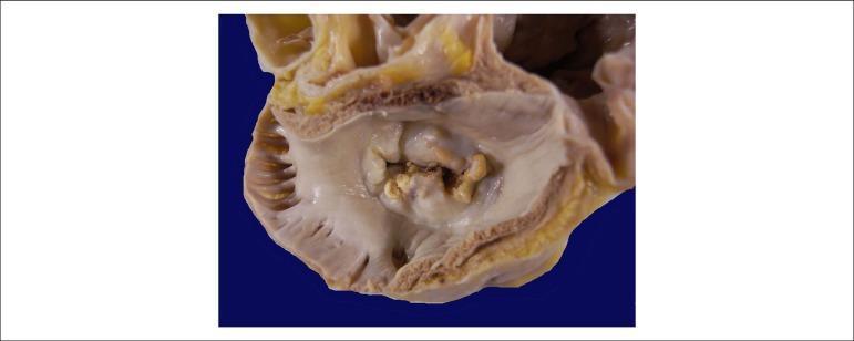
This clinical photograph shows a gross pathological specimen of a human heart, specifically an atrial view of the mitral valve. The image demonstrates hallmark signs of chronic rheumatic heart disease. Key features include significant thickening and fibrosis of the valve leaflets, resulting in a 'fish-mouth' or 'buttonhole' appearance of the valvular orifice. There is evidence of extensive commissural fusion and multiple focal areas of yellowish-white calcification along the leaflet margins and within the valve body. These structural changes characterize severe mitral stenosis, where the restricted opening of the valve impairs blood flow from the left atrium to the left ventricle. The surrounding atrial endocardium appears thickened, consistent with chronic pressure overload. This visual is highly representative for educational purposes in cardiology and pathology to illustrate the long-term sequelae of rheumatic fever on cardiac valves.
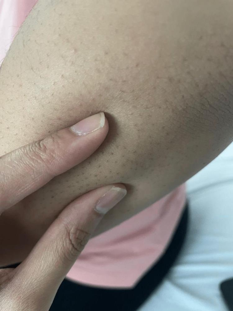
This clinical photograph displays a close-up view of the right upper extremity, specifically the extensor surface of the elbow region. The primary clinical finding is the presence of multiple, small subcutaneous nodules. These nodules appear as firm, rounded, non-tender-looking elevations beneath the skin surface, typically measuring less than 1 cm in diameter. The overlying skin is intact, displaying normal pigmentation and texture without visible erythema, warmth, or ulceration. Prominent hair follicles are visible as dark punctate spots across the skin. In a clinical context involving polyarthralgia and carditis, these nodules are a major Jones criterion for the diagnosis of acute rheumatic fever (ARF). This image serves as a teaching tool for medical students and clinicians to recognize the dermatological manifestations of systemic streptococcal-related diseases and rheumatological disorders.
"rheumatic fever" AND prevention
Rheumatic Fever - A Complete Teaching Guide
Overview
- Goldman-Cecil Medicine, 27e, p. 3043
1. Epidemiology
- Age: Most cases occur in children aged 5-15 years. ARF is rare before age 3 and infrequent after 15.
- Sex: Equal incidence in males and females, but RHD is more common in women.
- RHD peak: Third and fourth decades of life (due to cumulative damage from recurrent attacks).
- ARF develops in ~2.5% of untreated GAS pharyngitis cases in susceptible populations.
- Globally more common in tropical/developing countries, where it remains the leading cause of heart disease in young people.
- M protein types 1, 3, 5, 6, 18 are the most "rheumatogenic" strains.
- Key point: Only pharyngeal infection triggers ARF - cutaneous (skin) streptococcal infections do NOT cause rheumatic fever.
2. Pathophysiology - Molecular Mimicry
- GAS pharyngitis - The streptococcal M protein and Group A carbohydrate have molecular similarities to human tissue proteins.
- Antibody formation - The host generates antibodies against streptococcal antigens.
- Molecular mimicry - These antibodies cross-react with heart, brain, joints, and skin because streptococcal antigens resemble human proteins.
- Autoreactive T-cell infiltration - Inflammation is initiated at these organs.
- Acute manifestations (ARF) - Carditis, arthritis, chorea, erythema marginatum, subcutaneous nodules appear 2-4 weeks later.
- Chronic activation - Autoreactive T cells are continuously re-activated by cardiac proteins, leading to valvulitis → valvular scarring → RHD over years.
| Manifestation | Mechanism |
|---|---|
| Carditis | Antibody binding + T-cell infiltration of heart |
| Arthritis | Immune complex deposition in joints |
| Chorea | Antibody binding to basal ganglia |
| Skin (nodules, EM) | Delayed hypersensitivity reactions |
3. Clinical Manifestations (JONES mnemonic)
Major Manifestations:
| Manifestation | Frequency | Key Features |
|---|---|---|
| Joints (Arthritis) | 75% | Migratory polyarthritis of large joints - knees (76%), ankles (50%), elbows/wrists (12-15%). Sterile synovial fluid with lymphocyte predominance. Exquisitely tender, responds dramatically to aspirin/NSAIDs. |
| Oh! (Carditis) | 50-91% | Pancarditis - pericardium, myocardium, endocardium. Mitral valve involved in nearly ALL cases. Mitral regurgitation is the most common acute lesion. Carey-Coombs murmur (middiastolic flow murmur). PR prolongation on ECG. |
| Nodules (Subcutaneous) | <10% | 0.5-2 cm, painless, firm nodules over bony prominences or extensor tendons. |
| Erythema Marginatum | <10% | Pink, non-pruritic, blanching macules expanding centrifugally in a serpiginous pattern on trunk and proximal limbs. Never on the face. |
| Sydenham Chorea | 30% | Involuntary, non-rhythmic, purposeless movements of limbs, body, and face. More on one side. Stops during sleep. May appear weeks after the pharyngitis when other signs have resolved. |
- Mitral valve: almost all cases (papillary muscle involvement)
- Aortic valve: 20-30% of cases
- Tricuspid valve: histologically involved in 15-40%, rarely clinically significant
- Pulmonic valve: rarely involved
- Carey-Coombs murmur = middiastolic murmur due to mitral valve leaflet edema
- Heart failure in 5-10% of first attacks - the most life-threatening acute feature
Subcutaneous nodules (Jones criterion):
Minor Manifestations:
- Fever ≥38.5°C
- Elevated inflammatory markers (ESR, CRP)
- Prolonged PR interval on ECG (if not already a major criterion for carditis)
- Arthralgia (only if arthritis is NOT the major criterion)
4. Diagnosis - Revised Jones Criteria (AHA 2015)
Evidence of preceding GAS infection:
- Elevated or rising ASO (antistreptolysin O) titer - most useful (elevated in 80% of cases; culture often negative by the time RF develops)
- Anti-DNase B
- Positive throat culture for GAS (may be negative late)
Jones Criteria (2015 AHA Revision):
| Low-Risk Populations* | Moderate/High-Risk Populations | |
|---|---|---|
| Major: Carditis | Clinical ± echocardiographic | Clinical ± echocardiographic |
| Major: Arthritis | Polyarticular only | Mono- or polyarticular |
| Major: Chorea | Yes | Yes |
| Major: Erythema marginatum | Yes | Yes |
| Major: Subcutaneous nodules | Yes | Yes |
| Minor: Arthralgia | Polyarthralgia | Monoarthralgia |
| Minor: Fever | ≥38.5°C | ≥38.5°C |
| Minor: ESR/CRP | ESR ≥60 mm/hr and/or CRP ≥3.0 mg/dL | ESR ≥30 mm/hr and/or CRP ≥3.0 mg/dL |
| Minor: PR prolongation | Yes (if carditis not major criterion) | Yes |
Lab findings:
- Elevated ESR, CRP
- Leukocytosis
- Normochromic, normocytic anemia
- ECG: prolonged PR interval
- Echocardiogram: should be performed to detect subclinical valvulitis
5. Treatment
Hospitalization
A. Eradication of GAS (Primary Treatment):
- IM benzathine penicillin G 1.2 million units - single dose (preferred)
- Oral penicillin V 250 mg twice daily x 10 days
- Macrolide (azithromycin) if penicillin-allergic
B. Arthritis:
- Aspirin 80-100 mg/kg/day in divided doses (max 4-8 g/day) - first-line
- Naproxen 10-20 mg/kg/day (alternative, used in small studies)
- Continue NSAIDs 1-2 weeks until ALL symptoms resolve (not just joint pain)
- Dramatic NSAID response is almost diagnostic; lack of response should prompt reconsideration
C. Carditis:
- Heart failure management: diuretics, ACE inhibitors/ARBs, fluid restriction, bed rest
- Corticosteroids may be used in severe carditis (high-quality evidence lacking)
- Valve surgery rarely needed acutely (except acute chordal rupture)
D. Chorea:
- Usually requires no specific treatment; self-resolves in weeks to months
E. Secondary Prophylaxis (CRITICAL):
| Regimen | Dose |
|---|---|
| IM benzathine penicillin G (preferred) | 1.2 million units every 4 weeks |
| Oral penicillin V | 250 mg twice daily |
| Oral macrolide | Azithromycin 250 mg daily |
- ARF without carditis: 5 years or until age 21 (whichever is longer)
- ARF with carditis (no residual valve disease): 10 years or until age 21
- ARF with persistent RHD: Minimum 10 years, often lifelong
- A 2024 Cochrane systematic review (PMID 39312290) confirmed long-term antibiotic prophylaxis reduces RF recurrence and RHD progression.
6. Rheumatic Heart Disease (RHD) - Long-term Consequence
- 35-70% of ARF patients with carditis progress to chronic RHD
- Another 18% have subclinical echocardiographic carditis
- RHD affects:
- Mitral valve: ~100% of cases - mitral regurgitation initially, mitral stenosis with repeated attacks
- Aortic valve: 20-30% - regurgitation more common than stenosis
- Tricuspid and pulmonic rarely affected clinically
- RHD is progressive, with severe valvular disease typically appearing in the third or fourth decade of life
- In endemic regions, echocardiographic screening detects latent RHD and guides prophylaxis
7. PANDAS (Related Condition)
Key Memory Anchors
| Concept | Remember |
|---|---|
| Only pharyngitis triggers ARF (not skin infection) | Skin GAS → nephritis, NOT rheumatic fever |
| Latency | 2-3 weeks for arthritis/carditis; 4-8 weeks for chorea |
| Valve order | Mitral (100%) > Aortic (20-30%) > Tricuspid > Pulmonary |
| Aschoff bodies | Pathognomonic granulomas of rheumatic myocarditis |
| Carey-Coombs murmur | Middiastolic murmur in acute mitral valvulitis |
| Chorea stops during | Sleep |
| Secondary prophylaxis drug | IM benzathine penicillin G q4 weeks |
| Erythema marginatum | Trunk and proximal limbs, never on face |
- Goldman-Cecil Medicine, International Edition, Ch. 269, pp. 3043-3044
- Firestein & Kelley's Textbook of Rheumatology, Vol. 2, pp. 2572-2575
- Jawetz, Melnick & Adelberg's Medical Microbiology, 28e, pp. 225-226
- AHA 2015 Jones Criteria Revision (Gewitz et al., Circulation 131:1806-1818)
- Cochrane Review 2024: Long-term prophylaxis for RHD prevention (PMID 39312290)