
Qualitative platelet disorders
platelet aggregation disorders Glanzmann thrombasthenia Bernard-Soulier
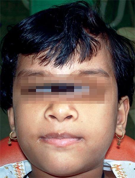
This clinical photograph shows the frontal facial view of a 6-year-old girl diagnosed with Glanzmann's thrombasthenia, a rare autosomal recessive bleeding disorder characterized by a defect in platelet aggregation. The patient exhibits a medium skin tone and short dark hair. While the eyes are obscured for patient anonymity, the visible facial features demonstrate a subtle, generalized puffiness. Clinically, the image serves to document the baseline physical appearance of a pediatric patient who presents with a significant medical history of spontaneous ecchymoses, easy bruising, and recurrent epistaxis. These manifestations are hallmark signs of the underlying qualitative platelet disorder involving the glycoprotein IIb/IIIa complex. In a dental context, identifying such patients is critical for pre-operative planning to prevent uncontrolled hemorrhage during procedures like extractions. The photograph highlights the importance of recognizing systemic hematological conditions in pediatric dentistry to ensure appropriate hematological consultation and hemostatic management.
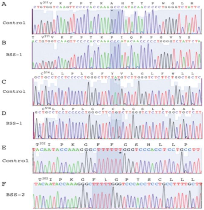
This diagnostic image displays DNA sequencing chromatograms (Sanger sequencing) comparing normal human control sequences with mutated GPIBA (glycoprotein Ib platelet subunit alpha) gene sequences from patients with Bernard-Soulier Syndrome (BSS). The visual consists of six panels (A-F) showing nucleotide peaks, corresponding DNA sequences, and translated amino acid sequences. Panels A and B compare a normal control with patient BSS-1 in the region V311–M326, highlighting a single nucleotide deletion (G*) at codon A318. Panels C and D show the region C514–L529, where patient BSS-1 exhibits a two-nucleotide deletion (A*T*) at codon Y521. Panels E and F illustrate the region T202–V214, where patient BSS-2 shows a single 'T' deletion within a homopolymeric tract of six thymine residues (F207–F208). Each mutation is indicated by an asterisk (*) and demonstrates a frame-shift mutation, leading to altered reading frames and premature stop codons. The image illustrates the molecular genetic basis of BSS, a bleeding disorder characterized by macrothrombocytopenia and defective platelet adhesion.
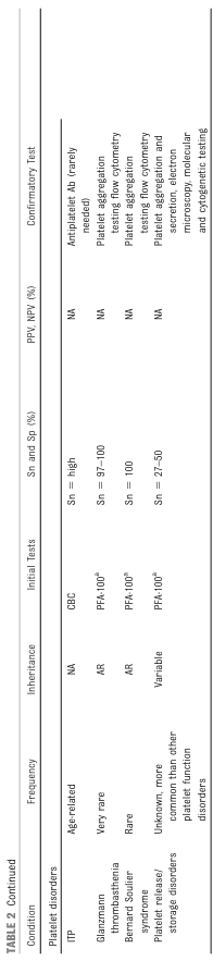
TABLE 2 Continued <table><thead><tr><th>Condition</th><th>Frequency</th><th>Inheritance</th><th>Initial Tests</th><th>Sn and Sp (%)</th><th>PPV, NPV (%)</th><th>Confirmatory Test</th></tr></thead><tbody><tr><td>Platelet disorders</td><td></td><td></td><td></td><td></td><td></td><td></td></tr><tr><td>ITP</td><td>Age-related</td><td>NA</td><td>CBC</td><td>Sn = high</td><td>NA</td><td>Antiplatelet Ab (rarely needed)</td></tr><tr><td>Glanzmann thrombasthenia</td><td>Very rare</td><td>AR</td><td>PFA-100ᵃ</td><td>Sn = 97-100</td><td>NA</td><td>Platelet aggregation testing flow cytometry</td></tr><tr><td>Bernard Soulier syndrome</td><td>Rare</td><td>AR</td><td>PFA-100ᵃ</td><td>Sn = 100</td><td>NA</td><td>Platelet aggregation testing flow cytometry</td></tr><tr><td>Platelet release/ storage disorders</td><td>Unknown, more common than other platelet function disorders</td><td>Variable</td><td>PFA-100ᵃ</td><td>Sn = 27-50</td><td>NA</td><td>Platelet aggregation and secretion, electron microscopy, molecular and cytogenetic testing</td></tr></tbody></table>
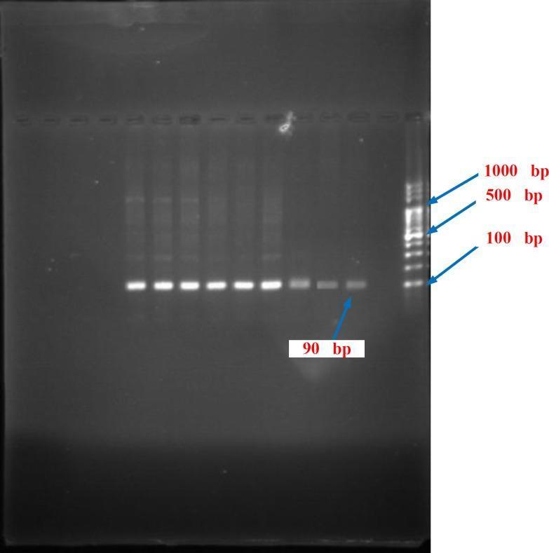
Diagnostic Image: Agarose gel electrophoresis of PCR products related to human platelet antigen (HPA-1) gene analysis. The image shows a dark rectangular gel with multiple vertical lanes. On the far right, a 100 bp DNA ladder (molecular weight standard) is present, with annotated bands at 1000 bp, 500 bp, and 100 bp providing reference markers for fragment size. To the left of the ladder, ten lanes contain clinical samples, each demonstrating a single, sharp, horizontal band of consistent intensity and migration distance. These sample bands are located just below the 100 bp marker and are explicitly labeled as 90 bp fragments. The visual illustrates the successful amplification and size-verification of HPA-1 gene products using Polymerase Chain Reaction (PCR), which is a standard molecular diagnostic technique for genotyping and studying platelet disorders like Glanzmann Thrombasthenia. The uniform migration across lanes suggests a consistent homozygous genotype among the samples shown.
Qualitative Platelet Disorders
Classification
- Defects of adhesion
- Defects of aggregation
- Disorders of platelet secretion (release reaction)
I. Inherited Qualitative Platelet Disorders
A. Defects of Adhesion
Bernard-Soulier Syndrome (BSS)
- Defect: Autosomal recessive deficiency/dysfunction of the GPIb/IX/V complex (the receptor for von Willebrand factor), due to mutations in GPIBA, GPIBB, or GP9
- Pathophysiology: GPIb/IX/V is required for platelet adhesion to the subendothelial matrix via vWF. Without it, platelets cannot tether to damaged vessel walls
- Phenotype: Macrothrombocytopenia (large platelets + low count) with mucocutaneous bleeding - often severe
- Aggregometry: Poor response to ristocetin; normal responses to ADP, arachidonic acid, collagen, and epinephrine
- Diagnosis: Flow cytometry confirms absent/reduced GPIb-IX-V on platelet surface; genetic testing
- Note: The monoallelic form is milder - large platelets, mildly low count; diagnosis requires flow cytometry or genetics
- Treatment: Platelet transfusion for bleeding; DDAVP may provide some benefit in mild to moderate bleeding
B. Defects of Aggregation
Glanzmann Thrombasthenia (GT)
- Defect: Autosomal recessive disorder caused by mutations in ITGA2B and ITGB3, encoding the fibrinogen receptor GPIIb/IIIa
- Pathophysiology: GPIIb/IIIa (integrin αIIbβ3) forms "bridges" between platelets by binding fibrinogen. Its absence/dysfunction means platelets cannot aggregate with each other
- Phenotype: Normal platelet count and platelet size; bleeding tends to be severe since childhood
- Aggregometry: Normal ristocetin response (GPIb/IX/V is intact); absent or markedly reduced responses to ADP, collagen, epinephrine, and arachidonic acid - the classic pattern
- Types:
- Type 1: Complete absence of GPIIb/IIIa (<5% of normal)
- Type 2: Moderate decrease (10-20% of normal)
- Type 3: GPIIb/IIIa is present but dysfunctional (variant form)
- Diagnosis: Flow cytometry for GPIIb/IIIa receptor density; genetic testing
- Treatment: Platelet transfusion. Due to risk of alloimmunization causing refractoriness, many experts prefer recombinant factor VIIa (rFVIIa) as first-line treatment, reserving platelets for life-threatening bleeding
C. Disorders of Platelet Secretion (Release Reaction / Storage Pool Diseases)
Dense Granule (Delta Granule) Deficiency - δ-Storage Pool Disease
- Most common intrinsic platelet function defect
- Dense granules store ADP, ATP, Ca²⁺, serotonin, and inorganic phosphate - key amplifiers of platelet activation
- Diagnosis: Mild bleeding, abnormal platelet aggregometry (absent secondary wave of aggregation), decreased dense granule number on wet mount or transmission electron microscopy (TEM)
- Associated syndromes:
- Hermansky-Pudlak Syndrome (HPS): Autosomal recessive; up to 10 different gene mutations causing subtypes. Dense granules severely decreased or absent. Accompanied by oculocutaneous albinism. Some subtypes also develop pulmonary fibrosis, granulomatous colitis, and immune defects
- Chediak-Higashi Syndrome: Dense granule disorder with oculocutaneous albinism, neutropenia, large cytoplasmic inclusions in neutrophils, severe immune deficiency, and risk of life-threatening lymphohistiocytosis
Alpha (α) Granule Deficiency - Gray Platelet Syndrome
- Defect: Autosomal recessive; complete absence of α-granules due to loss-of-function mutations in NBEAL2
- α-granules contain fibrinogen, vWF, factor V, platelet factor 4, P-selectin, and growth factors
- Phenotype: Platelets appear gray on peripheral blood smear (lack of granule contents); diagnosis confirmed by TEM
- Bleeding is usually mild to moderate
- Can be associated with splenomegaly and bone marrow fibrosis later in life
- Other rare α-granule syndromes: mutations in FLII, GFI1B, STIM1, GATA1
D. Other Rare Inherited Receptor Defects
- Defects in the thromboxane A2 receptor, ADP receptor (P2Y12), and collagen receptor - extremely rare, described in only a few families
- Over 75 genes have now been associated with inherited platelet disorders on comprehensive gene panels
- Some disorders (mutations in RUNX1, ETV6, ANKRD26) carry an increased risk of hematologic malignancies
II. Acquired Qualitative Platelet Disorders
A. Drug-Induced (Most Common Cause)
| Class | Examples |
|---|---|
| Antiplatelet agents | Aspirin, clopidogrel, prasugrel, ticagrelor, abciximab, eptifibatide, tirofiban |
| NSAIDs | Ibuprofen, naproxen, indomethacin, diclofenac |
| SSRIs | Fluoxetine, paroxetine, sertraline (deplete platelet serotonin) |
| Plasma expanders | Dextran, hydroxyethyl starch |
| Phosphodiesterase inhibitors | Cilostazol, dipyridamole, sildenafil |
| Foods & supplements | Vitamin E, fish oil, garlic, ginger, turmeric, alcohol |
| Antihistamines | Chlorpheniramine, diphenhydramine |
- Aspirin is a potent, irreversible inhibitor of cyclooxygenase (COX), blocking thromboxane A2 synthesis for the platelet's entire lifespan (~7-10 days). This is the basis for its use in preventing coronary thrombosis.
B. Uremia
- Second most common condition causing acquired platelet dysfunction
- Pathogenesis is complex: involves defects in adhesion, granule secretion, and aggregation
- Accumulated uremic toxins (guanidinosuccinic acid, phenolic acids) interfere with platelet function
- Treatment: DDAVP is effective for uremic bleeding; dialysis improves platelet function
C. Hematologic Disorders
- Paraproteinemias (multiple myeloma, Waldenström macroglobulinemia): abnormal immunoglobulins coat platelets, interfering with function
- Myeloproliferative neoplasms (PV, ET, MF): intrinsically abnormal megakaryocytes produce dysfunctional platelets; can cause both thrombosis and bleeding
- Myelodysplastic syndrome: defective platelet production
D. Liver Disease
- Reduced synthesis of vWF multimers and other factors; platelet dysfunction is a component of the complex hemostatic defect
E. Cardiopulmonary Bypass
- Transient platelet dysfunction due to mechanical trauma, hemodilution, and activation/exhaustion during bypass
III. Diagnosis Summary
| Test | BSS | Glanzmann GT | Storage Pool Disease |
|---|---|---|---|
| Platelet count | Low (macro) | Normal | Normal |
| Platelet size | Large | Normal | Normal |
| Ristocetin aggregation | Absent | Normal | Normal |
| ADP/collagen/epi aggregation | Normal | Absent | Reduced secondary wave |
| Flow cytometry | Low GPIb/IX/V | Low GPIIb/IIIa | Normal |
| TEM/electron microscopy | Normal | Normal | Decreased granules |
| Bleeding time / PFA-100 | Prolonged | Prolonged | Often prolonged |
IV. Treatment Principles
| Situation | Treatment |
|---|---|
| Most inherited/acquired disorders | Platelet transfusion (first-line for significant bleeding) |
| Glanzmann GT (alloimmunization) | rFVIIa (recombinant factor VIIa) preferred first-line |
| Uremia, storage pool, BSS (mild-moderate) | DDAVP (increases vWF release; caution - fluid retention, hyponatremia) |
| Mucosal/oral bleeding | Tranexamic acid or ε-aminocaproic acid (antifibrinolytics) |
| Platelet transfusion-refractory cases | rFVIIa: enhances thrombin generation at site of injury, recruits platelets |
- Goldman-Cecil Medicine (Specific Qualitative Platelet Disorders, Ch. 159)
- Robbins & Cotran Pathologic Basis of Disease (Bleeding Disorders Related to Defective Platelet Function)
- Schwartz's Principles of Surgery, 11th ed. (Platelet Functional Defects)
- Tietz Textbook of Laboratory Medicine, 7th ed. (Storage-Pool Deficiency; Molecular Classification of Hereditary Platelet Disorders)
Investigations and treatment of platelet disorders
Investigations and Treatment of Platelet Disorders
PART 1: INVESTIGATIONS
Step 1 - Initial Screening / History & CBC
- Always assess platelet count (CBC with differential) - to distinguish quantitative (thrombocytopenia) from qualitative (normal count, dysfunctional platelets) disorders
- Mean platelet volume (MPV): Large platelets suggest Bernard-Soulier syndrome, Gray platelet syndrome, MYH9-related disorders; small platelets suggest Wiskott-Aldrich syndrome
- Peripheral blood smear: Confirms count, evaluates platelet size, looks for platelet clumping, giant platelets, or gray (agranular) platelets
- Immature platelet fraction (IPF): Elevated in destructive causes (ITP); low/normal in production failure
- Coagulation screen (PT, aPTT, TT, fibrinogen): To distinguish platelet disorders (normal PT/aPTT) from coagulation factor deficiencies
- Drug & dietary history: NSAIDs, aspirin, clopidogrel, SSRIs, herbal supplements
- Bleeding history / Bleeding Assessment Tool (BAT) score: 99% probability of platelet function disorder if vWD is excluded
Step 2 - Platelet Function Screening Tests
A. Bleeding Time (BT)
- A skin puncture test measuring time for bleeding to stop
- Prolonged in thrombocytopenia, platelet dysfunction, and vWD
- Poorly reproducible and operator-dependent; largely replaced by PFA-100/200 in modern practice
B. PFA-100 / PFA-200 (Platelet Function Analyzer)
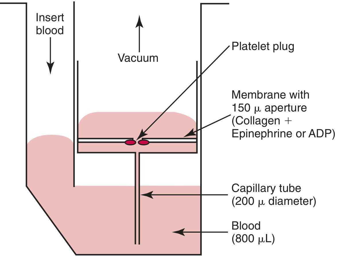
- Whole blood (citrated) is aspirated under high shear force through a capillary tube and over a membrane coated with collagen + epinephrine (CEPI) or collagen + ADP (CADP)
- Platelets adhere and aggregate at the aperture until it closes; closure time (CT) is measured
- A global test of platelet function and vWF under high shear conditions
| Condition | CEPI Cartridge | CADP Cartridge |
|---|---|---|
| Normal | Normal CT | Normal CT |
| Aspirin effect | Prolonged | Normal |
| Platelet function defect / vWD | Prolonged | Prolonged |
- Sensitive for Glanzmann thrombasthenia, Bernard-Soulier, vWD, and aspirin effect
- Sensitivity for storage pool disease is lower (27-50%)
Step 3 - Light Transmission Aggregometry (LTA) - Gold Standard
| Agonist | Receptor Activated |
|---|---|
| Collagen | GPVI, GPIa/IIa |
| ADP | P2Y₁ and P2Y₁₂ |
| Epinephrine | α₂ receptor |
| Arachidonic acid (AA) | COX pathway → TXA₂ |
| Ristocetin | vWF binding to GPIb/V/IX |
| TRAP (thrombin receptor-activating peptide) | PAR₁ and PAR₄ |
| Disorder | Ristocetin | ADP | Collagen | Epinephrine | AA |
|---|---|---|---|---|---|
| Bernard-Soulier syndrome | Absent | Normal | Normal | Normal | Normal |
| Glanzmann thrombasthenia | Normal | Absent | Absent | Absent | Absent |
| Storage pool disease (δ-SPD) | Normal | ↓ (no secondary wave) | ↓ | ↓ (no secondary wave) | ↓ |
| Aspirin / COX inhibitor | Normal | Normal (primary only) | ↓ | ↓ (no secondary wave) | Absent |
| P2Y₁₂ defect / clopidogrel | Normal | ↓ (no secondary wave) | Normal | Normal | Normal |
Step 4 - Simultaneous Secretion Measurement (Lumi-Aggregometry)
- A luciferin-luciferase reagent is added to PRP
- ATP released from dense granules reacts with luciferin-luciferase → bioluminescence is recorded simultaneously with aggregation
- Allows simultaneous, independent monitoring of aggregation AND dense granule secretion
- If aggregation is normal but secretion is reduced → isolated secretion defect
- If both aggregation and secretion are absent → severe aggregation defect (e.g., Glanzmann) or combined defect
Step 5 - Flow Cytometry
- Receptor expression: Confirms Bernard-Soulier (absent/reduced GPIb/IX/V) and Glanzmann thrombasthenia (absent/reduced GPIIb/IIIa)
- Can assess:
- GPIIb/IIIa conformational activation (PAC-1 binding)
- P-selectin expression (α-granule secretion marker)
- Annexin V binding (phosphatidylserine exposure - Scott syndrome)
- Dense granule content (mepacrine uptake)
- Platelet microparticle release
- Particularly valuable in children and neonates (requires only small blood volumes)
Step 6 - Electron Microscopy (TEM)
- Whole mount or thin section TEM: Quantifies dense granules (δ-granules) and α-granules
- Required for confirmation of storage pool diseases
- Gray platelet syndrome: absent α-granules on TEM
- Hermansky-Pudlak / Chediak-Higashi: absent/severely reduced dense granules
Step 7 - High-Throughput DNA Sequencing (Gene Panels)
- Over 75 genes now associated with inherited platelet disorders
- Targeted gene panels or whole-exome sequencing (WES)
- Provides genetic diagnosis in ~40-50% of patients with inherited platelet disorders
- Identifies mutations in ITGA2B/ITGB3 (Glanzmann), GPIBA/GPIBB/GP9 (BSS), NBEAL2 (Gray platelet syndrome), RUNX1, ETV6, ANKRD26 (malignancy risk), and many others
- Also detects large structural variants and novel mutations
Additional Specific Tests
| Test | Purpose |
|---|---|
| Bone marrow biopsy | ITP (normal/increased megakaryocytes), aplastic anemia, leukemia |
| Platelet antibody tests | ITP, drug-induced thrombocytopenia, neonatal alloimmune thrombocytopenia |
| Anti-PF4/heparin antibody + serotonin release assay | Heparin-induced thrombocytopenia (HIT) |
| ADAMTS13 activity | TTP (severely reduced <10%) |
| vWF antigen + activity (ristocetin cofactor) + multimer analysis | von Willebrand disease |
| Complement panel, factor H antibodies, genetic testing | Atypical HUS |
| Clot retraction | Delayed/incomplete in Glanzmann thrombasthenia and thrombocytopenia |
PART 2: TREATMENT
I. Quantitative Disorders (Thrombocytopenia)
A. Immune Thrombocytopenic Purpura (ITP)
| Severity | Management |
|---|---|
| Mild (skin only - petechiae/bruising) | Observation only |
| Mucosal / significant bleeding | IVIG, corticosteroids, or anti-Rh(D) immune globulin |
| Refractory / chronic | Rituximab (anti-CD20), thrombopoietin receptor agonists (TPO-RAs): eltrombopag, romiplostim |
| Splenectomy | For steroid-refractory chronic ITP |
| Emergency / life-threatening | IVIG + high-dose steroids + platelet transfusion |
B. TTP
- Early plasma exchange with fresh frozen plasma (FFP) + glucocorticoids
- Treat emergently on clinical suspicion before ADAMTS13 results return
- Add caplacizumab (anti-vWF nanobody) in refractory/severe cases
C. HUS (Typical - Shiga toxin)
- Supportive care with aggressive IV hydration, antihypertensives
- Platelet/RBC transfusions as needed
- Avoid antibiotics (may worsen Shiga toxin release)
D. Atypical HUS
- Eculizumab (complement C5 inhibitor) ± plasma exchange ± immunosuppression
E. HIT (Heparin-Induced Thrombocytopenia)
- Immediately stop heparin (including heparin flushes and heparin-coated lines)
- Start a non-heparin anticoagulant: argatroban, bivalirudin, fondaparinux
- Do NOT transfuse platelets (worsens thrombosis)
F. Drug-Induced Thrombocytopenia
- Permanently discontinue the offending drug
- Transfuse if severe thrombocytopenia with risk of intracranial or intrapulmonary hemorrhage
G. Neonatal Alloimmune Thrombocytopenia (NAIT)
- Head ultrasound to screen for intracranial hemorrhage (ICH)
- Transfuse platelets if count <30,000/μL or signs of bleeding
- IVIG if poor response to platelet transfusion
II. Qualitative Disorders (Platelet Dysfunction)
| Condition | Treatment |
|---|---|
| Most inherited qualitative disorders (first-line significant bleeding) | Platelet transfusion |
| Glanzmann thrombasthenia (alloimmunization / transfusion refractoriness) | Recombinant factor VIIa (rFVIIa) preferred first-line; reserve platelets for life-threatening bleeding |
| Uremic platelet dysfunction | DDAVP (desmopressin); dialysis improves platelet function |
| Storage pool disease / BSS (mild-moderate bleeding) | DDAVP may provide benefit |
| Mucosal / oral bleeding (all qualitative disorders) | Tranexamic acid (15-25 mg/kg orally TDS) or ε-aminocaproic acid (5 g PO then 1-1.25 g/hr × 8 hrs) |
| Aspirin-induced dysfunction | Hold aspirin (7-10 days for full platelet turnover); platelet transfusion if urgent surgery |
| Drug-induced (reversible - NSAIDs) | Stop drug |
Specific Treatment Notes
- Patients receiving multiple transfusions can develop alloimmunization to platelet-specific or HLA antigens → transfusion refractoriness
- In Glanzmann thrombasthenia, alloimmunization against GPIIb/IIIa is particularly problematic
- Releases vWF from endothelial Weibel-Palade bodies → increases circulating vWF → improves platelet adhesion
- Effective for uremic bleeding and some storage pool/release defects
- Caution: fluid retention and hyponatremia - monitor electrolytes, especially in children and the elderly
- Activates the extrinsic coagulation pathway at sites of vessel injury → massive thrombin generation → platelet recruitment and activation even when platelet adhesion/aggregation is impaired
- Dose: 90-120 mcg/kg IV; repeat every 2-3 hours as needed
- Inhibit plasminogen activation → prevent premature clot dissolution
- Particularly useful in mucosal and oral procedures (nose, mouth, throat, extraocular tissues) where local fibrinolytic activity is high
Summary Table: Investigations at a Glance
| Test | What it Detects | Level |
|---|---|---|
| CBC + smear | Count, platelet size (MPV), morphology | Screening |
| PT / aPTT / TT | Rules out coagulation factor defects | Screening |
| PFA-100 (closure time) | Global platelet function + vWD | Screening |
| LTA (aggregometry) - full agonist panel | Specific receptor/pathway defects | Specialized |
| Lumi-aggregometry (ATP secretion) | Dense granule secretion defects | Specialized |
| Flow cytometry | GPIb/IX/V, GPIIb/IIIa, P-selectin, annexin V | Specialized |
| TEM (electron microscopy) | Granule number/structure | Specialized |
| ADAMTS13 | TTP | Specific |
| Anti-PF4/heparin + SRA | HIT | Specific |
| Gene panel / WES | Inherited platelet disorders | Specialized |
- Goldman-Cecil Medicine (Qualitative Platelet Disorders, Diagnosis and Treatment - Ch. 159)
- Henry's Clinical Diagnosis and Management by Laboratory Methods (Platelet Aggregation and Secretion Studies, Flow Cytometry, DNA Sequencing - Ch. 41)
- Tietz Textbook of Laboratory Medicine, 7th ed. (PFA-100/200, Platelet Aggregation Tests - Ch. 80)
- The Harriet Lane Handbook, 23rd ed. (Management of Thrombocytopenia, Platelet Function Evaluation - Ch. 14)
- Robbins & Cotran Pathologic Basis of Disease (Bleeding Disorders - Defective Platelet Function)
Summerise platelet disorders in 5 most important points
Platelet Disorders - 5 Key Points
- Bernard-Soulier Syndrome - defective GPIb/IX/V → impaired adhesion → fails ristocetin, normal to other agonists; presents with macrothrombocytopenia
- Glanzmann Thrombasthenia - defective GPIIb/IIIa → impaired aggregation → normal ristocetin, fails ALL other agonists (ADP, collagen, epinephrine, AA); normal platelet count These two are the classic "spot diagnosis" disorders in any exam or clinical setting.
- Tier 1 (screening): CBC + smear, PT/aPTT, PFA-100 closure time
- Tier 2 (specialized): Light transmission aggregometry (LTA) with full agonist panel - the gold standard for identifying which specific step (adhesion/aggregation/secretion) is defective
- Tier 3 (confirmatory): Flow cytometry (receptor expression), electron microscopy (granule counts), and gene panels (>75 genes now identified)
- Platelet transfusion - mainstay for most bleeding episodes, but repeated transfusions cause alloimmunization
- rFVIIa - preferred first-line in Glanzmann thrombasthenia when transfusion refractoriness develops; works by driving thrombin generation directly at the injury site
- DDAVP - for uremia, mild storage pool disease, and Bernard-Soulier (mild-moderate bleeding); releases endothelial vWF stores
- Antifibrinolytics (tranexamic acid) - adjunct for mucosal bleeding in all qualitative disorders
- For ITP specifically: observation if mild, IVIG/steroids if significant bleeding, TPO receptor agonists (eltrombopag, romiplostim) for chronic/refractory disease
Some tables and flowcharts from Robbins or other standard books relavent to qualitative bleeding disorders
Tables and Flowcharts: Qualitative Bleeding Disorders
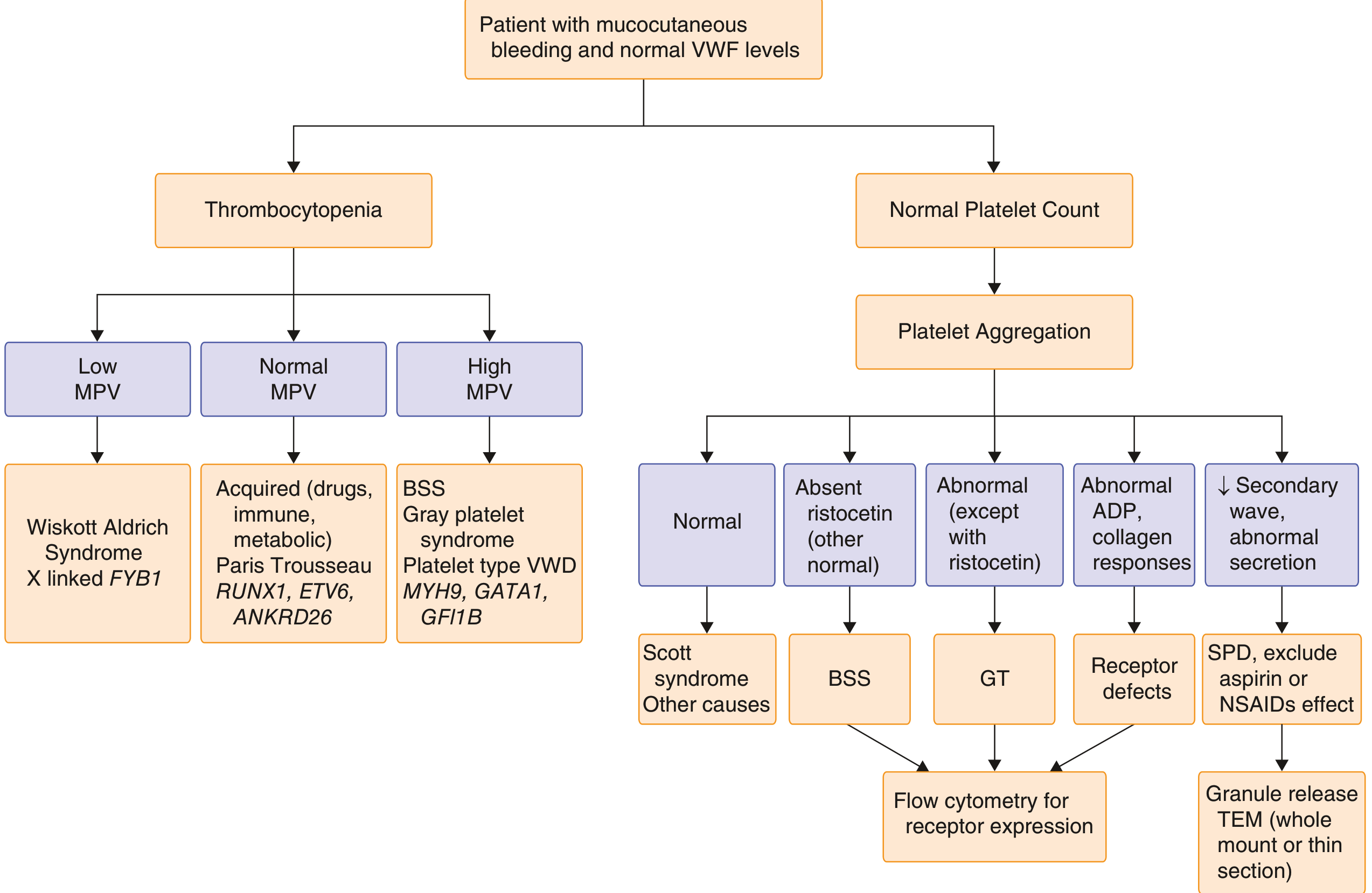
FLOWCHART 1 - Diagnostic Algorithm for Platelet Disorders
- Absent ristocetin only → BSS → Flow cytometry
- Abnormal except ristocetin → Glanzmann GT → Flow cytometry
- Abnormal ADP/collagen responses → Receptor defects → Flow cytometry
- Absent secondary wave + abnormal secretion → SPD → Granule release TEM
- Low MPV + thrombocytopenia → Wiskott-Aldrich syndrome / X-linked FYB1
- High MPV + thrombocytopenia → BSS, Gray platelet syndrome, MYH9-related disorders
FLOWCHART 2 - Platelet Molecular Signaling Pathways
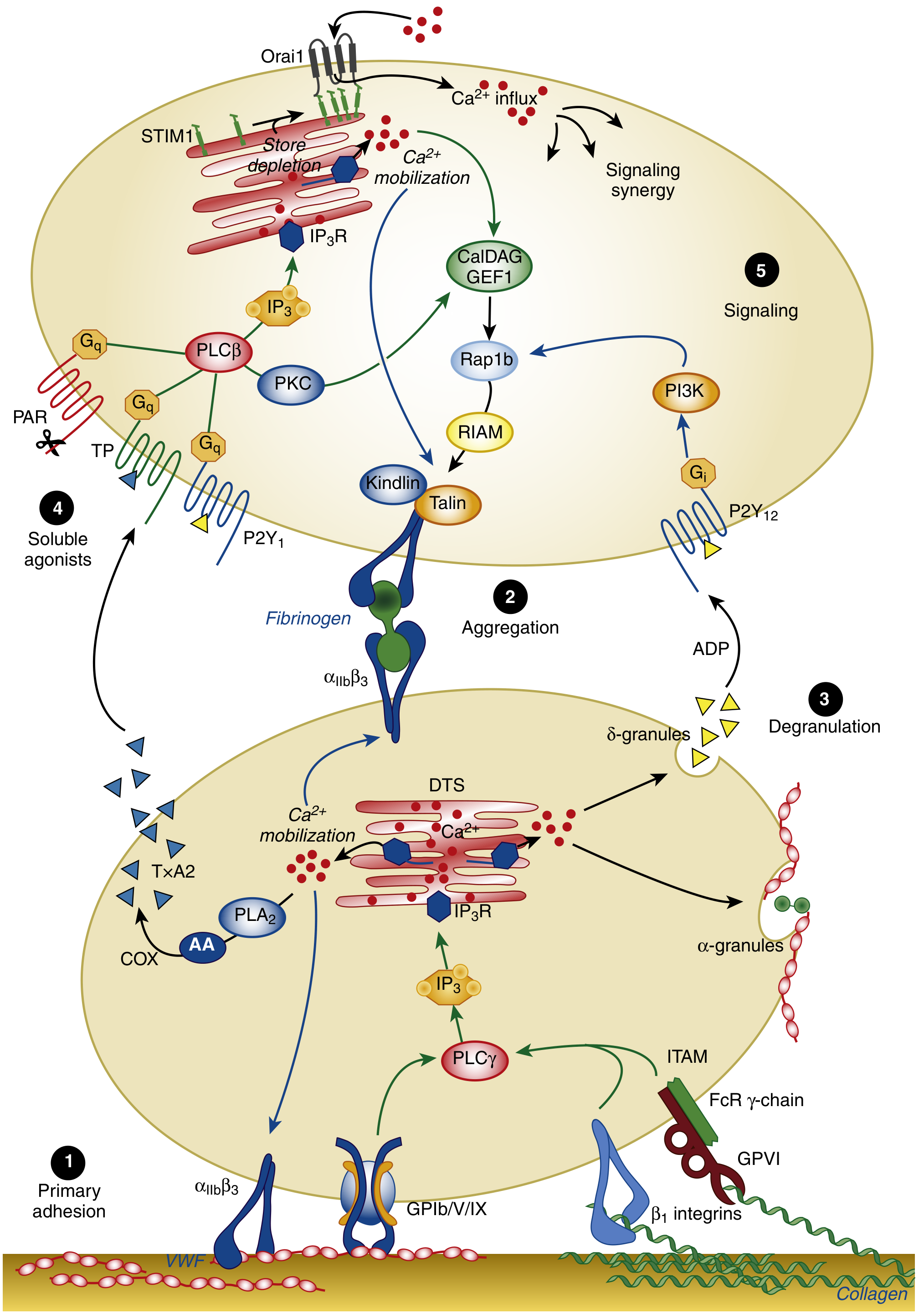
- Primary adhesion - GPIb/V/IX binds vWF on subendothelium (defective in BSS)
- Aggregation - αIIbβ₃ (GPIIb/IIIa) binds fibrinogen bridging platelets (defective in Glanzmann GT)
- Degranulation - δ-granules release ADP; α-granules release vWF, fibrinogen (defective in storage pool diseases)
- Soluble agonist signaling - TxA₂ via COX, PAR receptors (defective in aspirin, COX deficiency)
- Downstream signaling - P2Y₁₂, PI3K pathways (defective in clopidogrel effect, P2Y₁₂ deficiency)
TABLE 1 - Harrison's Classification of Qualitative Platelet Disorders
| Category | Disorders |
|---|---|
| Defects of Platelet Adhesion | Bernard-Soulier syndrome (absent GPIb/IX/V) |
| Defects of Platelet Aggregation | Glanzmann thrombasthenia (absent GPIIb/IIIa); Afibrinogenemia |
| Defects of Platelet Secretion | Decreased cyclooxygenase activity - drug-induced (aspirin, NSAIDs, thienopyridines); Inherited COX deficiency; Granule storage pool defects - inherited & acquired; Nonspecific secretory defects; Uremia; Platelet coating (paraprotein, penicillin) |
| Defect of Platelet Coagulant Activity | Scott syndrome |
TABLE 2 - Comprehensive Inherited Platelet Disorder Profiles
Part A: Surface Receptor Defects
| Disorder | Inheritance | Platelet Count | PBS | Aggregation Pattern | ATP Secretion | Flow Cytometry | EM | Gene |
|---|---|---|---|---|---|---|---|---|
| Glanzmann thrombasthenia | AR | NL (or mild ↓) | Normal platelets | AA ↓, ADP ↓, Epi ↓, Col ↓, Risto NL | NL | Absent GPIIb/IIIa | NL | ITGA2B, ITGB3 |
| Bernard-Soulier syndrome | AR | Mod-severe ↓ | Giant platelets | Risto absent, AA ↓, ADP NL, Epi ↓, Col ↓ | NL | Absent GPIb/IX/V | NL | GPIBA, GPIBB, GP9 |
Part B: Congenital Thrombocytopenias (with Qualitative Defects)
| Disorder | Inheritance | Platelet Count | PBS | Aggregation | ATP Secretion | Flow Cytometry | EM | Gene |
|---|---|---|---|---|---|---|---|---|
| Wiskott-Aldrich syndrome | XR | Mod-severe ↓ | Small platelets | AA ↓/NL, Epi ↓ | ↓ | ↓CD63 | Dense granule deficiency | WAS |
| MYH9-related disorders | AD | Mild-mod ↓ | Large platelets, leukocyte inclusions | Risto NL/↓ | NL | NL | Inclusions in WBCs | MYH9 |
Part C: Granule Defects (Storage Pool Diseases)
| Disorder | Inheritance | Platelet Count | PBS | Aggregation | ATP Secretion | Flow Cytometry | EM | Gene |
|---|---|---|---|---|---|---|---|---|
| Gray platelet syndrome | AR | Mild-mod ↓ | Gray platelets | AA NL↓, ADP ↓, Epi ↓, Col ↓ | ↓ | ↓P-selectin | Absent α-granules | NBEAL2 |
| Hermansky-Pudlak syndrome | AR | NL | NL | AA ↓/NL | ↓ | ↓CD63 after stimulation | Absent dense granules | HPS1-7, AP3B1 |
| Chediak-Higashi syndrome | AR | NL | Leukocyte inclusions | AA ↓/NL, ADP ↓, Epi ↓ 2nd W, Col NL | ↓ | ↓CD63 | Leukocyte lysosomal inclusions; ↓ dense granules | LYST |
| Combined αδ granule deficiency | AR/AD | NL | NL | All agonists NL↓ | ↓ | ↓P-selectin, ↓CD63 | Both α and δ granule deficiency | GFI1B |
| Paris-Trousseau/Jacobsen syndrome | AD | Mod-severe ↓ | NL or large platelets | ADP NL, Epi ↓ 2nd W | ↓ | N/A | Dense granule deficiency + giant α-granules | Del11q23 (FLI1) |
Part D: Signal Transduction Defects
| Disorder | Inheritance | Platelet Count | Aggregation Pattern | EM | Gene |
|---|---|---|---|---|---|
| G-protein activation defect | AD | NL | AA NL↓, ADP ↓, Epi ↓, Col ↓ | NL | GNAS |
| Thromboxane synthase deficiency | AD | NL | AA absent, ADP NL, Epi ↓, Col ↓/NL | NL | TBXAS1 |
| Cyclooxygenase (COX) deficiency | AD | NL | AA absent, ADP NL, Epi ↓, Col ↓/NL | NL | PTGS1 |
| Calcium mobilization defect | Unknown | NL | AA NL, ADP NL, Epi ↓ | NL | STIM1 |
TABLE 3 - Diagnostic Assay Comparison for Key Qualitative Disorders
| Disorder | Definition | Diagnostic Assays |
|---|---|---|
| Bernard-Soulier syndrome | Platelets lack GPIb-IX-V complex | Macrothrombocytopenia; reduced ristocetin on LTA; flow cytometry |
| Glanzmann thrombasthenia | Quantitative/qualitative deficiency of GPIIb/IIIa | Response to ristocetin only on LTA; flow cytometry |
| Platelet granule defects | Abnormal function/deficiency of α, δ, or both granules | Reduced LTA ± absent secondary wave with low-dose agonists (ADP, Epi, collagen); absent granules on EM |
| vWD Type I/III | Deficient/absent vWF | Low/absent vWF activity (<0.50 IU/mL) |
| vWD Type IIB | Spontaneous vWF-platelet binding; lacks HMW multimers | vWF activity/Ag ratio <0.7; enhanced RIPA; thrombocytopenia |
| Platelet-type vWD | Enhanced platelet-vWF interaction via mutant GPIb | Enhanced RIPA; multimeric analysis; GPIBA genetic testing |
TABLE 4 - Treatment of Congenital Platelet Disorders
| Disorder | Threshold for Treatment | Treatment |
|---|---|---|
| Bernard-Soulier syndrome | Bleeding; prophylaxis for delivery with bleeding history | HLA-matched platelet transfusion; DDAVP; rFVIIa |
| Glanzmann thrombasthenia | Bleeding; delivery; procedures | HLA-matched platelet transfusion; rFVIIa |
| Platelet granule defects | Bleeding | HLA-matched platelet transfusion; DDAVP |
TABLE 5 - Drugs Causing Acquired Platelet Dysfunction
| Class | Examples |
|---|---|
| NSAIDs | Aspirin, naproxen, ibuprofen, indomethacin |
| GPIIb/IIIa antagonists | Abciximab, eptifibatide, tirofiban |
| Thienopyridines | Clopidogrel, ticlopidine |
| cAMP/cGMP elevators | Iloprost, dipyridamole, prostacyclin |
| Volume expanders | Dextran, hydroxyethyl starch |
| Chemotherapeutic agents | Mitomycin, daunorubicin, carmustine |
| Psychotropic medications | Many (especially SSRIs) |
| Others | Antihistamines, clofibrate, radiocontrast agents |
TABLE 6 - PFA-100 Expected Results
| Condition | Collagen/Epinephrine CT | Collagen/ADP CT |
|---|---|---|
| Normal | Normal | Normal |
| Aspirin effect | Prolonged | Normal |
| Platelet function defect / vWD | Prolonged | Prolonged |
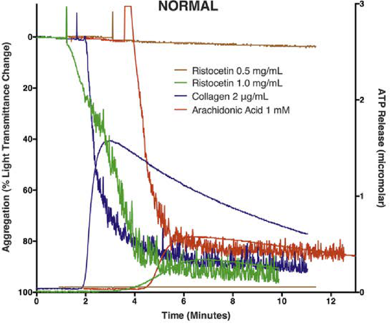
Normal Aggregometry Tracings (LTA) for Reference
- Goldman-Cecil Medicine (Diagnostic Algorithm, Fig. 159-2)
- Harrison's Principles of Internal Medicine, 22nd ed. (Classification table)
- Tietz Textbook of Laboratory Medicine, 7th ed. (Table 80.7, 80.8; PFA-100 Table 80.2)
- Henry's Clinical Diagnosis and Management by Laboratory Methods (Aggregometry tracings Fig. 41.2, 41.3)
- Creasy & Resnik's Maternal-Fetal Medicine (Tables 53.4 and 53.5)
- Robbins & Cotran Pathologic Basis of Disease (Qualitative platelet disorder classification)