
About cell death in pathology
Reading File
Cell death in pathology covers two major mechanisms - necrosis (uncontrolled, inflammatory) and apoptosis (programmed, non-inflammatory) - plus several other regulated death pathways like autophagy, pyroptosis, necroptosis, and ferroptosis. Let me pull comprehensive textbook coverage.
Searching sources...
Finding Sources
Finding Sources
Finding Sources
Finding Sources
Robbins Pathology has dedicated sections. Let me read those alongside a search for apoptosis.
Reading File
Finding Sources
Finding Sources
Now let me read the core cell death sections from Robbins Basic Pathology:
Reading File
Reading File
Excellent - rich content. Let me now read the apoptosis and autophagy sections:
Reading File
Reading File
Now let me read the Apoptosis mechanisms and Autophagy sections in detail:
Reading File
I now have comprehensive content. Let me also fetch the key diagrams to include in the answer:
Loading Image
Loading Image
I have all the material needed for a thorough answer. Let me now compile the complete response.
Cell Death in Pathology
(Robbins & Kumar Basic Pathology, 10th ed., Chapter 1)
Overview
Cell death is the end result of severe, irreversible cell injury. There are two principal forms - necrosis and apoptosis - and several other regulated pathways including autophagy, pyroptosis, necroptosis, and ferroptosis. The sequence of events from a healthy cell to death is shown below:
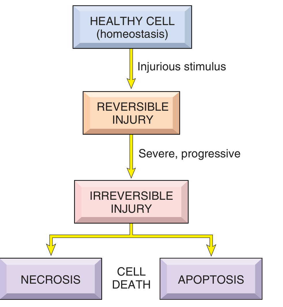
Fig 1.2 - Robbins & Kumar Basic Pathology
1. Reversible vs. Irreversible Injury
Before death, cells pass through a stage of reversible injury. Key features:
- Cellular swelling (hydropic change / vacuolar degeneration) from failure of energy-dependent Na⁺/K⁺-ATPase
- Fatty change (lipid vacuoles) in organs like the liver
- Plasma membrane blebbing, ER dilation, mitochondrial swelling, chromatin clumping
When the injury is severe enough to cross the "point of no return," cells die - typically by necrosis if the damage is overwhelming, or apoptosis if driven by specific signals.
An important concept: cellular function is lost well before morphologic evidence of death appears. Myocardial cells become noncontractile after 1-2 minutes of ischemia, yet morphologic features of death take 6-12 hours to appear by light microscopy.
2. Necrosis
Necrosis is the form of cell death associated with uncontrolled cellular digestion and inflammation. It is almost always pathologic.
Mechanisms
- Cellular membranes break down
- Lysosomal enzymes and leukocyte enzymes digest the cell contents
- Cellular contents leak into the extracellular space, triggering an inflammatory response
Morphologic Features (H&E)
Cytoplasmic changes:
- Increased eosinophilia (denatured proteins binding eosin; loss of RNA basophilia)
- Glassy, homogeneous appearance (loss of glycogen)
- Vacuolated, "moth-eaten" cytoplasm when organelles are digested
Nuclear changes (three patterns):
| Pattern | Description |
|---|---|
| Pyknosis | Nuclear shrinkage + increased basophilia; DNA condenses into a dark mass |
| Karyorrhexis | Fragmentation of the pyknotic nucleus |
| Karyolysis | Fading of basophilia due to DNase digestion of DNA; nucleus dissolves over 1-2 days |
Morphologic Patterns of Tissue Necrosis
| Type | Description | Example |
|---|---|---|
| Coagulative necrosis | Cell outlines preserved ("ghost cells"); denatured proteins resist enzymatic digestion | Ischemic infarct of heart, kidney |
| Liquefactive necrosis | Cell dissolved; creamy, liquid mass | Brain infarct; bacterial abscesses |
| Caseous necrosis | "Cheese-like" friable material; amorphous granular debris enclosed by granulomatous inflammation; no cell outlines | Tuberculosis |
| Fat necrosis | Outlines of necrotic fat cells with chalky-white calcium soap deposits | Acute pancreatitis (enzymatic); breast trauma |
| Fibrinoid necrosis | Bright pink, amorphous material in blood vessel walls; deposited immune complexes + plasma proteins | Immune vasculitis, malignant hypertension |
| Gangrenous necrosis | Coagulative necrosis of a limb + superimposed liquefaction (wet gangrene) | Ischemic limb |
Clinical Significance: Serum Markers
Necrosis causes leakage of intracellular proteins into the blood, forming useful diagnostic markers:
- Cardiac troponin - myocardial necrosis (MI)
- Transaminases (AST/ALT) - hepatocellular necrosis
- Alkaline phosphatase - biliary epithelial necrosis
3. Apoptosis
Apoptosis is programmed cell death - an orderly, energy-dependent process in which the cell activates its own destruction without triggering inflammation.
Key Features
- Caspase-mediated degradation of nuclear DNA and cytoplasmic proteins
- Plasma membrane remains intact throughout
- Cell fragments into apoptotic bodies that are rapidly phagocytosed by macrophages
- No inflammatory reaction (contents never leak out)
Causes of Apoptosis
Physiologic:
- Normal embryonic development (organ sculpting, cavitation)
- Turnover of proliferative tissues (intestinal epithelium, lymphocytes)
- Hormone-dependent involution (endometrial shedding)
- Deletion of self-reactive lymphocytes (prevention of autoimmunity)
- Resolution of immune responses (elimination of excess leukocytes)
Pathologic:
- DNA damage beyond repair (radiation, toxins) - via BH3-only protein activation
- Accumulation of misfolded proteins (ER stress)
- Viral infections - virally infected cells killed by cytotoxic T lymphocytes (CTLs)
Mechanisms of Apoptosis
Two distinct pathways converge on caspase activation:
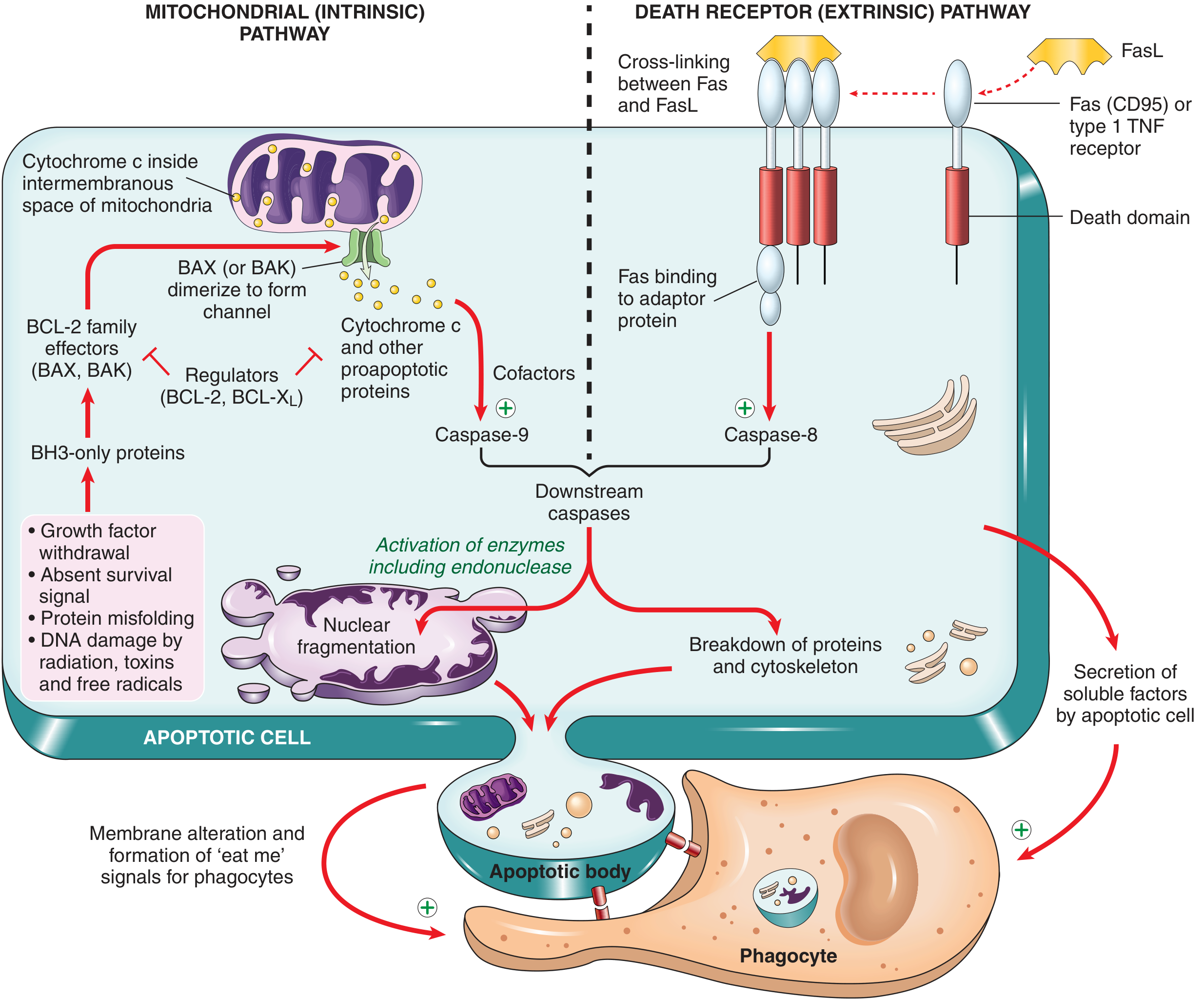
Fig 1.12 - Robbins & Kumar Basic Pathology
A. Mitochondrial (Intrinsic) Pathway
The main pathway in most physiologic and pathologic apoptosis.
- Stress signals (growth factor withdrawal, DNA damage, protein misfolding) activate BH3-only proteins
- BH3-only proteins shift the balance from anti-apoptotic BCL-2/BCL-XL toward pro-apoptotic BAX/BAK
- BAX/BAK dimerize, insert into the mitochondrial outer membrane, and form pores
- Cytochrome c and other proteins escape into the cytosol
- Cytochrome c + cofactors activate caspase-9
- Caspase-9 activates downstream effector caspases (caspase-3, -6, -7) - the executioner caspases
B. Death Receptor (Extrinsic) Pathway
Triggered by signals from the cell surface.
- Death receptors (e.g., Fas/CD95, TNF receptor-1) are engaged by their ligands (FasL, TNF)
- The death domains of receptor and adaptor proteins cluster to form the DISC (death-inducing signaling complex)
- Caspase-8 is recruited and activated
- Caspase-8 activates downstream effector caspases
- In some cells, caspase-8 cleaves BID (a BH3-only protein), which amplifies the signal through the mitochondrial pathway
End Result
Both pathways activate executioner caspases that cleave:
- CAD (caspase-activated DNase) - fragments nuclear DNA into ladder-like ~180 bp pieces
- Cytoskeletal proteins - causes cell shrinkage and membrane blebbing
- Phosphatidylserine is flipped to the outer leaflet - acts as an "eat me" signal for phagocytes
Morphologic Features of Apoptosis
- Cell shrinkage (vs. swelling in necrosis)
- Chromatin condensation and nuclear fragmentation (pyknosis, karyorrhexis)
- Cytoplasmic blebbing and formation of membrane-bound apoptotic bodies
- No inflammation (bodies are rapidly cleared by macrophages)
4. Necrosis vs. Apoptosis - Summary Comparison
| Feature | Necrosis | Apoptosis |
|---|---|---|
| Cause | Pathologic injury | Physiologic or pathologic |
| Mechanism | Uncontrolled enzymatic digestion | Caspase-mediated, programmed |
| Cell size | Swollen | Shrunken |
| Membrane integrity | Lost | Intact until phagocytosis |
| Contents | Leak out | Packaged in apoptotic bodies |
| Inflammation | Yes (always) | No |
| DNA fragmentation | Random, smear pattern | Internucleosomal, "ladder" pattern |
| Energy required | No | Yes (ATP-dependent) |
| Clinical significance | MI, stroke, bacterial infections | Normal development, cancer, immune regulation |
5. Autophagy
Autophagy ("self-eating") is a cell survival response to nutrient deprivation. Cytoplasmic components are sequestered in autophagosomes that fuse with lysosomes for digestion and recycling.
- Serves as a survival mechanism during starvation (recycling cellular components for energy)
- Can also trigger cell death if prolonged or excessive
- Plays roles in cancer (both tumor-suppressive and tumor-promoting), neurodegeneration, and immunity
6. Other Regulated Cell Death Pathways
Beyond necrosis and apoptosis, several other pathways have been defined:
| Pathway | Key Features |
|---|---|
| Necroptosis | "Programmed necrosis"; triggered by TNF when caspases are inhibited; involves RIPK1/RIPK3/MLKL; causes inflammation like necrosis |
| Pyroptosis | Caspase-1 or caspase-11 mediated; triggered by intracellular pathogens via the inflammasome; releases IL-1β and IL-18; highly inflammatory |
| Ferroptosis | Iron-dependent lipid peroxidation; driven by glutathione peroxidase 4 (GPX4) deficiency; implicated in ischemia-reperfusion injury and neurodegeneration |
| NETosis | Neutrophil-specific death forming neutrophil extracellular traps (NETs); antimicrobial but also drives tissue damage |
7. Mechanisms Underlying Cell Injury Leading to Death
The main biochemical mechanisms that drive cells from reversible injury to death include:
- Mitochondrial dysfunction - ATP depletion, mitochondrial permeability transition pore (mPTP) opening
- Oxidative stress - excess reactive oxygen species (ROS: O₂⁻, H₂O₂, •OH, ONOO⁻) damaging lipids, proteins, and DNA
- Calcium dysregulation - sustained ↑ intracellular Ca²⁺ activates phospholipases, proteases, ATPases, and endonucleases
- Membrane damage - lipid peroxidation by ROS, phospholipase activation, loss of selective permeability
- ER stress - accumulation of misfolded proteins triggers the unfolded protein response (UPR); if unresolved, triggers apoptosis via BH3-only proteins
- DNA damage - activation of p53 and BH3-only proteins leading to apoptosis, or necrosis if massive
Source: Robbins & Kumar Basic Pathology, 10th ed., Chapter 1 - Cell Injury, Cell Death, and Adaptations
This is a shared conversation. Sign in to Orris to start your own chat.