
Nephrotic and nephrotic syndrome every thing please
"nephrotic syndrome" AND treatment
nephrotic syndrome vs nephritic syndrome overview 2024
minimal change disease podocyte foot process effacement electron microscopy
crescentic glomerulonephritis histology crescent formation
nephrotic syndrome glomerulus diagram
glomerulonephritis kidney biopsy
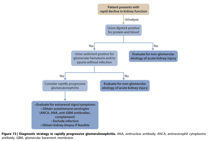
Summary : This flowchart outlines the diagnostic strategy for rapidly progressive glomerulonephritis in patients presenting with rapid decline in kidney function, focusing on the use of urinalysis and subsequent steps to differentiate glomerular from non-glomerular causes.
![Summary : This flowchart outlines the clinical decision-making process for managing patients with hepatitis C virus (HCV) and severe glomerulonephritis (such as rapidly progressive glomerulonephritis [RPGN] or nephrotic syndrome) who are undergoing direct-acting antiviral (DAA) treatment. It distinguishes between typical and atypical presentations and guides whether to proceed with kidney biopsy and immunosuppression.](https://cdn.orris.care/cdss_images/GLGCA_25111_1766507239572_bde5e5be-54b3-4100-b5a5-2458c4fb4b6b_d5f8af66-938c-4184-a9ca-102d5b698cc5.png)
Summary : This flowchart outlines the clinical decision-making process for managing patients with hepatitis C virus (HCV) and severe glomerulonephritis (such as rapidly progressive glomerulonephritis [RPGN] or nephrotic syndrome) who are undergoing direct-acting antiviral (DAA) treatment. It distinguishes between typical and atypical presentations and guides whether to proceed with kidney biopsy and immunosuppression.
![Summary : This flowchart outlines the clinical decision-making process for managing patients with hepatitis C virus (HCV) and severe glomerulonephritis (such as rapidly progressive glomerulonephritis [RPGN] or nephrotic syndrome) who are undergoing direct-acting antiviral (DAA) treatment. It distinguishes between typical and atypical presentations and guides whether to perform a kidney biopsy and consider immunosuppression.](https://cdn.orris.care/cdss_images/GLGCA_25111_1766507224845_e1ab8d2e-549f-4cc9-8ba0-f5c06e363f75_45930578-4b52-4dfa-9cea-fc8ce50b05cb.png)
Summary : This flowchart outlines the clinical decision-making process for managing patients with hepatitis C virus (HCV) and severe glomerulonephritis (such as rapidly progressive glomerulonephritis [RPGN] or nephrotic syndrome) who are undergoing direct-acting antiviral (DAA) treatment. It distinguishes between typical and atypical presentations and guides whether to perform a kidney biopsy and consider immunosuppression.
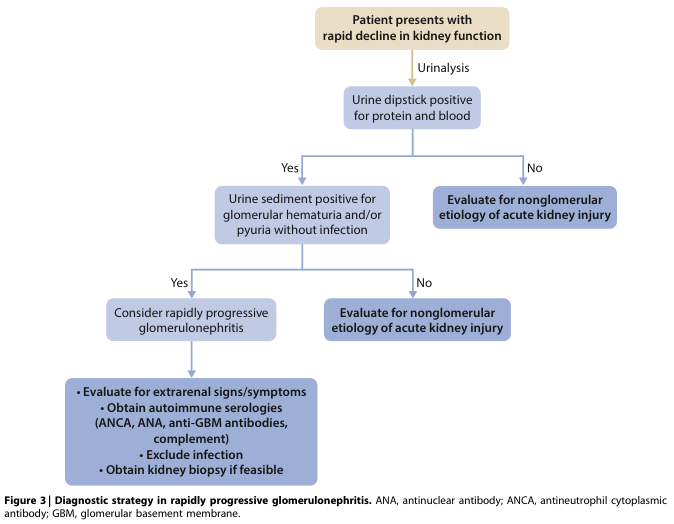
Summary : This flowchart outlines the diagnostic strategy for rapidly progressive glomerulonephritis in patients presenting with a rapid decline in kidney function, guiding clinicians through urinalysis findings and subsequent steps to differentiate glomerular from nonglomerular causes.
Nephrotic Syndrome and Nephritic Syndrome
1. Core Definitions
2. Side-by-Side Comparison
| Feature | Nephrotic Syndrome | Nephritic Syndrome |
|---|---|---|
| Onset | Insidious | Abrupt |
| Edema | Marked (++++) | Present but milder (++) |
| Blood pressure | Usually normal | Raised |
| Proteinuria | Heavy (++++, nephrotic-range) | Mild-moderate (non-nephrotic) |
| Hematuria | Absent or minor | Prominent, often with RBC casts |
| Serum albumin | Low | Normal or mildly reduced |
| GFR | Usually preserved (unless AKI supervenes) | Often reduced |
| Mechanism | Podocyte injury, non-inflammatory | Glomerular inflammation, cellular proliferation |
3. NEPHROTIC SYNDROME
Pathophysiology
Common Causes
- Minimal change disease (MCD) - ~15% of adult NS, 70-90% of pediatric NS; mechanism unclear, associated with NSAIDs, thymoma, Hodgkin lymphoma (T-cell mediated hypothesis)
- Focal segmental glomerulosclerosis (FSGS)
- Membranous nephropathy (MN) - notably higher hypercoagulability risk
- Secondary causes: diabetic kidney disease, amyloidosis, lupus, infections (Hep B/C, HIV), drugs, malignancy
Complications
- Iron-deficiency anemia (transferrin loss)
- Vitamin D deficiency (vitamin D-binding protein loss)
- Hypogammaglobulinemia -> increased infection/sepsis risk
- Hypercoagulability -> renal vein thrombosis, especially when proteinuria >10 g/day and albumin <2 g/dL (worst with membranous nephropathy and amyloidosis) - due to urinary loss of protein C, protein S, antithrombin III
- Acute kidney injury from intravascular volume depletion
Management
- Low-salt, protein-restricted diet (0.8-1 g/kg/day)
- ACE inhibitor or ARB (anti-proteinuric cornerstone)
- Diuretics for edema, statins for hyperlipidemia, anticoagulation if high thrombotic risk
- Disease-specific therapy: corticosteroids are first-line in MCD and childhood NS (prednisone ~60 mg/m²/day or 2 mg/kg/day); steroid-sparing agents (cyclophosphamide, mycophenolate, rituximab, calcineurin inhibitors) for frequently-relapsing or steroid-resistant disease
- In children, typical presentations are treated empirically with steroids without biopsy; biopsy is reserved for atypical features (age <1 year, macroscopic hematuria, hypertension, hypocomplementemia, extrarenal symptoms)
4. NEPHRITIC SYNDROME
Pathophysiology
Common Causes
- Postinfectious/poststreptococcal GN - 1-4 weeks after pharyngitis or impetigo, most common in children 6-10 years; immune complexes with streptococcal antigens
- IgA nephropathy - most common primary glomerulopathy worldwide; galactose-deficient IgA1 immune complexes in the mesangium, often triggered by mucosal (respiratory/GI) infection ("synpharyngitic" hematuria); can progress to Henoch-Schonlein purpura/IgA vasculitis
- Lupus nephritis (proliferative forms can be nephritic or mixed nephrotic-nephritic)
- ANCA-associated (pauci-immune) vasculitis
- Anti-GBM disease (Goodpasture syndrome) - can cause pulmonary-renal syndrome
- Infective endocarditis, abscess, shunt nephritis
Rapidly Progressive Glomerulonephritis (RPGN)
Clinical Picture
Management
- Postinfectious GN: usually supportive (salt/fluid restriction, diuretics, antihypertensives), self-limited in children
- IgA nephropathy: ACEi/ARB for proteinuria/BP control; corticosteroids or immunosuppression in progressive disease
- ANCA vasculitis / anti-GBM / lupus nephritis / crescentic GN: urgent immunosuppression (high-dose corticosteroids, cyclophosphamide or rituximab), plasma exchange in anti-GBM disease
5. Recent Evidence (PubMed)
- Corticosteroid therapy remains standard for childhood nephrotic syndrome, though optimal dose/duration is still being refined (Hahn et al., 2024, PMID: 39171624)
- For steroid-resistant nephrotic syndrome in children, calcineurin inhibitors and other agents are actively being compared for efficacy (Liu et al., 2025, PMID: 40337980)
- Nephrotic syndrome can rarely be paraneoplastic, most often linked to membranous nephropathy in the setting of solid tumors (Liu et al., 2024, PMID: 38965515)
Key Takeaway
Diagnosis