Create a photo of hilum addressing all the arteries veins and all the other parts
A medically accurate labeled anatomical illustration of the right and left lung hila shown side by side from the mediastinal surface. Clearly show and label the main bronchus and lobar bronchial branches, pulmonary artery, superior and inferior pulmonary veins, bronchial arteries and veins, lymph nodes and lymphatic vessels, pulmonary ligament, phrenic nerve anterior to the root, vagus nerve posterior to the root, and pleural reflection. Include the RALS relationship: on the right pulmonary artery lies Anterior to the bronchus; on the left pulmonary artery lies Superior to the bronchus. Use clear leader lines and readable labels.
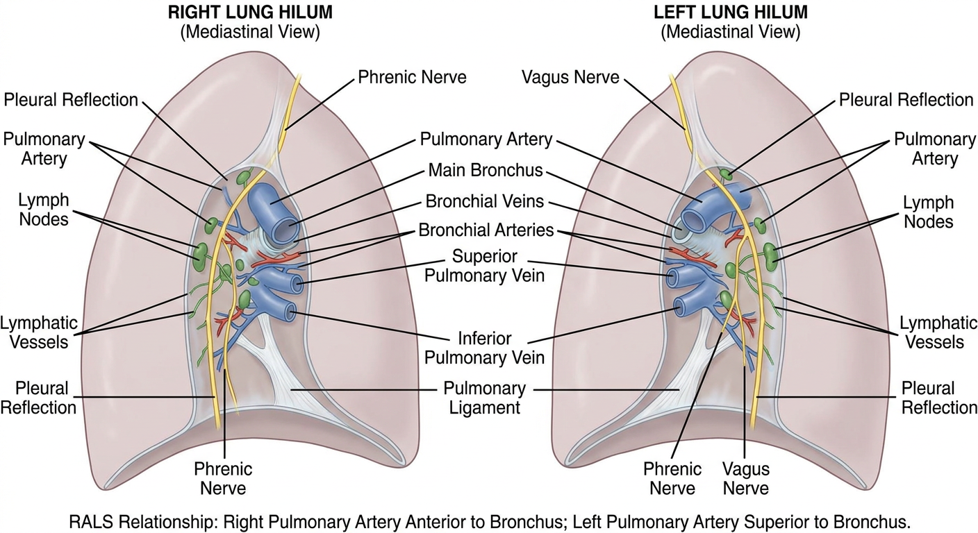
- Right pulmonary artery is Anterior to the bronchus.
- Left pulmonary artery is Superior to the bronchus.
Only the hilum part
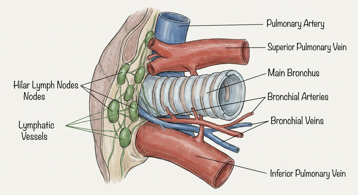
A close-up medically accurate labeled anatomical illustration of ONLY the lung hilum, isolated with no full lungs and no surrounding thorax. Show a single enlarged mediastinal surface hilum with the pulmonary artery, superior pulmonary vein, inferior pulmonary vein, main bronchus with cartilage rings, bronchial arteries, bronchial veins, hilar lymph nodes, and lymphatic vessels. Clearly label every structure with leader lines. Clean educational anatomy diagram.
How many arteries and veins are in the hilum
lung hilum anatomy
lung hilum pulmonary artery two pulmonary veins anatomy
- 1 pulmonary artery
- 2 pulmonary veins
- 1 superior pulmonary vein
- 1 inferior pulmonary vein
In the left hilum: 1 artery + 2 veins.
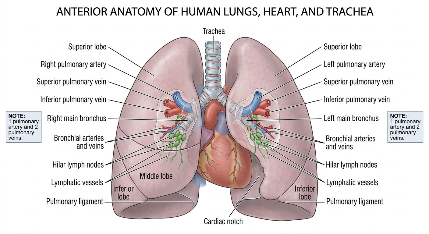
Give a broader photo of the lungs addressing this parts
A broad, medically accurate, clearly labeled anterior anatomical illustration of both human lungs with the heart and trachea for orientation. Emphasize the hila and pulmonary roots on both lungs. Label right and left pulmonary artery, superior pulmonary vein, inferior pulmonary vein, main bronchi, bronchial arteries and veins, hilar lymph nodes, lymphatic vessels, pulmonary ligament, and right and left lung lobes. Include a small clear note at each hilum: 1 pulmonary artery and 2 pulmonary veins. Clean educational anatomy diagram with readable leader-line labels.