
Congeital rubella syndrome psm mbbs
congenital rubella syndrome features blueberry muffin rash cataract
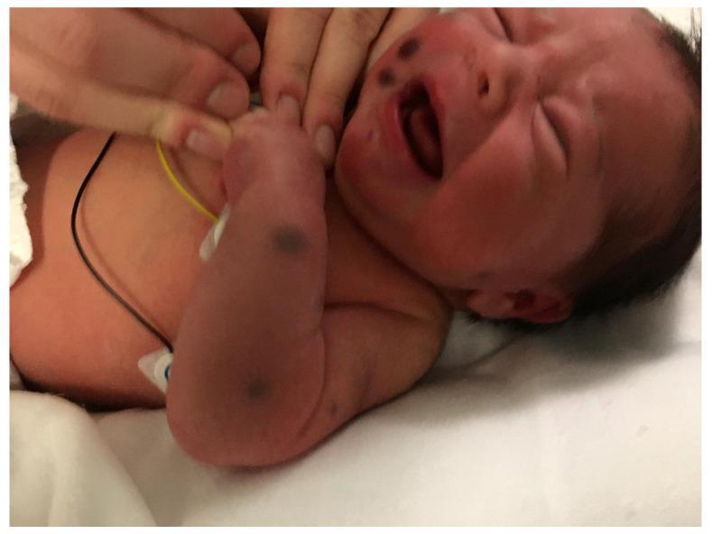
This clinical photograph displays a neonate presenting with a 'blueberry muffin' rash. The image shows multiple discrete, raised, violaceous (bluish-red) nodules and papules. These lesions are distributed across the face, specifically on the right cheek, as well as on the right upper extremity and torso. The nodules appear firm and non-blanching, ranging from a few millimeters to approximately one centimeter in diameter. The infant is shown in a clinical setting with medical monitoring leads visible on the chest. This physical finding is classically associated with extramedullary hematopoiesis (dermal erythropoiesis) and can be a sign of congenital TORCH infections (such as rubella or cytomegalovirus) or systemic neonatal malignancies, including congenital leukemia and metastatic neuroblastoma. The educational focus is on identifying these characteristic cutaneous markers to prompt further diagnostic investigation for serious underlying systemic conditions.
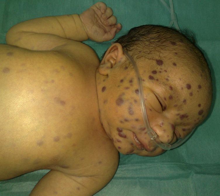
A clinical photograph of a neonate presenting with 'Blueberry Muffin Syndrome.' The image shows multiple, non-blanching, infiltrative papulo-nodular skin lesions. These lesions are erythematous-blue to violaceous in color, ranging approximately 4–8 mm in diameter. They are distributed diffusely across the body, with a significant concentration on the face, forehead, and scalp. Morphologically, these represent extramedullary hematopoiesis or neoplastic infiltration. The neonate is also seen with a nasal cannula in place, indicating concurrent respiratory distress or support requirements. This clinical presentation is a critical diagnostic sign often associated with congenital infections (such as rubella, CMV, or toxoplasmosis) or hematologic/oncologic disorders (including congenital leukemia, neuroblastoma, or Langerhans cell histiocytosis). The photograph serves as a high-yield educational resource for pediatric dermatology and neonatal intensive care, illustrating the characteristic 'blueberry muffin' appearance used in differential diagnosis of neonatal purpura.
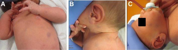
A series of clinical photographs (panels A, B, and C) showing a newborn male with a disseminated 'blueberry muffin' skin rash. Panel A demonstrates multiple oval and circular macules and papules on the trunk and chest, ranging in color from faint magenta to deep purple-livid. Panels B and C highlight similar lesions on the neck, ear, and facial region. The lesions are approximately 0.5 to 1 cm in diameter, non-blanching, and characteristic of dermal erythropoiesis or infiltrative histiocytic disorders. This visual presentation is a classic clinical sign often associated with TORCH infections (such as rubella or cytomegalovirus) or hematologic/histiocytic conditions. In this specific educational case, the lesions are representative of congenital juvenile xanthogranuloma. The distribution is widespread, involving the face, trunk, and extremities, serving as a primary diagnostic indicator for neonatal skin pathology evaluation.
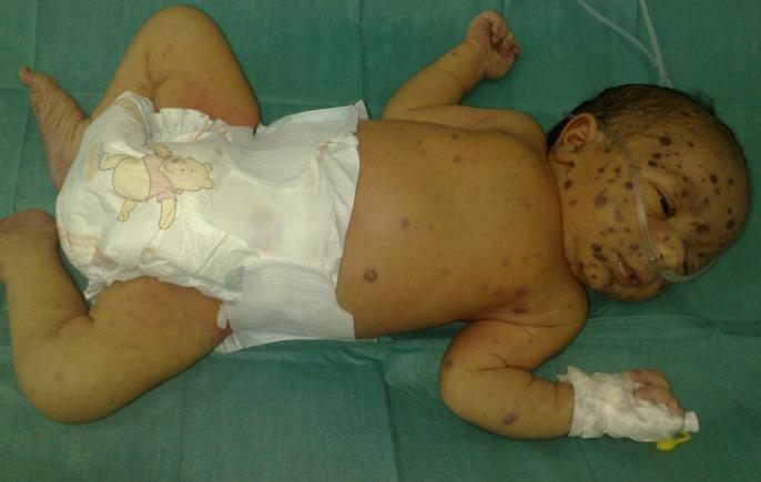
This clinical photograph shows a neonate exhibiting the classic presentation of 'blueberry muffin syndrome.' The image demonstrates numerous disseminated, non-blanching, violaceous to bluish-red macules, papules, and firm nodules. These cutaneous lesions are widely distributed across the infant's body, including the face, scalp, trunk, and extremities, with a high density particularly noted on the face and forehead. Such lesions represent extramedullary hematopoiesis and are classically associated with congenital infections (like Rubella, CMV, or Toxoplasmosis) or neonatal malignancies (such as neuroblastoma or leukemia). The infant is also seen with a nasal cannula and a peripheral intravenous line, indicating acute clinical care. This visual is an essential diagnostic example for pediatric dermatology and neonatology, illustrating the characteristic 'blueberry muffin' appearance used to trigger investigations into underlying systemic pathology.
Congenital Rubella Syndrome (CRS) - PSM (MBBS)
1. Overview / Introduction
Historical milestone: In 1941, Norman Gregg, an Australian ophthalmologist, first described an epidemic of congenital cataracts with other birth defects in children born to mothers who had rubella during pregnancy - revolutionizing understanding of the disease. The virus was isolated in 1962, and an attenuated vaccine developed in 1967.
2. Definition of CRS
-
Presence of IgM rubella antibodies shortly after birth (IgM does NOT cross the placenta, so their presence confirms fetal synthesis in utero), OR
-
IgG antibodies persisting >6 months (maternally derived IgG would have disappeared by then)
-
Park's PSM, p.172; Jawetz Medical Microbiology
3. Epidemiological Determinants (PSM Focus)
Agent Factors
| Factor | Details |
|---|---|
| Agent | RNA virus, Togavirus family |
| Source | Clinical or subclinical cases (large proportion of infections are subclinical - major difference from measles) |
| No carrier state | For postnatally acquired rubella |
| CRS infants | Can shed virus for up to 12-18 months after birth |
| Vaccine virus | Not communicable |
Period of Communicability
- Extends from 1 week before symptoms to ~1 week after rash appears
- Infectivity greatest 1-5 days after appearance of rash
- Less communicable than measles (no coughing in rubella)
Host Factors
- Age: Mainly children aged 3-10 years; but in developed countries with immunization, >70% of cases now occur in those older than 15 years
- Immunity: One attack gives lifelong immunity; second attacks are rare
- Maternal antibody protection: Infants of immune mothers are protected for 4-6 months
- 10-40% of populations may reach adulthood without infection if unimmunized
Environmental Factors
- Seasonal pattern: late winter and spring (temperate zones)
- Epidemics every 4-9 years (some sources say 5-9 years) in pre-vaccination era
Transmission
- Person-to-person via droplets and droplet nuclei (aerosols) from nose and throat
- Portal of entry: respiratory route
4. Pathogenesis
-
Maternal viremia → infection of placenta → fetal viremia
-
Only a limited number of fetal cells become infected, but growth rate is reduced → fewer cells in affected organs at birth → structural anomalies
-
Inapparent (subclinical) maternal infection can still produce all the anomalies
-
Jawetz Medical Microbiology
5. Risk by Gestational Age (Critical PSM Point)
| Gestational Age | Risk of Defects |
|---|---|
| Just before conception to first 8-10 weeks | Multiple abnormalities in up to 90% |
| First trimester (up to 12 weeks) | ~85% risk of abnormalities |
| Second trimester | ~16% risk of detectable defects |
| After 16th week | Congenital anomalies rare (except hearing loss) |
| After 20th week | Birth defects uncommon |
| Up to 20th week | Sensorineural hearing loss may still occur |
- Park's PSM; The Developing Human (Moore's Embryology)
6. Clinical Features of CRS
Classic Triad (Gregg's Triad)
Cataracts + Cardiac defects + Sensorineural deafness
Three Categories of Manifestations (Jawetz classification)
- Growth retardation / low birth weight
- Thrombocytopenic purpura (blueberry muffin rash - due to dermal erythropoiesis)
- Jaundice / hepatitis
- Hepatosplenomegaly
- Meningoencephalitis
- Interstitial pneumonitis
| System | Features |
|---|---|
| Ophthalmic | Cataracts, microphthalmia, glaucoma, pigmentary retinopathy, chorioretinitis |
| Auditory | Sensorineural deafness (most common permanent defect) |
| Cardiac | Patent ductus arteriosus (PDA), ventricular septal defect (VSD), peripheral pulmonary artery stenosis |
| Craniofacial | Microcephaly |
- Mental retardation (moderate to profound) - most common developmental manifestation
- Autism
- Problems with balance and motor skills
- Visual and hearing impairment
- Developmental delay
- Radiolucency in long bones - a characteristic radiological pattern of CRS
-
Progressive rubella panencephalitis - develops in the second decade of life, severe neurologic deterioration progressing inevitably to death
-
Park's PSM, pp.171-172; Jawetz Medical Microbiology
7. "Blueberry Muffin" Rash
8. Viral Shedding in CRS Infants
- Pharyngeal secretions
- Multiple organs
- Cerebrospinal fluid
- Urine
- Rectal swabs
9. Prevention and Control (PSM Core)
Vaccine
- Strain: Live attenuated RA 27/3 strain
- Formulations: Monovalent, MR (Measles-Rubella), MMR (Measles-Mumps-Rubella), MMRV (with Varicella)
- Dose: 0.5 ml, subcutaneous injection
- Site: Anterolateral thigh (infants) or outer aspect of upper arm (older children/adults)
- Minimum potency: ≥1000 plaque-forming units (PFU) per dose
- Storage: 2-8°C, protected from light; shelf life 2-3 years
- Efficacy: >95% with single dose (even at 9 months)
- Immunity: Vaccine-induced immunity is lifelong
Dosing Schedule
- Rubella is less infectious than measles, so only 1 dose required for elimination if high coverage achieved
- Often given as 2nd dose combined with measles (MR/MMR) for programmatic convenience
Vaccine Interactions
- Can be given concurrently with inactivated vaccines
- Other live vaccines: give simultaneously or ≥4 weeks apart
- Exception: OPV can be given at any time relative to RCV without interference
- MMR + Yellow fever vaccine: may interfere if given simultaneously to children <2 years
Contraindications
- Severe allergic reaction to previous dose or vaccine component
- Active tuberculosis
- Severe immunodeficiency (symptomatic HIV/AIDS, congenital immune disorders, malignancies, aggressive immunosuppressive therapy)
- Pregnancy (theoretical teratogenic risk, though never demonstrated)
- Advise avoidance of pregnancy for 1 month post-vaccination
- Inadvertent vaccination during pregnancy is NOT an indication for termination
- Recent blood products: wait ≥3 months before RCV vaccination; avoid blood products for 2 weeks after vaccination
Target Group for Direct Protection
- All non-pregnant women of reproductive age who are unvaccinated or seronegative should receive one dose of RCV
10. Global Context
- WHO Global Framework 2021-2030: Aims for elimination of measles and rubella
- India introduced MR vaccine under the Universal Immunization Programme (UIP)
- In pre-vaccination era: epidemics every 4-9 years
11. High-Yield Summary Table for Exams
| Feature | Details |
|---|---|
| Causative agent | RNA virus, Togavirus family |
| Classic triad | Cataracts + Cardiac defects + Deafness |
| Most common permanent defect | Sensorineural deafness |
| Most dangerous gestational period | First 8-10 weeks (up to 90% risk) |
| After 16 weeks | Anomalies rare (only deafness up to week 20) |
| Mechanism of teratogenicity | Inhibition of cell division |
| Diagnosis in neonate | IgM antibodies at birth OR IgG persisting >6 months |
| Viral shedding in CRS infants | 12-18 months |
| Characteristic X-ray finding | Radiolucency of long bones |
| Blueberry muffin rash | Extramedullary hematopoiesis (dermal erythropoiesis) |
| Late CNS complication | Progressive rubella panencephalitis (2nd decade) |
| Vaccine strain | RA 27/3 (live attenuated) |
| Vaccine efficacy | >95% with single dose |
| Vaccine immunity | Lifelong |
| Post-vaccination pregnancy avoidance | 1 month |
| Norman Gregg's discovery | 1941 (cataracts + birth defects after maternal rubella) |