
Shillohet sign cause
"Shillohet sign" cause medical
Shillohet
- Pneumonia or lung consolidation
- Atelectasis (collapse)
- Lung mass
- Pleural or mediastinal disease next to the structure
- Loss of the right heart border suggests right middle lobe disease.
- Loss of the left heart border suggests lingular disease.
- Loss of a hemidiaphragm can indicate lower-lobe consolidation.
Explain the silhouette sign with a labeled chest X-ray diagram
labeled chest x ray silhouette sign right middle lobe consolidation loss of right heart border
"silhouette sign" AND chest radiography
silhouette sign chest radiograph loss of right heart border right middle lobe authoritative radiology
https://pmc.ncbi.nlm.nih.gov/articles/PMC7517573/
An educational labeled frontal PA chest X-ray style diagram demonstrating the silhouette sign from right middle lobe consolidation. Include normal black aerated lungs, white heart and diaphragms, a white opacity adjacent to the right heart border, an arrow labeling “Right middle lobe consolidation”, and labels “Right heart border lost”, “Right hemidiaphragm preserved”, “Left heart border”, and “Patient's right”. Ensure anatomically correct side orientation and legible labels.
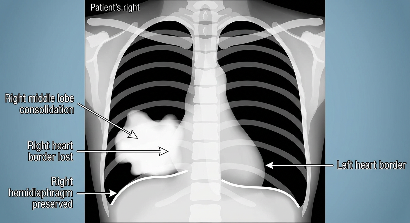
Silhouette sign
- Air-filled lung appears dark.
- The heart, diaphragm, and mediastinum are soft-tissue density and appear relatively white.
- The dark lung beside these white structures creates a clear outline.
Example in the diagram
- The right middle lobe lies against the right atrium, forming the right cardiac border.
- Right middle lobe consolidation makes that interface white-on-white, so the border is lost.
- Because the right lower lobe is not primarily involved, the right hemidiaphragm can remain visible.
Quick localization table
| Lost normal border | Likely adjacent location |
|---|---|
| Right heart border | Right middle lobe |
| Left heart border | Lingula of left upper lobe |
| Right hemidiaphragm | Right lower lobe |
| Left hemidiaphragm | Left lower lobe |
| Aortic knuckle | Left upper lobe |
| Descending aortic border | Left lower lobe |