
Pathophysiology clinical feature investigation and management of torsion testis
I'll start by reading the medical library skill, then search for comprehensive information on testicular torsion.
Reading File
Now let me run parallel searches across the library and PubMed simultaneously.
Finding Sources
Finding Sources
Searching PubMed
"testicular torsion"[MeSH Terms] AND management
Excellent. I have the key textbook locations. Now let me read the most comprehensive sources in parallel.
Reading File
Reading File
Reading File
Excellent content. Now let me read more from Bailey & Love (the main torsion section) and fetch the bell-clapper diagram image, plus get the Campbell-Walsh section on management.
Reading File
Reading File
Loading Image
I now have comprehensive content from multiple textbook sources. Let me read the remaining section of Bailey & Love for clinical features and investigations, and search for a Doppler ultrasound image.
Reading File
Finding Sources
I now have all the key content needed. Let me read the Doppler/investigation section and the Rosen's EM section for the ED management perspective.
Reading File
Reading File
I now have all the comprehensive information needed from multiple authoritative textbooks. Let me compile the full detailed answer.
Torsion of the Testis
Definition
Testicular torsion is the twisting of the spermatic cord and its contents such that the testicular blood supply becomes compromised. It is a surgical emergency - if left untreated, blood flow to the testicle ceases and the testicle undergoes ischaemic necrosis.
- Incidence: 3.8-4.5 per 100,000 males under 18 years annually
- Accounts for 10-15% of acute scrotal disease in children
- Most common between ages 10 and 25 years
Pathophysiology
Types of Torsion
1. Extravaginal Torsion
- Seen almost exclusively in neonates
- Occurs due to increased mobility of the testicle before it descends into the scrotum and becomes attached to the scrotal wall via the tunica vaginalis
- The entire spermatic cord, including the tunica vaginalis, twists
2. Intravaginal Torsion
- The predominant type beyond the neonatal period
- Results from one or more anatomical predispositions:
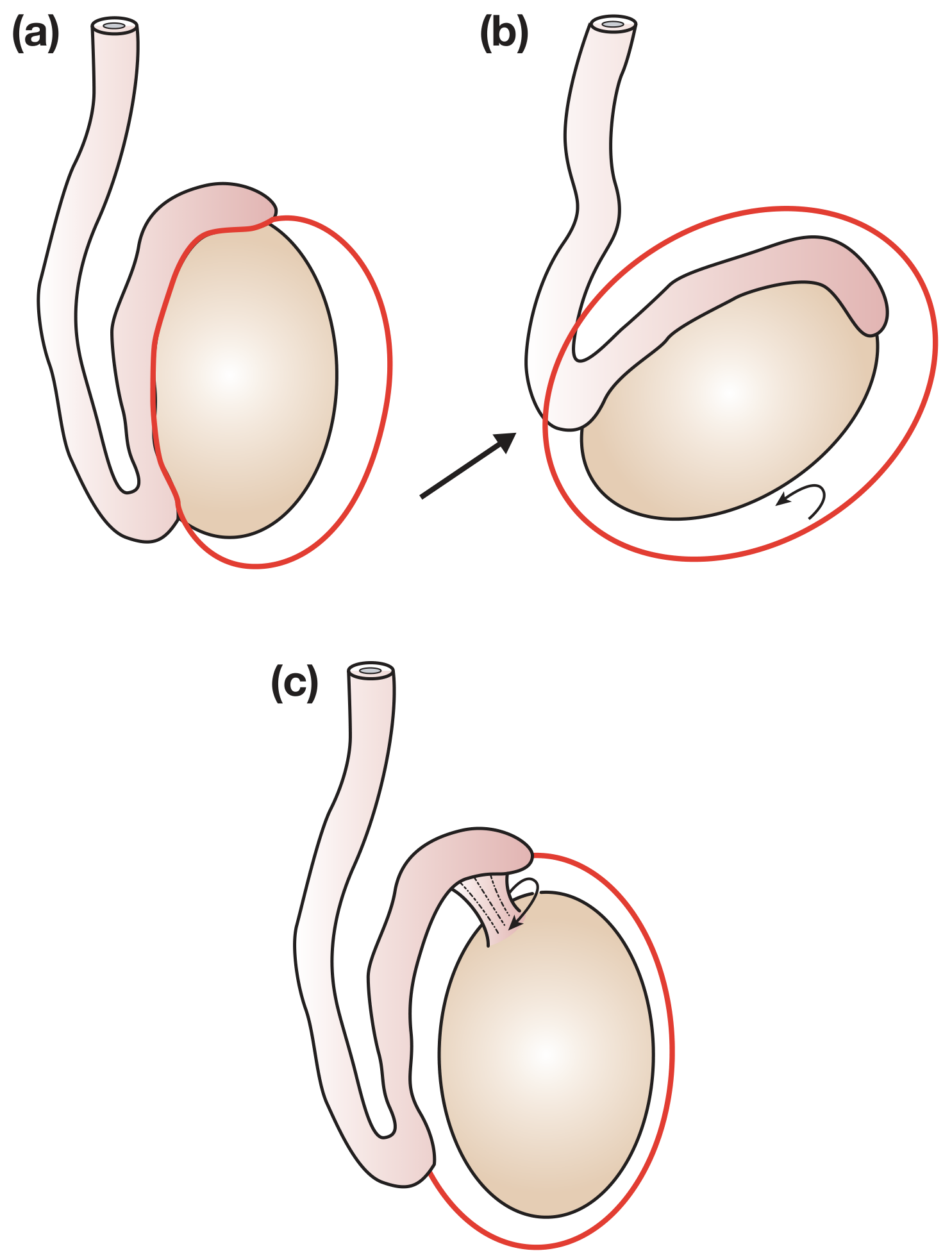
a) Bell-Clapper Deformity (most common cause in adolescents)
High investment of the tunica vaginalis causes the testis to hang freely within the tunica - like a clapper in a bell. This is typically a bilateral anatomical abnormality, which is why the contralateral testis must always be fixed.
b) Inversion of the testis - the testis lies transversely or upside down
c) Separation of the epididymis from the body of the testis, permitting torsion of the testis on the pedicle connecting them
Precipitating Factors
Normally, cremasteric contraction pulls the testis upward. In the presence of the anatomical abnormalities above, the spiral attachment of the cremaster favours rotation around the vertical axis. Precipitants include:
- Physical activity (straining, lifting, sport)
- Sexual activity
- Thermal stimulation
- Occasionally occurs during sleep (cremasteric reflex while waking)
Degree of Torsion and Ischaemia
- Twists of 720° cause more rapid ischaemia than twists of 360° or less
- Duration-dependent damage: venous outflow obstructed first, then arterial inflow
- Contralateral testicular biopsy findings are abnormal in 57-88% of males when torsion occurs, suggesting underlying bilateral spermatogenic dysfunction
Consequence of Torsion
- Up to 50% of men develop adverse spermatogenic effects after torsion
- 36-39% will have sperm concentrations below 20 million/mL
- Up to 11% develop antisperm antibodies due to disruption of the blood-testis barrier (tight junctions between Sertoli cells)
Clinical Features
Classic presentation in a 10-25-year-old male:
| Feature | Detail |
|---|---|
| Onset of pain | Sudden, severe - patient can often state exact time it started |
| Location of pain | Groin and lower abdomen (can mimic appendicitis) |
| Associated symptoms | Nausea and vomiting (sudden onset, distinguishes from epididymo-orchitis) |
| Scrotal appearance | Swollen, firm, tense, reddened - not erythematous early on |
| Testicular position | High-riding testis (pulled upward by twisted cord) |
| Cord palpation | Tender, thickened twisted cord often palpable above the testis |
| Temperature | Apyrexial (fever favours epididymo-orchitis) |
| Cremasteric reflex | Absent (important sign - reflex is present in epididymo-orchitis and appendage torsion) |
| Elevation test (Prehn's sign) | Pain worsens with testicular elevation (in epididymo-orchitis, elevation relieves pain) |
Important note: Torsion may also occur in neonates - presenting as a hard, painless scrotal mass with skin discolouration (often detected at birth).
Intermittent torsion: Acute, short-duration (<2 hours) scrotal pain with rapid, spontaneous resolution. Nausea and vomiting in ~25% of cases. Must be taken seriously and treated with orchiopexy.
Differential Diagnosis
| Condition | Distinguishing Features |
|---|---|
| Epididymo-orchitis | Gradual onset, fever, dysuria, urethral discharge, normal cremasteric reflex, pain relieved by elevation |
| Appendage torsion (Hydatid of Morgagni) | Milder pain, gradual onset, upper pole tenderness, "blue dot sign" (in fair-skinned children early), cremasteric reflex intact |
| Mumps orchitis | Cord not thickened, often bilateral, parotitis history |
| Idiopathic scrotal oedema | Age 4-12, bilateral swelling with minimal pain, extends to perineum/groin/penis |
| Strangulated inguinal hernia | Inguinal mass, bowel obstruction symptoms |
Investigations
Clinical Priority First
The management decision is primarily clinical. Do NOT delay surgical exploration awaiting investigation if clinical suspicion is high.
1. Doppler Ultrasound (most commonly used)
- Can confirm absence of blood flow to the affected testis
- Sensitivity 92-100% for confirming epididymitis
- Absence of blood flow = torsion until proven otherwise
- Caveat: False-positive results (apparent flow) can occur, so it is not routinely recommended as a reason to withhold surgery. A positive Doppler (flow present) does not absolutely exclude torsion.
2. Radionuclide Scanning (Technetium-99m scan)
- Uptake of tracer into the centre of the testis rules out torsion
- Less available, rarely used in acute setting
3. Urinalysis
- Normal in testicular torsion (WBCs and bacteria suggest epididymo-orchitis)
4. FBC/Bloods
- Leukocytosis suggests infection rather than torsion
- No specific blood test for torsion
Key principle from Smith & Tanagho's General Urology:
"The presence of blood flow in the testis on Doppler ultrasonography or uptake of tracers into the center of the testis on radionuclide scanning rules out torsion."
Management
Time is Testis - Salvage Rates by Duration
| Time from Onset | Testicular Salvage Rate |
|---|---|
| < 6 hours | ~100% |
| 6-12 hours | ~70% |
| 12-24 hours | ~20% |
| > 24 hours | Very low / near 0% |
Step 1: Immediate Urological Consultation
Upon presentation, early urology consultation is critical - even before confirmatory imaging in high-suspicion cases.
Step 2: Manual Detorsion (can be attempted in ED)
- Testis usually rotates medially (inward), so manual detorsion involves rotating it outward (laterally), like opening a book
- If successful, pain relief is immediate and vascular flow restored
- This is a temporising measure only - does not negate need for surgical fixation
Step 3: Emergency Surgical Exploration
Incision: Transverse or midline scrotal incision
Intraoperative decision-making:
a) If testis is viable after detorsion:
- Fix with 3 non-absorbable sutures between the tunica albuginea and the scrotal raphe (orchiopexy)
- Absorbable sutures must NOT be used (risk of recurrent torsion as suture dissolves)
- Fix the contralateral testis as well - the anatomical predisposition is bilateral
b) If viability is uncertain:
- Wrap the testis in a warm swab and observe for several minutes
- Make a small incision in the tunica albuginea - if bright red arterial bleeding is seen, the testis may survive
- Fix as above if viable
c) If testis is infarcted (necrotic):
- Orchiectomy is performed - the dead testis must be removed
- Counsel the patient about testicular prosthesis later
- Still fix the contralateral testis to prevent future torsion
Step 4: Contralateral Orchiopexy
Always performed simultaneously with ipsilateral fixation/orchiectomy. The bell-clapper deformity is bilateral in the majority of cases.
Step 5: Cases With Prolonged History (Several Days)
- The affected testis will already be infarcted
- Little can be gained (other than pain relief) by immediate exploration of the affected side
- However, urgent contralateral fixation is still necessary
Orchiopexy Techniques (for intermittent/elective cases)
- Suture fixation - 3-0 non-absorbable sutures fixing tunica albuginea to dartos muscle or scrotal raphe
- Dartos pouch procedure - testis placed in a subcutaneous pouch between dartos and external spermatic fascia, secured by purse-string suture; preferred by some as it avoids intratesticular vessel injury
Summary Algorithm
Acute Scrotal Pain
      ↓
High clinical suspicion for torsion?
      ↓
YES → Emergency surgical exploration (do not delay)
      ↓
Viable testis? → Bilateral orchiopexy (non-absorbable sutures)
Infarcted?    → Orchiectomy + contralateral orchiopexy

NO / Uncertain → Doppler ultrasound
      ↓
No flow → Emergency exploration
Flow present + confident alternative diagnosis → Treat accordingly
Sources:
- Bailey and Love's Short Practice of Surgery, 28th Ed., pp. 1582-1584 (Torsion of the Testis)
- Campbell-Walsh-Wein Urology, 3-Volume Set, pp. 1612-1614; 1180-1184 (Testis Torsion; Intermittent Torsion)
- Smith and Tanagho's General Urology, 19th Ed., p. 787 (Distinguishing Torsion)
- Rosen's Emergency Medicine: Concepts and Clinical Practice, p. 1595 (Disposition and Management)
What is laplace law and how it is imp in a surgeons pony of view
Laplace's Law states that the wall tension (T) in a hollow organ is proportional to the transmural pressure (P) and radius (r), and inversely proportional to wall thickness (w): T = P × r / w. From a surgical standpoint, this explains why large, thin-walled hollow organs (dilated bowel, aneurysms, gravid uterus) are at greatest risk of rupture, and why small-radius structures can withstand high pressures with less wall stress.
Searching sources...
Finding Sources
Finding Sources
I have the key sources. Let me read the Ganong physiology section and the Fischer's Surgery surgical context in parallel.
Reading File
Reading File
Finding Sources
Now let me read the Rosen's EM aneurysm section and the Fischer's Surgery section for the surgical applications.
Reading File
Reading File
Reading File
I now have comprehensive content from multiple authoritative textbooks. Let me also fetch the alveolar diagram and search for bowel obstruction + bladder surgical Laplace applications.
Finding Sources
Loading Image
I now have everything needed to write a comprehensive, textbook-backed answer. Let me compile it.
Laplace's Law - Physics, Physiology, and Surgical Significance
The Formula
For a hollow cylinder (e.g., blood vessel, bowel):
T = P × r / w
For a sphere (e.g., heart, bladder, aneurysm):
P = 2HT / r (rearranged: T = P × r / 2w)
Where:
- T = Wall tension (force per unit length of wall)
- P = Transmural pressure (internal minus external pressure)
- r = Radius of the hollow structure
- w / H = Wall thickness
Plain English: The wall of a hollow organ must generate more tension to contain the same internal pressure as it gets larger (bigger radius). Conversely, a thin wall at large radius = catastrophically high tension for any given pressure.
Core Concepts
| Variable | Effect on Wall Tension |
|---|---|
| Radius increases (dilation) | Tension increases - higher rupture risk |
| Pressure increases | Tension increases |
| Wall thickness increases (hypertrophy) | Tension decreases - protective |
Surgical Applications - Organ by Organ
1. Aortic and Arterial Aneurysms (Most Direct Surgical Application)
This is the most important surgical implication. As Rosen's Emergency Medicine states:
"Aneurysmal dilation accelerates, increasing the risk of rupture as diameter increases, as described by Laplace's law: tension in the wall of a hollow viscus varies directly with its radius."
- A normal aorta (radius ~1 cm) generates ~170,000 dynes/cm of wall tension
- As the aorta dilates to 5-6 cm (AAA), wall tension rises proportionally - and each millimetre of further growth disproportionately increases rupture risk
- This explains why the 5.5 cm threshold for elective AAA repair is used clinically: beyond this, annual rupture risk (~10-20%) overtakes operative mortality
- A dilating aneurysm is a self-reinforcing cycle: dilation → increased tension → further dilation - a vicious positive feedback loop
- The same principle applies to popliteal, splenic, and visceral artery aneurysms
Tintinalli's EM: "In aortic true aneurysm, the dilatation and increased wall force are intertwined, creating more dilatation (Laplace law: wall tension = pressure × radius)."
2. Bowel Obstruction and Cecal Perforation
This is a classic surgical emergency driven entirely by Laplace's Law.
- The cecum has the largest diameter in the colon (~9 cm normal, can reach 12-15 cm in obstruction)
- Under the same intraluminal pressure as the sigmoid colon, the cecum generates far greater wall tension
- This is why the cecum perforates first in large bowel obstruction, even when the obstructing lesion is in the left colon
- The "safe" radiological threshold is cecal diameter > 12 cm (increasing perforation risk)
From Fischer's Mastery of Surgery (8th ed):
A decompressing stoma "avoids acute cecal perforation based on the law of Laplace" - this is the explicit surgical rationale for urgent defunctioning in left-sided colonic obstruction.
3. The Heart in Cardiac Failure and Hypertrophy
As Costanzo's Physiology explains:
For a sphere: P = 2HT / r
- In systemic hypertension: elevated aortic pressure → left ventricle compensates by wall hypertrophy (increasing H/thickness) → this reduces wall tension for any given pressure, protecting the myocardium
- In dilated cardiomyopathy (increased radius r): the ventricle must generate more wall tension to produce the same ejection pressure → increases myocardial O₂ consumption, accelerates decompensation
- Ganong: "When the radius of a cardiac chamber is increased, a greater tension must be developed in the myocardium to produce any given pressure; consequently, a dilated heart must do more work than a nondilated heart"
- This explains why afterload reduction (ACE inhibitors, ARBs) is so beneficial in dilated cardiomyopathy - reducing wall tension reduces myocardial work
4. The Urinary Bladder
- Normal bladder wall compliance maintains low intravesical pressure despite increasing volume (large radius, but thin compliant wall compensated by accommodation)
- In chronic urinary retention with a massively distended bladder: Laplace's Law predicts enormous wall tension - predisposing to:
- Ischaemia of the detrusor muscle
- Loss of contractility (overflow incontinence)
- Risk of bladder rupture (rare, but seen with trauma on a full bladder)
- Rapid decompression of an acutely distended bladder (>1 litre) should be done carefully to avoid haematuria from sudden drop in transmural pressure
5. Alveoli and Respiratory Surgery
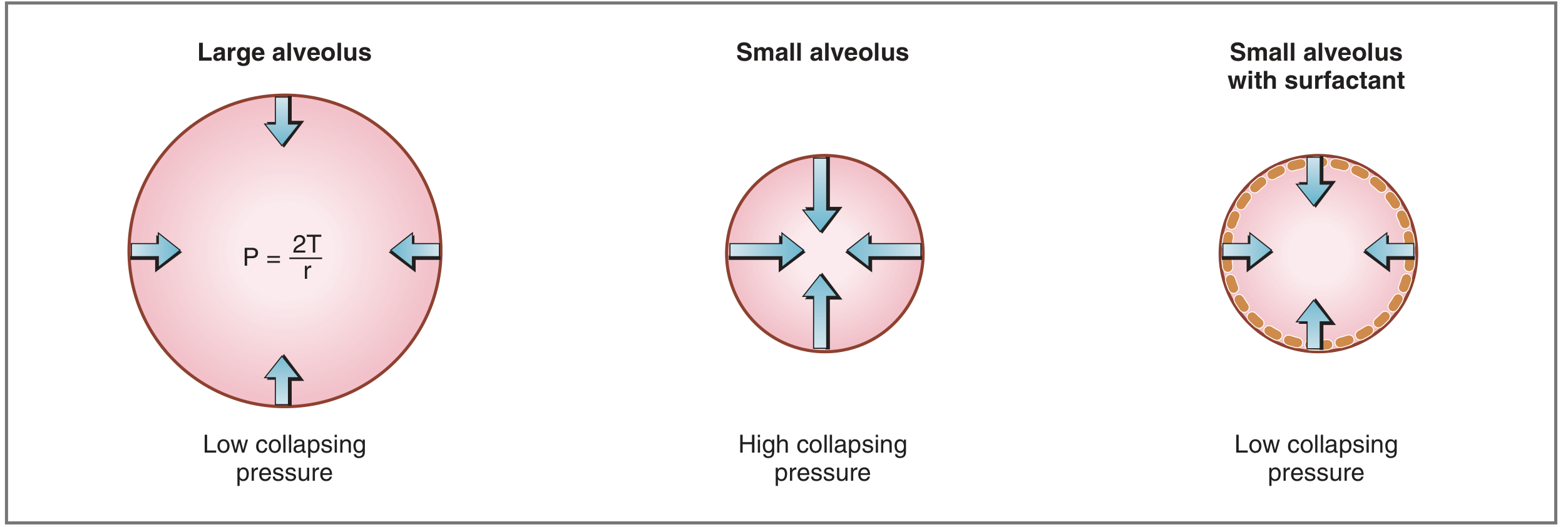
For a sphere: P (collapsing) = 2T / r
- Small alveoli have high collapsing pressure - they tend to collapse (atelectasis)
- Surfactant reduces surface tension (T), lowering collapsing pressure - this is the biological solution
- In neonatal respiratory distress syndrome (surfactant deficiency), small stiff alveoli collapse
- Surgically: post-operative atelectasis occurs in collapsed, surfactant-depleted small alveoli - managed with PEEP (positive end-expiratory pressure), deep breathing, and physiotherapy
- After pneumonectomy or lung resection, the remaining lung must expand: larger radius → lower wall tension per Laplace, which is actually beneficial
6. Capillaries - Why They Don't Burst
Despite being exposed to the same systemic blood pressure as arteries, capillaries don't rupture because of their tiny radius (~4-5 µm).
From Ganong: "In the human aorta, wall tension at normal pressures is about 170,000 dynes/cm; in capillaries it is approximately 16 dynes/cm" - a 10,000-fold difference explained entirely by the radius term in Laplace's Law.
Summary Table: Surgical Implications at a Glance
| Clinical Scenario | Laplace Mechanism | Surgical Relevance |
|---|---|---|
| AAA repair threshold (5.5 cm) | Large radius → high wall tension → rupture risk | Elective repair before catastrophic rupture |
| Cecal perforation in L bowel obstruction | Cecum = widest segment → highest tension | Urgent decompression / defunctioning stoma |
| Dilated cardiomyopathy | Large radius → high myocardial wall stress | ACE inhibitors reduce afterload/wall tension |
| LV hypertrophy in hypertension | Thick wall lowers tension for given pressure | Adaptive response, explains why hypertrophy develops |
| Atelectasis / neonatal RDS | Small alveoli → high collapsing pressure | PEEP, surfactant therapy |
| Bladder distension | Large radius → ischaemia, risk of rupture | Careful catheterisation, gradual decompression |
| Capillary integrity | Tiny radius → minimal wall tension | Capillaries are paradoxically protected |
Sources:
- Ganong's Review of Medical Physiology, 26th Ed., p. 576 (Law of Laplace - capillaries and vessels)
- Costanzo Physiology, 7th Ed., pp. 159, 210 (Heart, alveoli)
- Fischer's Mastery of Surgery, 8th Ed., p. 3603 (Cecal perforation and Laplace)
- Rosen's Emergency Medicine, p. 1178 (Aneurysmal dilation)
- Tintinalli's Emergency Medicine, p. 3104 (Aortic aneurysm)
This is a shared conversation. Sign in to Orris to start your own chat.