
Classification of cardiac arrhythmias and their management 20 mark answer
classification of cardiac arrhythmias diagram
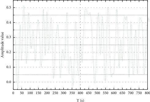
This diagnostic image displays a schematic diagram of a continuous segment of an electrocardiogram (ECG) signal waveform, utilized in the context of cardiac rhythm analysis and machine learning classification. The graph plots the amplitude value (on the y-axis, ranging from 0.0 to 0.5) against time in seconds (T (s) on the x-axis, spanning 0 to 800 seconds). The waveform demonstrates typical physiological oscillations consisting of repeated peaks and troughs that correspond to cardiac electrical activity. Gridlines are provided at 0.1 intervals for amplitude and 50-second intervals for time to facilitate precise measurement. Such continuous data segments are essential in medical informatics for preprocessing tasks like heartbeat segmentation, denoising, and the identification of arrhythmias, such as right bundle-branch blocks or atrial premature contractions (APC). The visual representation serves as a foundational tool for understanding signal morphology and the temporal distribution of heartbeats in cardiovascular diagnostic studies.
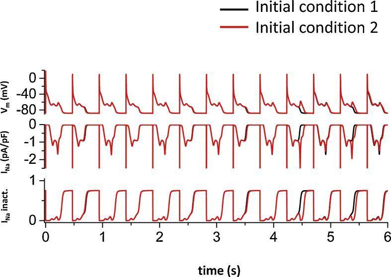
This pathophysiology diagram consists of three stacked electrophysiological simulation traces over a 6-second period, illustrating cellular cardiac arrhythmias. The Y-axes represent membrane potential (Vm in mV), sodium current (INa in pA/pF), and sodium inactivation (INa inact.). The simulation compares two starting states: 'Initial condition 1' (black line) and 'Initial condition 2' (red line), where the latter begins with a membrane potential 1 mV higher than the former. The Vm trace demonstrates the development of early afterdepolarizations (EADs) during the repolarization phase of the action potentials. Initially, the two simulation traces are identical, but they eventually diverge, illustrating sensitivity to initial conditions and chaotic dynamics in cardiac electrical signaling. The corresponding INa traces show that during EADs, there is a reactivation of sodium currents, which correlates with partial recovery in the INa inactivation variable. This visual evidence supports the role of sodium channel window currents in triggering irregular EADs under pathological conditions, such as long QT syndrome or heart failure.
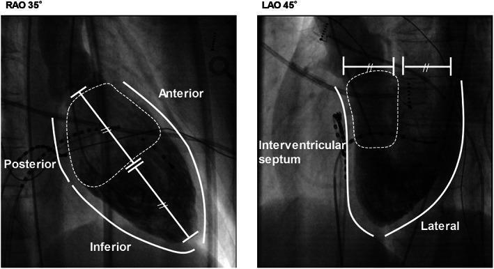
Two grayscale fluoroscopic images illustrate cardiac anatomy and classification criteria for proximal versus non-proximal ventricular arrhythmias. The left panel shows a Right Anterior Oblique (RAO 35°) projection, with labels for the anterior, posterior, and inferior aspects of the heart. A solid white line outlines the cardiac silhouette, while a dashed region defines the proximal half of the heart, where structures like the His bundle and proximal fascicles are located. A bisecting line with tick marks indicates the division between proximal and distal sections. The right panel shows a Left Anterior Oblique (LAO 45°) projection, highlighting the interventricular septum and lateral wall. A similar dashed area delineates the targeted septal and proximal anterior wall regions. These images serve as a fluoroscopic guide for identifying successful ablation sites in patients with focal Purkinje ventricular arrhythmias, emphasizing anatomical landmarks used to differentiate arrhythmia origins based on septal and proximal proximity.
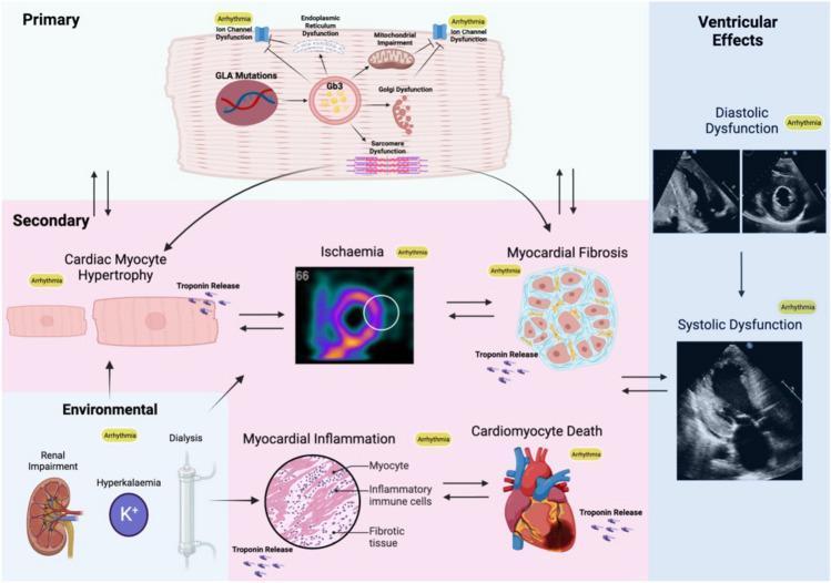
This pathophysiology diagram illustrates the multi-level mechanisms leading to arrhythmia in Fabry Disease (FD), categorized into primary, secondary, environmental, and ventricular effects. The 'Primary' section focuses on cellular dysfunction, showing GLA mutations leading to Gb3 accumulation, which triggers endoplasmic reticulum dysfunction, mitochondrial impairment, Golgi dysfunction, sarcomere dysfunction, and direct ion channel dysfunction. The 'Secondary' section details macro-level pathological changes: cardiac myocyte hypertrophy, myocardial ischemia (represented by a perfusion scan), myocardial fibrosis, and cardiomyocyte death, all associated with troponin release and designated as pro-arrhythmic. 'Environmental' factors include renal impairment, hyperkalemia (K+), and dialysis, which contribute to myocardial inflammation (depicted via histopathology showing myocytes, inflammatory cells, and fibrotic tissue). The final column, 'Ventricular Effects,' displays diagnostic ultrasound images correlating these processes with clinical stages of diastolic and systolic dysfunction. The diagram uses arrows to establish bidirectional relationships between hypertrophy, ischemia, fibrosis, and inflammation, highlighting the complex, interrelated substrate for both atrial and ventricular arrhythmias in FD.
ECG atrial fibrillation ventricular tachycardia heart block
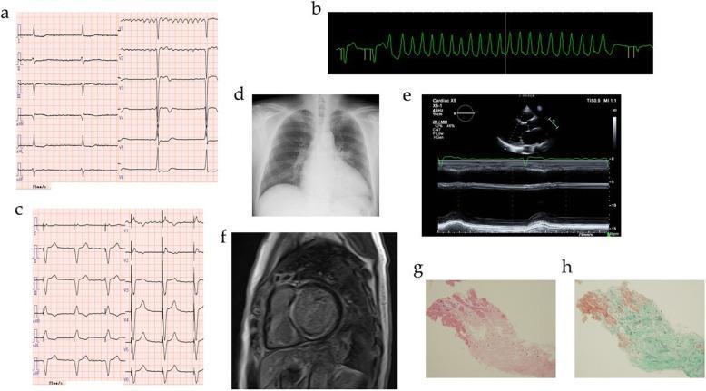
This composite figure presents a comprehensive clinical workup for dilated cardiomyopathy (DCM) and associated arrhythmias. Panels (a-c) display cardiovascular monitoring findings: a standard 12-lead ECG (a) showing atrial fibrillation with complete atrioventricular (AV) block (bradycardia at 40 bpm); a rhythm strip (b) capturing non-sustained ventricular tachycardia (NSVT); and a post-intervention ECG (c) demonstrating a regular paced rhythm following cardiac resynchronization therapy defibrillator (CRTD) implantation. Radiographic and imaging studies include a chest X-ray (d) showing cardiomegaly with a cardiothoracic ratio of 60.4% and pulmonary congestion; an echocardiogram with M-mode (e) indicating left ventricular systolic dysfunction; and a cardiovascular magnetic resonance (CMR) image (f) revealing late gadolinium enhancement (LGE) within the left ventricle, suggestive of myocardial fibrosis. Histopathological examination of endomyocardial biopsy specimens (g, h) at 50x magnification shows interstitial fibrosis via Hematoxylin and Eosin (g) and Elastica-Masson (h) staining. The collection illustrates the diagnostic progression from primary electrical disturbances to structural heart disease and definitive histopathological confirmation in a patient with an LMNA mutation.
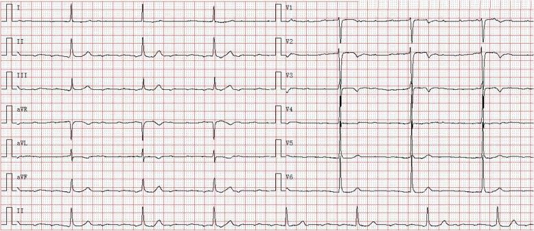
A 12-lead baseline electrocardiogram (ECG) recorded at 25 mm/s and 10 mm/mV, demonstrating complex cardiac conduction abnormalities. The tracing shows a rapid, irregular atrial arrhythmia, likely atrial fibrillation or high-frequency atrial tachycardia, with an atrial rate of approximately 180 beats per minute. Distinct P waves are absent, replaced by fibrillatory waves visible in the baseline, particularly in lead V1. Concurrent with this is a complete (third-degree) atrioventricular (AV) block, evidenced by the total dissociation between atrial activity and ventricular response. The ventricles are driven by a junctional escape rhythm, resulting in a slow, regular ventricular rate of approximately 43 beats per minute. The QRS complexes are relatively narrow (approximately 90 ms), consistent with a supra-Hisian escape focus. The tracing includes limb leads (I, II, III, aVR, aVL, aVF) and precordial leads (V1-V6), with a continuous lead II rhythm strip at the bottom. This visual record is characteristic of advanced conduction system disease, often associated with LMNA-related cardiomyopathies or other structural heart diseases.
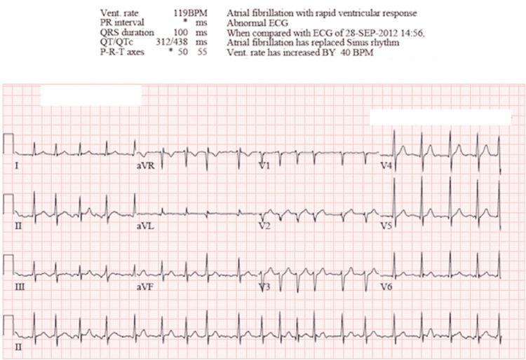
A standard 12-lead diagnostic electrocardiogram (ECG) demonstrating atrial fibrillation with rapid ventricular response (RVR). The tracing shows a classic irregularly irregular rhythm with narrow QRS complexes (100 ms duration). There is a total absence of discernible P waves, which are replaced by baseline fibrillatory oscillations, most visible in lead V1. The ventricular rate is approximately 119 beats per minute, indicating tachycardia. The ECG includes the limb leads (I, II, III, aVR, aVL, aVF) and precordial leads (V1-V6), with a continuous rhythm strip of lead II at the bottom. Automated measurements provided at the top of the tracing confirm a QTc of 438 ms and a heart rate increase compared to prior records. This diagnostic image serves as a clinical example of supraventricular tachycardia originating from disorganized atrial electrical activity, necessitating evaluation for hemodynamic stability and rate/rhythm control.
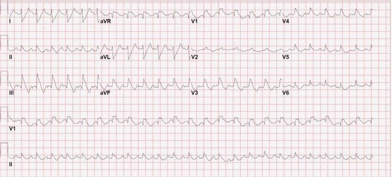
A 12-lead electrocardiogram (ECG) demonstrating an irregularly irregular rhythm with a rapid ventricular response, characteristic of atrial fibrillation. Notable features include the absence of distinct P waves and varying R-R intervals. The ventricular rate is tachycardia, estimated at over 150 bpm. A widening of the QRS complex is observed, exceeding 120 ms. In the precordial leads V1-V3, there is a prominent RSR' pattern, where the terminal R wave is taller than the initial R wave, coupled with secondary ST-segment depression and T-wave inversion. Leads V5 and V6 exhibit characteristic slurred S waves. These morphological findings are diagnostic of a right bundle branch block (RBBB). This diagnostic image is clinically significant for identifying acute right ventricular strain or conduction disturbances in the context of emergency presentations such as syncope or suspected pulmonary embolism. It is suitable for medical students and clinicians studying cardiovascular diagnostics and acute care.
antiarrhythmic drug Vaughan Williams classification table
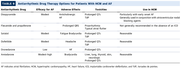
TABLE 9 Antiarrhythmic Drug Therapy Options for Patients With HCM and AF <table><thead><tr><th>Antiarrhythmic Drug</th><th>Efficacy for AF</th><th>Adverse Effects</th><th>Toxicities</th><th>Use in HCM</th></tr></thead><tbody><tr><td>Disopyramide</td><td>Modest</td><td>Anticholinergic<br>HF</td><td>Prolonged QTc<br>TdP</td><td>Particularly with early onset AF<br>Generally used in conjunction with atrioventricular nodal blocking agents</td></tr><tr><td>Flecainide and propafenone</td><td>...</td><td>Prolonged QRS</td><td>Proarrhythmia<br>Typical atrial flutter</td><td>Not generally recommended in the absence of an ICD</td></tr><tr><td>Sotalol</td><td>Modest</td><td>Fatigue Bradycardia</td><td>Prolonged QTc<br>TdP</td><td>Reasonable</td></tr><tr><td>Dofetilide</td><td>Modest</td><td>Headache</td><td>Prolonged QTc<br>TdP</td><td>Reasonable</td></tr><tr><td>Dronedarone</td><td>Low</td><td>HF</td><td>Prolonged QTc</td><td>...</td></tr><tr><td>Amiodarone</td><td>Modest-high</td><td>Bradycardia</td><td>Liver, lung, thyroid, skin, neurologic<br>Prolonged QTc</td><td>Reasonable</td></tr></tbody></table> AF indicates atrial fibrillation; HCM, hypertrophic cardiomyopathy; HF, heart failure; ICD, implantable cardioverter-defibrillator; and TdP, torsades de pointes.
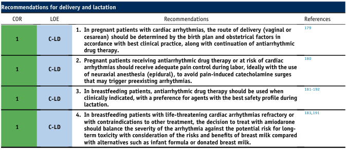
Recommendations for delivery and lactation <table><thead><tr><th>COR</th><th>LOE</th><th>Recommendations</th><th>References</th></tr></thead><tbody><tr><td>1</td><td>C-LD</td><td>1. In pregnant patients with cardiac arrhythmias, the route of delivery (vaginal or cesarean) should be determined by the birth plan and obstetrical factors in accordance with best clinical practice, along with continuation of antiarrhythmic drug therapy.</td><td>179</td></tr><tr><td>1</td><td>C-LD</td><td>2. Pregnant patients receiving antiarrhythmic drug therapy or at risk of cardiac arrhythmias should receive adequate pain control during labor, ideally with the use of neuraxial anesthesia (epidural), to avoid pain-induced catecholamine surges that may trigger preexisting arrhythmias.</td><td>180</td></tr><tr><td>1</td><td>C-LD</td><td>3. In breastfeeding patients, antiarrhythmic drug therapy should be used when clinically indicated, with a preference for agents with the best safety profile during lactation.</td><td>181-192</td></tr><tr><td>1</td><td>C-LD</td><td>4. In breastfeeding patients with life-threatening cardiac arrhythmias refractory or with contraindications to other treatment, the decision to treat with amiodarone should balance the severity of the arrhythmia against the potential risk for long-term toxicity with consideration of the risks and benefits of breast milk compared with alternatives such as infant formula or donated breast milk.</td><td>183,191</td></tr></tbody></table>
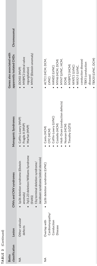
TABLE 3 (Continued) <table><thead><tr><th>Botto classification</th><th>Lesion</th><th>CNVs and CNV syndromes</th><th>Monogenic Syndromes</th><th>Genes also associated with apparently isolated CHDs</th><th>Chromosomal</th></tr></thead><tbody><tr><td>NA</td><td>Other vascular defects</td><td>• 1p36 deletion syndrome (Ebstein anomaly)<br>• 7q11.23 deletion/Williams Syndrome (SVAS)<br>• 11q terminal deletion syndrome/ Jacobsen syndrome (mitral stenosis)<br>• 1p36 deletion syndrome (LVNC)</td><td>• Coffin-Lowry (MVP)<br>• Fragile X (MVP)<br>• Marfan (MVP)</td><td>• DCHS1 (MVP)<br>• MYBPC3 (mitral valve regurgitation)<br>• MYH7 (Ebstein anomaly)</td><td rowspan="2"></td></tr><tr><td>NA</td><td>Overlap with Cardiomyopathy/ Conduction Disease</td><td></td><td>• Cantu (HCM)<br>• CFC (HCM)<br>• Coffin-Lowry (LVNC)<br>• Costello (HCM)<br>• Holt-Oram (conduction defects)<br>• Noonan (HCM)<br>• Timothy (LQTS)</td><td>• ACTC1 (HCM, DCM, LVNC)<br>• HAND2 (LVNC)<br>• MYH6 (HCM, DCM)<br>• MYH7 (LVNC, HCM)<br>• DCM)<br>• MYBPC3 (HCM)<br>• NFATC1 (LVNC)<br>• NKX2-5 (LVNC, conduction disease)<br>• PRDM16 (cardiomyopathy, conduction defects)<br>• TBX5 (disease)<br>• TBX20 (LVNC, DCM)</td></tr></tbody></table>
Classification of Cardiac Arrhythmias and Their Management
1. INTRODUCTION
2. CLASSIFICATION OF CARDIAC ARRHYTHMIAS
A. By Rate
| Category | Heart Rate |
|---|---|
| Bradyarrhythmias | < 60 beats/min |
| Tachyarrhythmias | > 100 beats/min |
| Normocardic arrhythmias | Normal rate but abnormal rhythm |
B. By Site of Origin
I. SUPRAVENTRICULAR ARRHYTHMIAS
- Sinus bradycardia: Rate < 60/min. Occurs with excess vagal tone, drugs (beta-blockers, calcium channel blockers, digoxin, amiodarone), hypothyroidism, hypothermia, inferior MI (10-15% of cases). Benign in athletes.
- Sinus tachycardia: Rate 100-180/min. Physiologic response to fever, pain, hypovolemia, hyperthyroidism, anxiety.
- Sinus arrhythmia: Phasic variation in sinus cycle length >120 ms; respiratory type is normal in the young.
- Sick sinus syndrome (SSS): Inappropriate bradycardia, sinus pauses, SA exit block, or alternating bradycardia-tachycardia (brady-tachy syndrome). Results from fibrosis/infiltration of the SA node.
- Sinoatrial (SA) exit block: Sinus impulse forms but fails to exit the node; recognized by pauses on ECG.
- Premature atrial complexes (PACs): Early P waves of different morphology; usually benign.
- Atrial tachycardia (AT): Sustained ectopic atrial rate 100-250/min; often due to triggered activity or micro-re-entry.
- Atrial flutter: Regular atrial rate ~300/min with saw-tooth flutter waves (best in inferior leads); typically 2:1 or 4:1 AV conduction; macro-re-entry in the right atrium (cavotricuspid isthmus).
- Atrial fibrillation (AF): Chaotic atrial activity >350/min; no discrete P waves; irregularly irregular ventricular response. Most common sustained arrhythmia. Classified as paroxysmal (<7 days), persistent (>7 days), long-standing persistent (>1 year), or permanent.
- Multifocal atrial tachycardia (MAT): >3 P-wave morphologies, irregular rhythm; associated with COPD and hypokalemia.
- AV nodal re-entrant tachycardia (AVNRT): Most common paroxysmal SVT; dual AV nodal pathways; retrograde P waves buried in or just after QRS; rate 150-250/min.
- AV re-entrant tachycardia (AVRT): Involves an accessory pathway (e.g., WPW syndrome). Orthodromic (narrow QRS, retrograde P after QRS) or antidromic (wide QRS).
- Junctional rhythm: Escape rhythm at 40-60/min when SA node fails.
II. VENTRICULAR ARRHYTHMIAS
- Premature ventricular complexes (PVCs): Wide QRS (>120 ms), no preceding P wave; compensatory pause. Frequent PVCs (>10,000/day or >10% of beats) can cause PVC-induced cardiomyopathy.
- Ventricular tachycardia (VT):
- Non-sustained VT (NSVT): ≥3 consecutive ventricular beats at >100/min, lasting <30 seconds.
- Sustained VT: Duration ≥30 seconds or requiring termination due to hemodynamic compromise.
- Monomorphic VT: Identical QRS morphology; usually re-entrant; associated with ischemic scar.
- Polymorphic VT: Varying QRS morphology; seen in ischemia, long QT, and Brugada syndrome.
- Torsades de pointes (TdP): A form of polymorphic VT occurring in the setting of QT prolongation; characteristic "twisting of points" around the isoelectric axis.
- Bidirectional VT: Beat-to-beat QRS axis alternation; associated with digoxin toxicity and catecholaminergic polymorphic VT (CPVT).
- Ventricular flutter: Sinusoidal pattern at ~300/min; hemodynamically unstable.
- Ventricular fibrillation (VF): Disorganized chaotic ventricular activity, no effective cardiac output; leads to cardiac arrest within seconds.
- Accelerated idioventricular rhythm (AIVR): 40-100/min; occurs in acute MI reperfusion ("reperfusion arrhythmia"); benign.
- Idiopathic VT: VT in structurally normal hearts; e.g., right ventricular outflow tract (RVOT) VT (LBBB, inferior axis), left ventricular fascicular VT (verapamil-sensitive).
III. CONDUCTION DISTURBANCES (Heart Block)
| Degree | ECG Features | Site of Block |
|---|---|---|
| First degree | PR > 200 ms; all P waves conducted | AV node (usually) |
| Second degree - Mobitz I (Wenckebach) | Progressive PR lengthening until P wave not conducted; grouped beating | AV node |
| Second degree - Mobitz II | Sudden non-conduction of P wave without prior PR change; fixed PR in conducted beats | Below AV node (His-Purkinje) |
| 2:1 AV block | Alternate P waves not conducted; cannot be classified as Mobitz I or II by ECG alone | Variable |
| High-grade AV block | Two or more consecutive non-conducted P waves | AV node or His-Purkinje |
| Third degree (complete) AV block | Complete AV dissociation; independent atrial and ventricular rates; ventricular rate <40/min in acquired | AV node, His bundle, or infranodal |
- RBBB: RSR' in V1, wide S in V5/V6; can be normal variant
- LBBB: Broad notched R in I, aVL, V5/V6; always pathological
- Bifascicular block (RBBB + left anterior or posterior fascicular block) and trifascicular block carry risk of progression to complete AV block.
C. By Mechanism of Arrhythmogenesis
- Enhanced/abnormal automaticity: Accelerated spontaneous phase 4 depolarization in ectopic foci. Examples: sinus tachycardia, AIVR, junctional tachycardia.
- Triggered activity:
- Early afterdepolarizations (EADs): During phase 2/3; cause TdP and polymorphic VT in long QT syndrome.
- Delayed afterdepolarizations (DADs): During phase 4; cause digitalis-induced arrhythmias, CPVT.
- Re-entry: Most common mechanism. Requires two pathways with unidirectional block and differential conduction velocity. Examples: AF, atrial flutter, AVNRT, AVRT, ischemic VT.
3. MANAGEMENT OF CARDIAC ARRHYTHMIAS
A. Antiarrhythmic Drugs - Vaughan-Williams Classification
| Class | Mechanism | Drugs | Key Adverse Effects |
|---|---|---|---|
| IA | Na+ channel block (intermediate kinetics); prolongs action potential | Quinidine, Procainamide, Disopyramide | QT prolongation, TdP, lupus-like syndrome (procainamide), cinchonism (quinidine), anticholinergic effects |
| IB | Na+ channel block (fast kinetics); shortens action potential | Lidocaine, Mexiletine, Phenytoin | CNS toxicity (tremor, seizures, confusion); effective only at fast rates |
| IC | Na+ channel block (slow kinetics); markedly slows conduction; minimal effect on repolarization | Flecainide, Propafenone | Proarrhythmic in structural heart disease (CAST trial); avoid in post-MI patients |
| II | Beta-adrenoceptor blockade; reduces automaticity, slows AV nodal conduction | Metoprolol, Atenolol, Esmolol, Carvedilol | Bradycardia, bronchospasm, fatigue, hypotension |
| III | K+ channel block; prolongs repolarization and refractory period | Amiodarone, Sotalol, Dofetilide, Dronedarone, Ibutilide | QT prolongation, TdP; amiodarone: pulmonary toxicity, thyroid dysfunction, hepatotoxicity, corneal microdeposits, photosensitivity |
| IV | Ca2+ channel block (L-type); slows AV node conduction | Verapamil, Diltiazem | Negative inotropy, bradycardia, hypotension; contraindicated in pre-excitation + AF (WPW) |
| Other | Adenosine: activates K+ channels in AV node; terminates AV-nodal-dependent SVT | Adenosine (6-12 mg IV bolus) | Transient asystole, flushing, bronchospasm |
| Other | Digoxin: vagotonic effect; slows AV node | Digoxin | Narrow therapeutic index; toxicity: nausea, bidirectional VT, AV block |
Note: The Sicilian Gambit is a more nuanced classification based on specific ion channel/receptor effects and vulnerability of arrhythmia mechanisms, but Vaughan-Williams remains the clinical standard.
B. Management by Arrhythmia Type
1. Sinus Bradycardia
- No treatment unless hemodynamically compromised or symptomatic.
- Acute: Atropine 0.5 mg IV (repeat as needed up to 3 mg).
- Remove offending drugs (beta-blockers, digoxin, calcium channel blockers).
- Persistent symptomatic: Permanent pacemaker implantation.
2. Sick Sinus Syndrome
- Permanent pacemaker (typically dual-chamber, DDD) when symptomatic.
- Brady-tachy syndrome: Pacemaker for bradycardia + antiarrhythmic drug (e.g., beta-blocker) for tachycardia component.
3. Atrial Flutter
- Rate control: Beta-blockers, calcium channel blockers, digoxin.
- Rhythm control: DC cardioversion (50-100 J biphasic); ibutilide IV for chemical cardioversion.
- Radiofrequency catheter ablation of the cavotricuspid isthmus: highly effective (>95% success), curative for typical flutter.
- Anticoagulation as per CHA2DS2-VASc score (same principles as AF).
4. Atrial Fibrillation
- First-line: beta-blockers or non-dihydropyridine calcium channel blockers (verapamil/diltiazem).
- Refractory: digoxin (especially in heart failure), amiodarone; AV node ablation + pacemaker as last resort.
- DC cardioversion (200 J biphasic synchronized).
- Antiarrhythmic drugs: Class IC (flecainide, propafenone - only in structurally normal hearts); Class III (sotalol, dofetilide, dronedarone, amiodarone - broadest spectrum).
- Catheter ablation: pulmonary vein isolation (PVI) is highly effective for paroxysmal AF; increasingly used for persistent AF.
- CHA2DS2-VASc score guides therapy (C=CHF, H=HTN, A2=Age≥75, D=DM, S2=Stroke/TIA, V=Vascular disease, A=Age 65-74, Sc=Sex category female).
- Score ≥2 (men), ≥3 (women): DOAC preferred (apixaban, rivaroxaban, dabigatran, edoxaban) over warfarin.
- Score 1 (men), 2 (women): anticoagulation should be considered.
- Score 0: no anticoagulation.
- AF-flutter cardioversion: If duration >48h or unknown - anticoagulate for ≥3 weeks before and ≥4 weeks after cardioversion, OR perform TOE-guided early cardioversion.
5. AVNRT / AVRT (SVT)
- Acute termination:
- Vagal maneuvers first (Valsalva, carotid sinus massage).
- Adenosine 6 mg IV rapid bolus (12 mg if ineffective); drug of choice.
- If adenosine fails: IV verapamil or diltiazem.
- Hemodynamically unstable: synchronized DC cardioversion.
- Long-term prevention:
- Beta-blockers or calcium channel blockers.
- Radiofrequency catheter ablation is curative (>95% success for AVNRT; approaches 100% for accessory pathways in WPW).
- WPW with pre-excited AF: AVOID digoxin, verapamil, and adenosine (can accelerate ventricular response via accessory pathway and precipitate VF). Use procainamide IV or DC cardioversion.
6. Ventricular Premature Complexes (PVCs)
- In structurally normal hearts: reassurance; treat only if highly symptomatic (beta-blockers, mexiletine, or flecainide).
- Frequent PVCs causing PVC-induced cardiomyopathy: catheter ablation is highly effective.
- In structural heart disease: treat the underlying cause; avoid Class IC drugs.
7. Ventricular Tachycardia
- Hemodynamically unstable VT: Immediate unsynchronized DC cardioversion (defibrillation).
- Hemodynamically stable VT:
- IV procainamide (10 mg/kg over 20 min) - superior to amiodarone for termination of wide-complex tachycardia.
- IV amiodarone (150 mg over 10 min): better for preventing recurrence.
- IV lidocaine (50 mg bolus): effective during acute ischemia.
- Avoid IV calcium channel blockers unless idiopathic left VT is confirmed.
- Electrical storm (recurrent VT/VF): Treat reversible causes (ischemia, electrolyte imbalance); IV beta-blockers (esmolol); IV amiodarone or lidocaine infusion; intra-aortic balloon pump; catheter ablation for refractory cases; bilateral cardiac sympathetic denervation.
- Long-term prevention:
- ICD (implantable cardioverter-defibrillator): first-line for secondary prevention; primary prevention in EF <35% despite optimal medical therapy.
- Amiodarone as adjunct to ICD or when ICD is declined/contraindicated.
- Catheter ablation for monomorphic scar-related VT.
- Idiopathic RVOT VT: catheter ablation or beta-blockers/CCBs.
8. Ventricular Fibrillation
- Immediate unsynchronized DC defibrillation (200 J biphasic) following ACLS protocol.
- CPR until defibrillator is ready; minimize interruptions in compressions.
- IV adrenaline (epinephrine) 1 mg every 3-5 min.
- IV amiodarone (300 mg) or lidocaine for shock-refractory VF.
- Identify and reverse reversible causes (4H's and 4T's).
- ICD implantation for survivors of VF (secondary prevention).
9. AV Block
- First-degree AV block: No treatment; correct precipitating cause if drug-induced.
- Second-degree Mobitz I (Wenckebach): Usually benign; treat if symptomatic or related to inferior MI (atropine/pacing if hemodynamically unstable).
- Second-degree Mobitz II: Higher risk of progression to complete heart block; pacemaker implantation recommended even if asymptomatic.
- Complete (third-degree) AV block:
- Acute: IV atropine (usually ineffective for infranodal block); temporary transcutaneous or transvenous pacing as bridge.
- Definitive: Permanent pacemaker (dual-chamber DDD preferred; VVI for chronic AF).
- Exception: Lyme disease-related complete heart block may resolve with antibiotics.
C. Non-Pharmacological / Interventional Therapies
| Modality | Indications |
|---|---|
| Synchronized DC cardioversion | Unstable SVT, AF, atrial flutter, hemodynamically stable VT |
| Defibrillation (unsynchronized) | VF, pulseless VT |
| Radiofrequency catheter ablation | AVNRT, AVRT (WPW), atrial flutter, AF (PVI), idiopathic VT, ischemic VT |
| Permanent pacemaker | Symptomatic bradyarrhythmias, SSS, AV block (Mobitz II, complete AV block) |
| ICD | Secondary prevention of SCD; primary prevention in high-risk (EF <35%) |
| CRT (cardiac resynchronization therapy) | Heart failure with LBBB and EF <35% |
| Surgical ablation (Maze procedure) | AF at time of other cardiac surgery |
4. SPECIAL CONSIDERATIONS
- Proarrhythmic risk: All antiarrhythmic drugs can cause arrhythmias. Class IA and III agents prolong QT and risk TdP. Class IC agents are proarrhythmic in structural heart disease (CAST trial showed increased mortality with flecainide/encainide post-MI).
- Electrolyte disturbances: Hypokalemia and hypomagnesemia potentiate QT prolongation and TdP; must be corrected before and during antiarrhythmic therapy.
- Reversible causes of VF/VT (4H4T): Hypoxia, Hypovolemia, Hypo/Hyperkalemia, Hypothermia; Tension pneumothorax, Tamponade, Toxins, Thromboembolism.
SUMMARY TABLE
| Arrhythmia | First-line Acute Treatment | Long-term/Definitive Treatment |
|---|---|---|
| Sinus bradycardia (symptomatic) | Atropine 0.5 mg IV | Permanent pacemaker |
| SVT (AVNRT/AVRT) | Adenosine IV | Catheter ablation |
| Atrial flutter | Cardioversion / rate control | Cavotricuspid isthmus ablation |
| AF (unstable) | DC cardioversion | DOAC + rate/rhythm control |
| Stable VT | Procainamide/Amiodarone IV | ICD + catheter ablation |
| Unstable VT | DC cardioversion | ICD |
| VF | Defibrillation + CPR | ICD |
| Complete AV block | Temporary pacing | Permanent pacemaker |
| Mobitz II | Pacemaker (even asymptomatic) | Permanent pacemaker |
Make proper flow digram of classification part
A detailed medical flow diagram showing the classification of cardiac arrhythmias. The diagram starts at the top with "CARDIAC ARRHYTHMIAS" as the root node, then branches into three main categories: "BRADYARRHYTHMIAS (HR <60 bpm)", "TACHYARRHYTHMIAS (HR >100 bpm)", and "CONDUCTION DISTURBANCES". Under TACHYARRHYTHMIAS, branch into two groups: "SUPRAVENTRICULAR" and "VENTRICULAR". Under SUPRAVENTRICULAR, show sub-branches: "Sinus Node" (listing Sinus Tachycardia, Sick Sinus Syndrome), "Atrial" (listing PACs, Atrial Tachycardia, Atrial Flutter, Atrial Fibrillation, MAT), and "AV Nodal/Junctional" (listing AVNRT, AVRT/WPW, Junctional Tachycardia). Under VENTRICULAR, show sub-branches: "PVCs", "Ventricular Tachycardia" (with sub-nodes: Monomorphic VT, Polymorphic VT, Torsades de Pointes, Sustained vs Non-sustained), "Ventricular Flutter", "Ventricular Fibrillation", "AIVR". Under BRADYARRHYTHMIAS, show: "Sinus Bradycardia", "Sinus Arrest/Pause", "SA Exit Block", "Sick Sinus Syndrome". Under CONDUCTION DISTURBANCES, show "AV Block" with sub-branches: "1st Degree (PR>200ms)", "2nd Degree" (further splitting into "Mobitz I - Wenckebach" and "Mobitz II"), "High-Grade AV Block", "3rd Degree - Complete AV Block". Also show "Bundle Branch Block" with RBBB and LBBB. Also show a separate branch for "BY MECHANISM" with three nodes: "Abnormal Automaticity", "Triggered Activity (EADs/DADs)", "Re-entry". Use a clean white background, color-coded boxes (blue for main categories, green for supraventricular, red/orange for ventricular, purple for conduction, yellow for mechanism), clear connecting arrows, and professional medical typography. Make it comprehensive and easy to read.
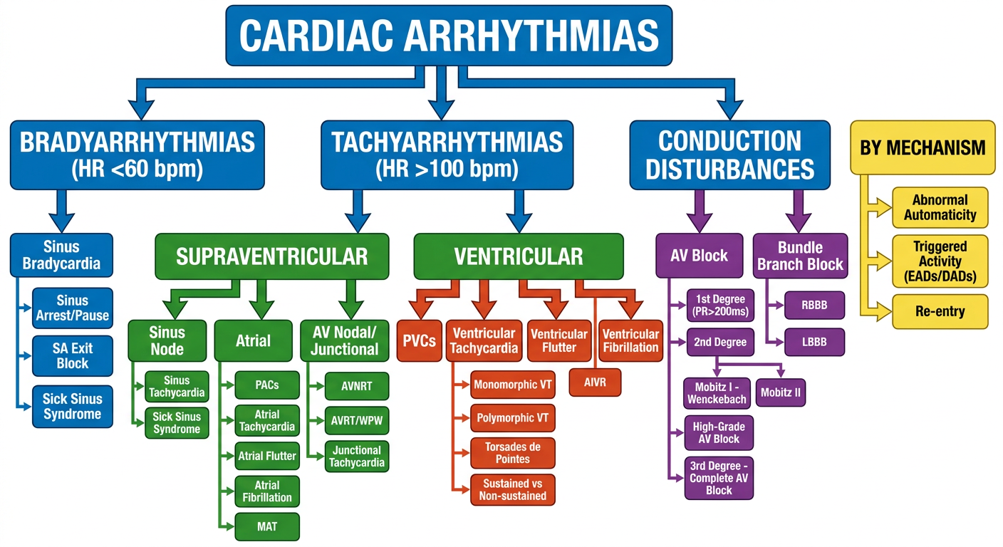
- Blue (top) - Root node: Cardiac Arrhythmias
- Green - Supraventricular arrhythmias (Sinus node, Atrial, AV Nodal/Junctional types)
- Red/Orange - Ventricular arrhythmias (PVCs, VT subtypes, VF, AIVR)
- Purple - Conduction disturbances (AV blocks 1st/2nd/3rd degree, BBB)
- Teal/Blue - Bradyarrhythmias (Sinus bradycardia, SSS, SA exit block)
- Yellow - Mechanism-based classification (Re-entry, Automaticity, Triggered activity with EADs/DADs)