Muscles of the oral diaphragm
Reading File
Finding Sources
Finding Sources
Finding Sources
Finding Sources
Finding Sources
Finding Sources
Reading File
Reading File
Reading File
Reading File
Loading Image
Loading Image
Loading Image
Loading Image
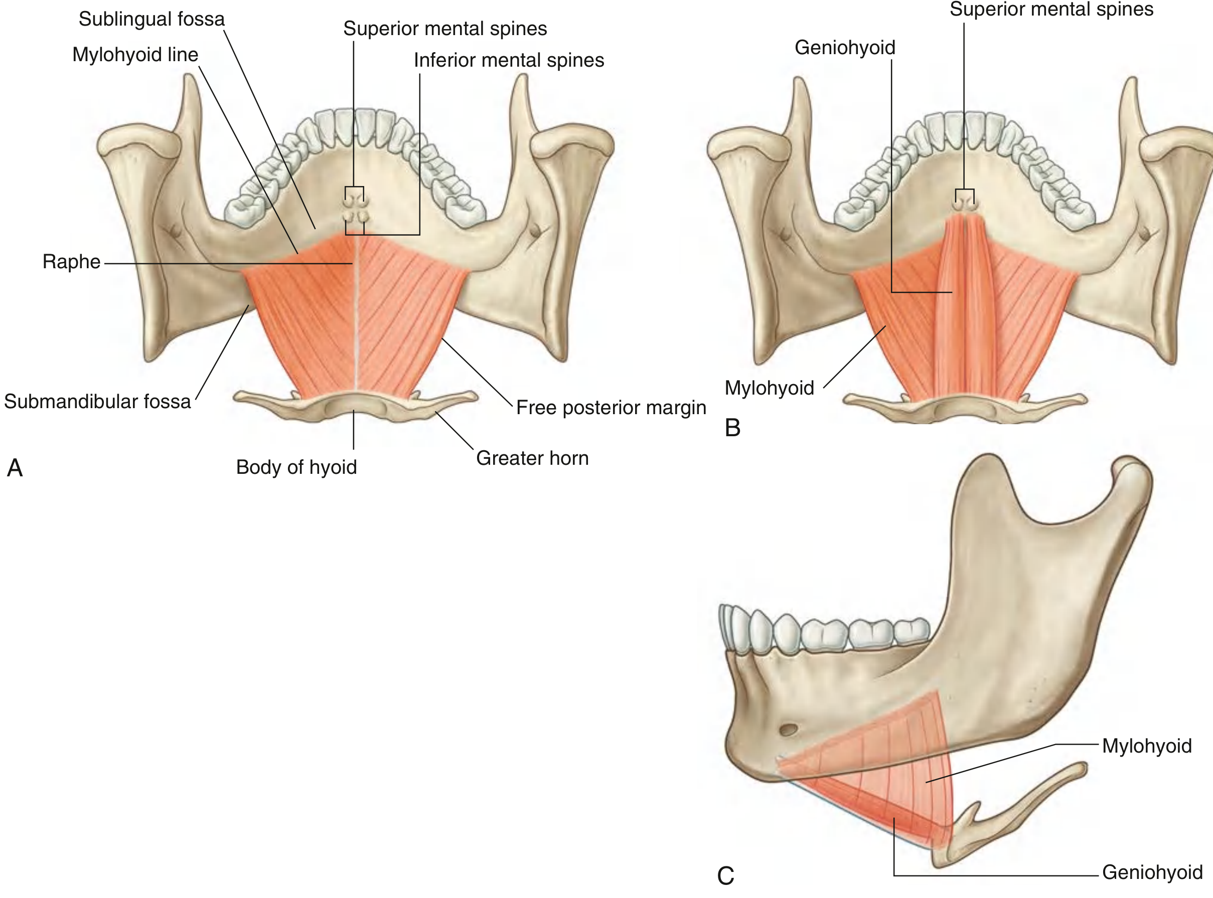
The oral diaphragm (also called the diaphragma oris) is the muscular floor of the oral cavity formed by two principal muscles: the mylohyoid and the geniohyoid. Additional muscles associated with the floor (the suprahyoid group) are described below.
Primary Muscles of the Oral Diaphragm
1. Mylohyoid (the true "diaphragm")
The paired mylohyoid muscles are the structural foundation of the oral diaphragm. Each is a flat, triangular muscle that together form a muscular sheet across the floor of the mouth — the diaphragma oris.
| Feature | Detail |
|---|---|
| Origin | Mylohyoid line on the medial surface of the body of the mandible |
| Insertion | Median fibrous raphe (midline) running from mandibular symphysis to hyoid bone; posterior fibers attach directly to the body of the hyoid |
| Innervation | Nerve to mylohyoid — a branch of the inferior alveolar nerve (from mandibular nerve, V₃) |
| Actions | (1) Supports and elevates floor of oral cavity; (2) elevates and pulls hyoid forward during swallowing; (3) depresses mandible when hyoid is fixed |
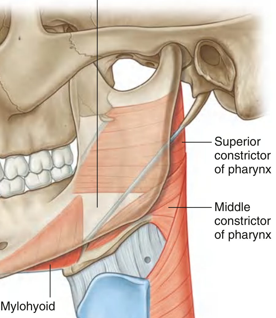
The posterior margin of each mylohyoid is free, and this free border — together with the superior and middle pharyngeal constrictors — forms the triangular oropharyngeal aperture, the major gateway through which vessels, nerves, and muscles (e.g., lingual artery, hypoglossal nerve, hyoglossus) pass between the floor of the mouth and the neck.
2. Geniohyoid
The geniohyoid muscles lie immediately superior to the mylohyoid (on its upper surface), running as paired cord-like muscles on either side of the midline.
| Feature | Detail |
|---|---|
| Origin | Inferior mental spines (genial tubercles) on the posterior surface of the mandibular symphysis |
| Insertion | Anterior surface of the body of the hyoid bone |
| Innervation | C1 fibers that hitchhike along the hypoglossal nerve (CN XII) |
| Actions | (1) Pulls hyoid forward and upward during swallowing; (2) depresses mandible when hyoid is fixed |
Associated Suprahyoid Muscles
All muscles above the hyoid bone contribute to the functional oral diaphragm complex. The complete suprahyoid group includes:
| Muscle | Origin | Insertion | Nerve | Action |
|---|---|---|---|---|
| Mylohyoid | Mylohyoid line of mandible | Median raphe + hyoid | Nerve to mylohyoid (V₃) | Forms floor; elevates hyoid; depresses mandible |
| Geniohyoid | Inferior mental spines of mandible | Body of hyoid | C1 via CN XII | Pulls hyoid forward; depresses mandible |
| Digastric (anterior belly) | Digastric fossa of mandible | Intermediate tendon at hyoid | Nerve to mylohyoid (V₃) | Depresses mandible; elevates hyoid |
| Digastric (posterior belly) | Mastoid notch of temporal bone | Intermediate tendon at hyoid | Facial nerve (CN VII) | Elevates and retracts hyoid |
| Stylohyoid | Styloid process | Body/greater horn of hyoid | Facial nerve (CN VII) | Elevates and retracts hyoid |
| Mylohyoid (as above) | — | — | — | — |
Key Functional Points
- The mylohyoid is the structural diaphragm; it divides the floor into a sublingual space above and a submental/submandibular space below.
- During swallowing, the mylohyoid and geniohyoid elevate and advance the hyoid, raising the larynx and initiating the pharyngeal phase.
- The submandibular gland is "hooked" around the free posterior margin of the mylohyoid — its deep part lies above it (sublingual space) and its superficial part below (submandibular space).
- Ludwig's angina (oral cellulitis) is a life-threatening infection that spreads through these spaces, exploiting the anatomical boundaries defined by the mylohyoid diaphragm.
Sources: Gray's Anatomy for Students, p. 1232–1234; Color Atlas of Human Anatomy Vol. 2, p. 258–259
This is a shared conversation. Sign in to Orris to start your own chat.