


Everything about it treatment danger
gallstone cholelithiasis ultrasound diagram
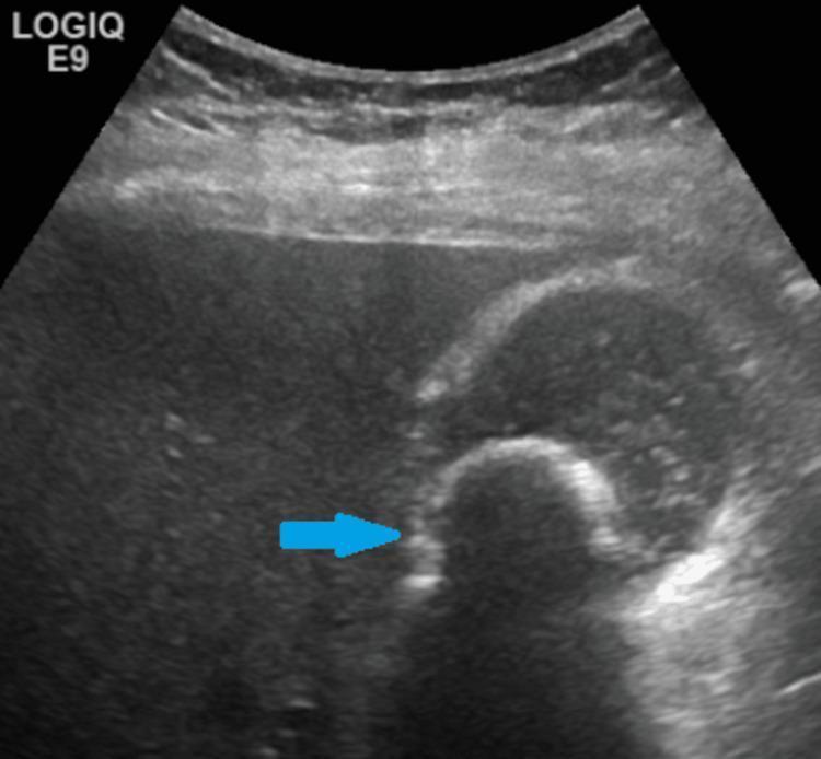
This abdominal ultrasound image displays a diagnostic view of the gallbladder containing a prominent 2.7-cm gallstone (cholelithiasis), indicated by a blue arrow. The gallstone appears as a curvilinear, highly hyperechoic structure within the gallbladder lumen. Characteristically, there is a clear, well-defined area of posterior acoustic shadowing distal to the calculus, resulting from the attenuation of ultrasound waves by the dense calcified object. The gallbladder itself appears as a sac-like structure with primarily anechoic (fluid-filled) contents, although scattered low-level internal echoes are present, suggesting the presence of biliary sludge. The surrounding liver parenchyma shows a uniform echotexture. This image is a classic representation used in radiology and gastroenterology for teaching the sonographic diagnosis of uncomplicated cholelithiasis, emphasizing the 'bright' appearance of stones and the diagnostic importance of acoustic shadowing.
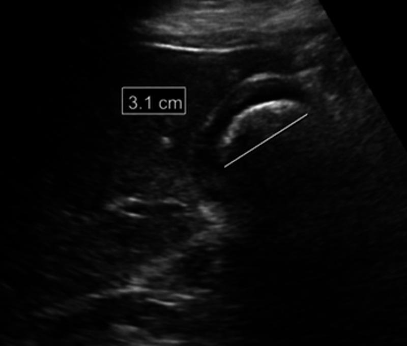
A diagnostic ultrasound image of the gallbladder in the right upper quadrant demonstrating cholelithiasis. The primary finding is a large, hyperechoic (bright) structure with a curvilinear surface, identified as a gallstone, which occupies a significant portion of the gallbladder lumen. The stone measures approximately 3.1 cm, as indicated by the calipers and on-screen measurement label. A prominent diagnostic artifact known as posterior acoustic shadowing (a clean, dark anechoic region beneath the stone) is visible, resulting from the near-complete reflection and absorption of ultrasound waves by the dense calculus. The surrounding liver parenchyma appears homogeneous and relatively hypoechoic compared to the stone's surface. This imaging is characteristic of cholelithiasis and is used to assess for complications such as cholecystitis or biliary obstruction.
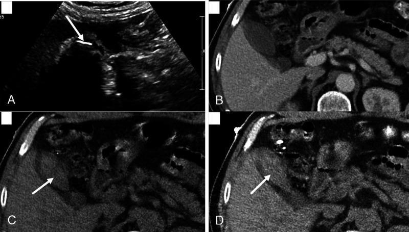
This four-panel image provides a comparison of cholelithiasis (gallstones) across different imaging modalities. Panel A: Abdominal ultrasound displays a gallbladder containing a large, hyperechoic structure (arrow) with a prominent posterior acoustic shadow, diagnostic of a gallstone. Panel B: A standard Portal-Venous Phase (PC) CT scan shows the gallbladder lumen with fluid density, but the stone is iso-attenuating and thus invisible. Panel C: A Virtual Non-Contrast (VNC) reconstruction from Dual-Energy CT (DECT) successfully delineates the stone (arrow), which appears hyperdense relative to the surrounding bile. Panel D: A 50-keV Virtual Monoenergetic Image (VMC) reconstruction further highlights the gallstone (arrow) by enhancing the contrast between the cholesterol-rich stone and the gallbladder fluid. The composite illustrates how specialized DECT reconstructions (VNC and low-keV VMC) can overcome the limitations of conventional CT in detecting radiolucent or iso-attenuating gallstones, approaching the sensitivity of ultrasound for biliary pathology.
Miss Yogita's Ultrasound Report - Cholelithiasis (Gallstone)
What the Report Found
Why the Location Matters (Neck Stone)
- Every time the gallbladder contracts (especially after eating fatty/heavy food), the stone can temporarily block the outflow of bile, causing pain
- A stone here carries a higher risk of causing acute cholecystitis (infection/inflammation) than one sitting quietly in the gallbladder body
Symptoms to Watch For
- Biliary colic: Severe, dull, continuous right upper quadrant or epigastric pain, often radiating to the back/shoulder. Typically starts at night, lasts minutes to hours, with nausea and vomiting
- Worsening after fatty meals: Fried food, ghee, butter, rich curries
- Fever + pain: This combination means the gallstone has caused acute infection (cholecystitis) - this is an emergency
Dangers / Complications
| Complication | What Happens | Severity |
|---|---|---|
| Biliary Colic | Stone blocks neck temporarily, severe pain | Common, not life-threatening |
| Acute Cholecystitis | Sustained blockage causes gallbladder infection and inflammation | Serious - requires urgent treatment |
| Empyema of gallbladder | Gallbladder fills with pus | Very serious |
| Gallbladder Perforation | Infected wall ruptures, causing peritonitis | Emergency |
| Choledocholithiasis | Stone slips into the common bile duct (CBD), blocking it | Causes jaundice, severe pain |
| Acute Cholangitis | CBD stone causes bile duct infection - fever, jaundice, pain (Charcot's triad) | Life-threatening if untreated |
| Gallstone Pancreatitis | Stone blocks the pancreatic duct, causing severe pancreatitis | Potentially life-threatening |
| Gallstone Ileus | Rare - stone erodes through gallbladder wall into intestine, causes bowel obstruction | Surgical emergency |
| Mucocele of gallbladder | Blocked neck fills with mucus, distended gallbladder | Requires surgery |
| Gallbladder Cancer | Long-standing gallstones, especially large ones, are a risk factor | Rare but serious |
Treatment
- This is the gold standard treatment worldwide - Maingot's Abdominal Operations, Schwartz's Principles
- Small incisions (3-4 tiny holes), camera-guided surgery
- Short hospital stay (1-2 days), quick recovery (1-2 weeks)
- Removes the whole gallbladder - prevents ALL future stone-related problems
- Converting to open surgery (larger incision) may be needed if complications arise
- Symptomatic stones (pain, vomiting, or complications): Surgery is recommended
- Asymptomatic stones (no symptoms): The standard approach is watchful waiting with dietary precautions, since only ~20% develop symptoms over 20 years - Current Surgical Therapy 14e
- Stone in the neck (like in this report) has a higher risk of causing blockage and symptoms, so most surgeons will recommend surgery sooner rather than later
- Oral dissolution therapy: Ursodeoxycholic acid (UDCA/ursodiol) can dissolve small cholesterol stones over months - but stones recur when stopped
- Extracorporeal shockwave lithotripsy (ESWL): Sound waves break stones - only for small solitary stones, rarely used now
What the Radiologist Recommends Next
- CECT abdomen-pelvis - a CT scan with contrast to better evaluate
- S. Amylase & Lipase - to check if the stone is affecting the pancreas
- Thyroid profile, CBC, LFT - full baseline blood work
- Urine examination
- APD evaluation - to rule out acid-related stomach problems (which can mimic gallstone symptoms)
Immediate Danger Signs - Go to Hospital NOW If:
- Fever above 38.5°C + right-sided abdominal pain (acute cholecystitis)
- Yellowing of eyes or skin (jaundice) (CBD stone)
- Severe, uncontrollable vomiting
- Pain that does not settle in 6 hours
- Rapid heart rate, low blood pressure, confusion (sepsis/cholangitis emergency)
Diet Advice While Waiting for Surgery
- Avoid fatty, oily, fried foods, ghee, butter, full-fat dairy
- Eat small, frequent, low-fat meals
- Stay well hydrated
- Avoid long gaps between meals (bile stasis promotes stone growth)
You examin and will what will doctor say She has several episodes of nausea vomiting
🩺 What the Doctor Will Say - A Full Consultation
Step 1: Doctor Asks Questions (History Taking)
"Where exactly is the pain?" → Right upper abdomen (below the right rib), sometimes going to the back or right shoulder"When does it come?" → Mostly after eating, especially fatty or oily food; often at night"How long does each episode last?" → Minutes to a few hours, then subsides"How many times has this happened?" → Several episodes (she has had multiple)"Any fever with these episodes?" → Important to know"Any yellowing of eyes/skin (jaundice)?" → Critical to rule out CBD stone"Are the episodes getting more frequent or worse?" → Likely yes - this confirms progression
Step 2: Doctor Examines (Physical Examination)
| Sign | What It Means |
|---|---|
| Murphy's Sign - pain when pressing under the right rib while the patient takes a deep breath | Positive = gallbladder inflammation (cholecystitis) |
| Right upper quadrant tenderness | Present in both biliary colic and cholecystitis |
| Guarding or rigidity | Suggests peritonitis - more serious |
| Yellow eyes (scleral icterus) | Jaundice = stone may be in bile duct |
Step 3: What the Doctor Will CONCLUDE
"Miss Yogita has SYMPTOMATIC CHOLELITHIASIS. She is no longer an 'asymptomatic' patient. She has had multiple episodes of biliary colic. Surgery is now indicated."
Step 4: What the Doctor Will ORDER
- CBC - check for infection (high WBC = cholecystitis)
- LFT (Liver Function Tests) - check if liver is affected or if bile duct is blocked
- S. Amylase & Lipase - rule out gallstone pancreatitis
- Thyroid profile - routine pre-op
- Blood sugar, kidney function - routine pre-op
- The CECT (CT scan) the radiologist suggested - to check the CBD more clearly, since the distal CBD was not visualized on ultrasound
Step 5: What the Doctor Will PRESCRIBE NOW (While Waiting for Surgery)
| Medicine | Purpose |
|---|---|
| Antispasmodic (Dicyclomine / Drotaverine) | Relaxes the gallbladder muscle, relieves colic pain |
| Antiemetic (Ondansetron / Domperidone) | Controls nausea and vomiting |
| Painkiller (Diclofenac / Ketorolac injection in hospital) | Pain relief during acute episodes |
| Low-fat diet strict advice | Reduce frequency of attacks until surgery |
| Avoid all oily/fatty food | Prevents gallbladder contractions that trigger pain |
Step 6: The Doctor's Final Recommendation
"You need a Laparoscopic Cholecystectomy - keyhole removal of the gallbladder."
- 3-4 tiny cuts (less than 1 cm each) on the abdomen
- A camera (laparoscope) is inserted, the gallbladder is clipped and removed
- Hospital stay: 1 day usually
- Recovery: 5-7 days at home, back to normal in 2 weeks
- Risk of conversion to open surgery is low (~5%) but possible
Why Surgery Should NOT Be Delayed
- Cause acute cholecystitis (gallbladder infection) - requiring emergency surgery
- Slip into the bile duct (choledocholithiasis) - causing jaundice
- Block the pancreatic duct - causing acute pancreatitis (very serious)
In Simple Words - What the Doctor Will Say to the Family:
"The ultrasound confirms a gallstone stuck at a dangerous location. She has already had several painful episodes with vomiting - this means the stone is causing problems. We cannot wait any longer. I am referring her for a laparoscopic cholecystectomy (keyhole surgery to remove the gallbladder). In the meantime, she must strictly avoid oily food and come to the emergency if she gets fever, severe pain, or yellow eyes."
⚠️ Disclaimer: This is educational information based on her ultrasound report and stated symptoms. Only her treating doctor who examines her in person can make the final clinical decision. Please visit a General Surgeon as soon as possible.