
Brief me on diabetic nephropathy leading to kidney failure with raised creatinine and pleural effusion
diabetic nephropathy progression end stage renal disease
diabetic nephropathy Kimmelstiel-Wilson nodular glomerulosclerosis histology
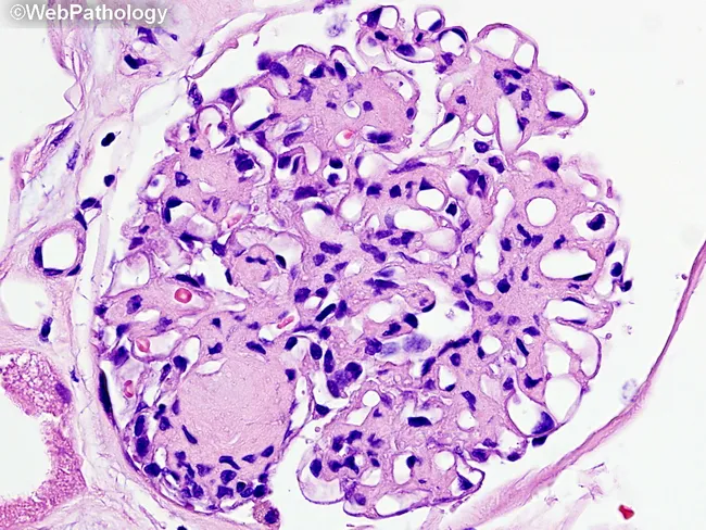
This is a renal biopsy histology slide examined under brightfield light microscopy. The primary subject is diabetic nephropathy with nodular glomerulosclerosis (Kimmelstiel-Wilson nodules) and diffuse mesangial sclerosis within the glomerular tuft of a renal cortex specimen. The glomerulus shows nodular mesangial expansion forming rounded, eosinophilic nodules that disrupt capillary loops, together with diffuse increased mesangial matrix. Capillary lumina are variably compressed, and arterioles frequently exhibit hyaline thickening consistent with diabetic microangiopathy. The cellular morphology is characterized by expanded mesangial cells with increased extracellular matrix and relatively sparse inflammatory infiltrate. The basement membrane appears thickened in cross-sectional elements. In this image, Masson’s trichrome is not visible; a subsequent stain would color collagen in blue, highlighting fibrotic remodeling. Clinically, these histologic features correlate with progressive proteinuria and reduced glomerular filtration rate in long-standing diabetes. Diagnostic significance rests on the combination of nodular sclerosis (Kimmelstiel-Wilson nodules) and diffuse mesangial sclerosis as hallmarks of diabetic nephropathy. Differential considerations include focal segmental glomerulosclerosis and hypertensive nephrosclerosis, but the nodular pattern is characteristic. This image is valuable for pathology education, nephrology training, and research into diabetic kidney disease progression and glycemic control effects. Representative for teaching clinics, this image supports correlating histology with clinical nephrology.
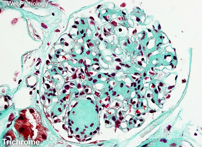
Masson's trichrome-stained renal cortex histology imaged at light microscopy reveals nodular glomerulosclerosis characteristic of diabetic nephropathy. The glomerulus shows circumscribed Kimmelstiel-Wilson nodules-eosinophilic mesangial nodules that expand the tuft and encroach on capillary loops. Diffuse mesangial sclerosis is evident as widespread mesangial matrix expansion, with increased basophilic mesangial tissue and accentuated blue-green collagen deposition in the extracellular matrix on Masson stain. The stain differentiates collagen (blue/green) from cytoplasm and nuclei (red/purple), highlighting the fibrotic nodules and mesangial expansion. Within the mesangium, there is progressive accumulation of matrix with variable cellularity; some capillary lumina appear narrowed due to nodular compression. The surrounding tubulointerstitial tissue may show mild chronic changes, consistent with chronic diabetic nephropathy. The overall pattern is typical of long-standing diabetes mellitus with nephropathy and correlates with progressive proteinuria, reduced glomerular filtration rate, and eventual renal insufficiency. Clinically, this finding supports diagnosis of diabetic kidney disease (diabetic nephropathy) and is essential for grading disease stage and guiding management. Differential diagnoses include nodular glomerulosclerosis due to amyloidosis or hypertensive nephrosclerosis, but the classic KW nodules and mesangial expansion favor diabetes. This image is valuable for medical education, pathology training, and research into diabetic renal pathology.
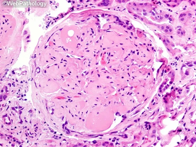
Imaging modality: brightfield histopathology. This slide shows a renal glomerulus from a biopsy specimen of diabetic nephropathy. The mesangial matrix is markedly expanded, producing round to nodular consolidations within the tuft. Several Kimmelstiel‑Wilson nodules are evident as acellular, hyaline nodules that distend the mesangial stalks and encroach on capillary loops. The glomerular basement membranes appear thickened on corresponding PAS‑positive and silver‑stained sections (referenced in adjacent images), consistent with nodular diabetic glomerulosclerosis. The surrounding cortex exhibits mild chronic interstitial changes and arteriolar hyalinosis in keeping with chronic diabetic kidney disease. The glomerulus displays relatively extensive mesangial deposition with focally patent capillary lumina, creating a characteristic nodular pattern. Overall, the morphology is diagnostic for diabetic microangiopathy with progressive nodular sclerosis. Clinically, these findings correlate with long‑standing hyperglycemia, proteinuria, and reduced GFR. Pathological significance: KW nodules indicate advanced disease and higher risk of progression to end‑stage renal disease. Differential considerations include non‑diabetic nodular glomerulosclerosis and other glomerulopathies, but the classic KW nodules and mesangial expansion strongly support diabetic nephropathy. This image is valuable for education, pathology review, and correlating histology with clinical diabetes management. It highlights key features for learners: mesangial expansion, KW nodules, PAS positivity, and altered capillary luminal flow in practice.
Diabetic Nephropathy Leading to Kidney Failure: Raised Creatinine and Pleural Effusion
Overview
1. Pathology: What Happens in the Kidney
A. Glomerular Lesions (the core injury)
B. Vascular Lesions
C. Tubulointerstitial and Papillary Lesions
2. Stages of Progression
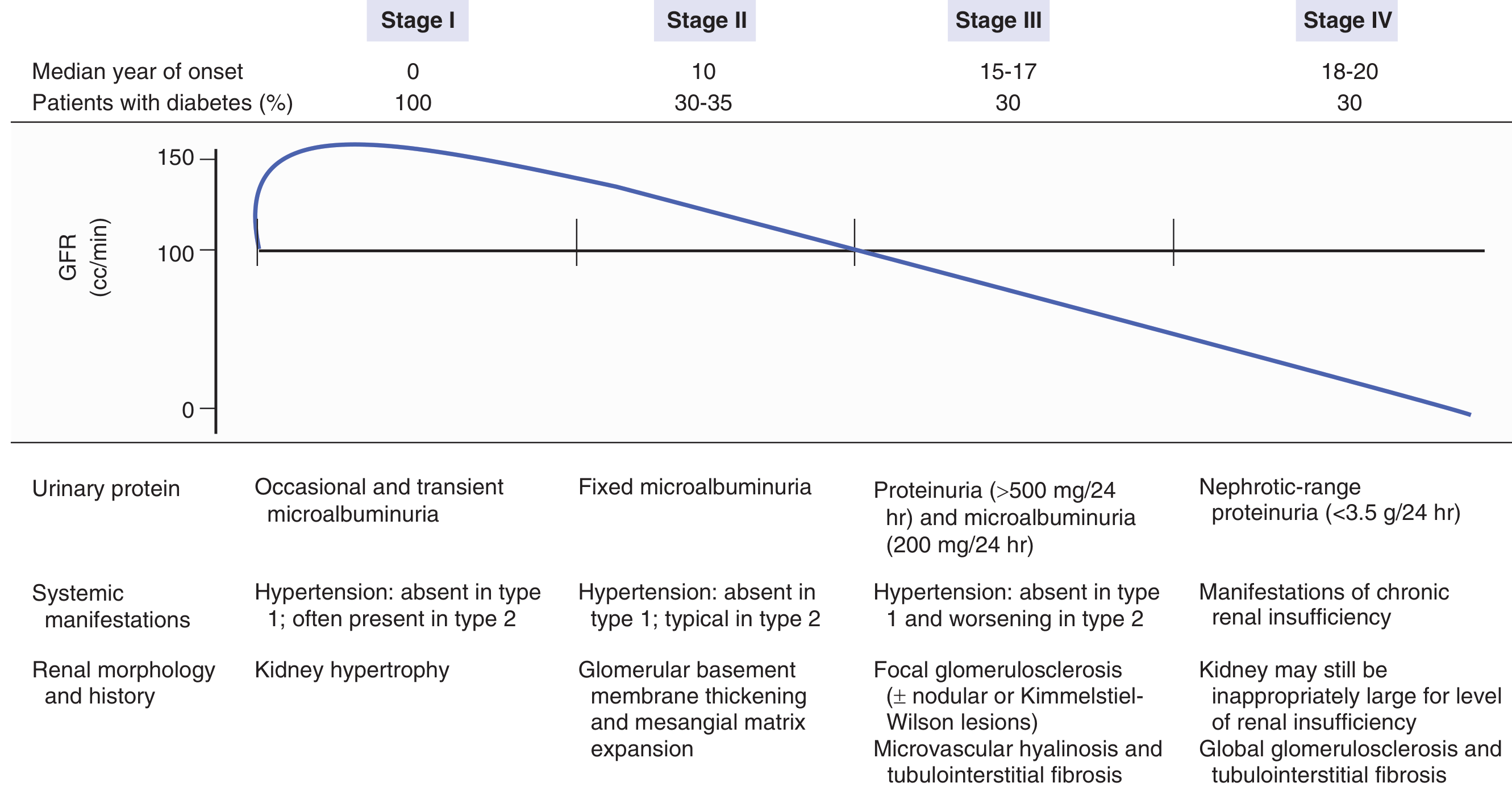
| Stage | Onset | GFR | Urinary Protein | Morphology |
|---|---|---|---|---|
| I | Year 0 | Elevated (hyperfiltration, ~150 cc/min) | Occasional/transient microalbuminuria | Kidney hypertrophy |
| II | ~Year 10 | Still normal or mildly reduced | Fixed microalbuminuria | GBM thickening, mesangial expansion |
| III | Year 15-17 | Declining (crosses 100 cc/min) | Proteinuria >500 mg/24h | Focal glomerulosclerosis, KW nodules, microvascular hyalinosis |
| IV | Year 18-20 | Relentless decline toward 0 | Nephrotic-range proteinuria (>3.5 g/24h) | Global glomerulosclerosis, tubulointerstitial fibrosis |
3. Raised Creatinine: What It Signals
- A creatinine >1.4 mg/dL at pregnancy onset carries a >40% risk of progression to ESRF within 5-6 years.
- The rate of creatinine rise predicts time to ESRD. ACE inhibitors decreased the likelihood of doubling of serum creatinine or kidney failure by 48% in diabetic nephropathy (Lewis et al., 1993 captopril trial).
- Other causes of acute creatinine rise in a diabetic patient to exclude: renal artery stenosis (especially after ACE inhibitor initiation), papillary necrosis with ureteral obstruction, bladder dysfunction from autonomic neuropathy, contrast nephropathy, prerenal azotemia from gastroparesis/vomiting, and AKI superimposed on CKD.
4. Pleural Effusion in This Context
Causes in Uremia/ESRD (from a study of 100 effusions in hospitalized patients with uremia on hemodialysis):
- Heart failure: 46% (most common)
- Uremic pleuritis: 16%
- Parapneumonic: 15%
- Atelectasis: 11%
- Miscellaneous: 12%
Uremic Pleuritis Specifically:
- Incidence of pleural effusion with uremia: ~3% (in non-dialyzed patients)
- With chronic hemodialysis: CT scan reveals effusion in ~50% of patients
- More than half of patients with uremic pleuritis also have uremic pericarditis (analogous pathogenesis - fibrinous inflammation)
- Effusion can occupy >50% of the hemithorax; bilateral in ~20%
- Symptoms: fever (50%), chest pain (30%), cough (35%), dyspnea (20%)
- Pleural fluid: exudate, frequently serosanguineous or bloody, predominantly lymphocytes on differential
- Biopsy: invariably shows chronic fibrinous pleuritis
- No close relationship between degree of uremia and presence of effusion
5. Management Framework
Slowing Progression
| Intervention | Evidence |
|---|---|
| ACE inhibitors / ARBs | Reduce CKD progression 48% (captopril); losartan (RENAAL), irbesartan (IDNT) trials confirmed benefit |
| SGLT2 inhibitors | Canagliflozin (CREDENCE, 4401 pts): substantial reduction in progression to kidney failure or CV mortality. Dapagliflozin (DAPA-CKD): significant reduction in CKD progression in both diabetic and non-diabetic CKD |
| GLP-1 receptor agonists | Recent 2026 meta-analysis (PMID 40982219) supports renal outcome benefit |
| BP control | Target <130/80 mmHg in diabetics with albuminuria. SPRINT subgroup: intensive BP lowering reduced all-cause mortality in CKD stages 3-5 |
| Glycemic control | Foundational - delays onset and slows progression of all microvascular complications |
When Creatinine is Rising Rapidly - Rule Out
- ACE inhibitor/ARB-induced AKI (check for renal artery stenosis)
- Volume depletion (gastroparesis, autonomic diarrhea)
- Contrast exposure
- Obstructive uropathy (neurogenic bladder, papillary sloughing)
- Superimposed AKI on background CKD
Managing Pleural Effusion
- Identify the dominant cause (CHF vs uremic pleuritis vs infection)
- CHF-related: optimize fluid balance, diuretics, dialysis ultrafiltration
- Uremic pleuritis: adequate dialysis resolves ~75% within 4-6 weeks; resistant cases may need thoracentesis or decortication
- Diagnostic thoracentesis warranted if cause uncertain (exudate with lymphocytic differential suggests uremic; bloody exudate is classic for uremic pleuritis)
6. Diagnosis of Diabetic Nephropathy
- Proteinuria within the appropriate time frame (significant proteinuria appears 11-23 years after type 1 diabetes onset; microalbuminuria always precedes overt proteinuria in progressors)
- Diabetic retinopathy present in 90-95% of type 1, 60-65% of type 2 diabetic nephropathy
- Absence of other causes of nephrotic syndrome or renal insufficiency
Recent Evidence Updates
- Treatment of diabetic kidney disease (2023) - network meta-analysis comparing therapies
- GLP-1 agonists on renal outcomes (2026) - supports GLP-1 RA benefit on kidney endpoints
- Inflammatory markers in type 2 DN (2025) - inflammatory markers (e.g. IL-6, TNF-alpha) as predictors of nephropathy; this may open future therapeutic targets