
Plural effusion
Reading File
Pleural effusion is an abnormal accumulation of fluid in the pleural space between the visceral and parietal pleura. It is classified as a transudate (low protein, due to hydrostatic/oncotic pressure changes) or an exudate (high protein/LDH, due to inflammation, infection, or malignancy). Common causes include heart failure, pneumonia, malignancy, TB, and hypoalbuminemia.
Searching sources...
Finding Sources
Searching PubMed
"pleural effusion" AND management
Good results. Now let me read the most comprehensive sections in parallel - Murray & Nadel's (respiratory medicine specialist textbook) and Rosen's (emergency medicine, clinical management), plus the radiology textbook for imaging.
Reading File
Reading File
Reading File
Now let me get the main comprehensive pleural effusion sections - the dedicated chapter in Murray & Nadel and Rosen's emergency medicine.
Reading File
Reading File
Reading File
Good content from Rosen's and Grainger's radiology. Let me now get more from Rosen's (the clinical management/diagnosis section) and fetch the key radiology image.
Reading File
Loading Image
Excellent - I now have comprehensive content. Let me get the remaining part of Rosen's covering management/treatment and also get the radiology imaging section continuation.
Reading File
Reading File
I now have comprehensive information from multiple authoritative sources. Let me compile the full answer.
Pleural Effusion
Definition and Normal Physiology
Pleural effusion is an abnormal collection of fluid in the pleural space. Under normal circumstances, a thin layer of fluid lies between the visceral and parietal pleura. Pleural fluid is produced from systemic capillaries at the parietal pleural surface and absorbed into the pulmonary capillaries at the visceral pleural surface. Lymphatics also contribute to drainage. Any imbalance in these forces - increased hydrostatic pressure, decreased oncotic pressure, or impaired lymphatic drainage - can result in fluid accumulation. - Rosen's Emergency Medicine, p. 988
Classification: Transudate vs. Exudate
All types of pleural effusion are radiographically identical. Differentiation requires pleural fluid analysis, most reliably using Light's Criteria. - Grainger & Allison's Diagnostic Radiology
Light's Criteria (Exudate if ANY one is met):
| Criterion | Threshold |
|---|---|
| Pleural fluid protein / Serum protein | > 0.5 |
| Pleural fluid LDH / Serum LDH | > 0.6 |
| Pleural fluid LDH | > 2/3 upper limit of normal serum LDH |
- Rosen's Emergency Medicine, p. 990
Causes
Transudates
- Congestive heart failure (most common in the USA)
- Cirrhosis with ascites (hepatic hydrothorax)
- Nephrotic syndrome
- Hypoalbuminemia
- Myxedema
- Peritoneal dialysis
- Glomerulonephritis
Exudates
-
Bacterial pneumonia / parapneumonic effusion / empyema
-
Malignancy (lung, breast metastases are most common causes of massive effusion)
-
Tuberculosis (classic cause in endemic regions)
-
Pulmonary embolism
-
Collagen vascular disease (rheumatoid, SLE)
-
Pancreatitis (typically left-sided)
-
Esophageal rupture (rare but highly morbid)
-
Ovarian hyperstimulation syndrome
-
Rosen's Emergency Medicine, p. 988
Laterality Clues
-
Right-sided: ascites, heart failure, liver abscess
-
Left-sided: pancreatitis, pericarditis, esophageal rupture, aortic dissection
-
Bilateral: most often transudates (heart failure, nephrotic, hypoalbuminemia); bilateral exudates suggest metastatic disease, lymphoma, PE, SLE, myxedema
-
Massive effusion: most commonly malignancy (lung or breast metastases), but also heart failure, cirrhosis, TB, empyema, trauma
-
Grainger & Allison's Diagnostic Radiology, p. 62
Pleural Fluid Analysis
| Test | Significance |
|---|---|
| pH < 7.3 | Parapneumonic, malignancy, TB, rheumatoid, systemic acidosis |
| pH < 7.0 | Strongly suggests empyema or esophageal rupture - indication for tube thoracostomy |
| Bloody fluid (Hct >50% peripheral blood) | Hemothorax |
| Bloody (non-traumatic tap) | Trauma, neoplasm, pulmonary infarction |
| Cytology | Submit if malignancy suspected |
| Gram stain + culture | Empyema / parapneumonic |
| AFB smear/culture | TB (low sensitivity - culture positive in only 25-30%) |
- Rosen's Emergency Medicine, pp. 990-991
In tuberculous pleural effusion: WBC typically 500-2500 cells/mL; exudate with protein >50% serum protein; glucose may be normal to low; AFB smear rarely positive; pleural biopsy confirms diagnosis in most cases. - Rosen's Emergency Medicine, p. 2710
Imaging
Chest X-Ray
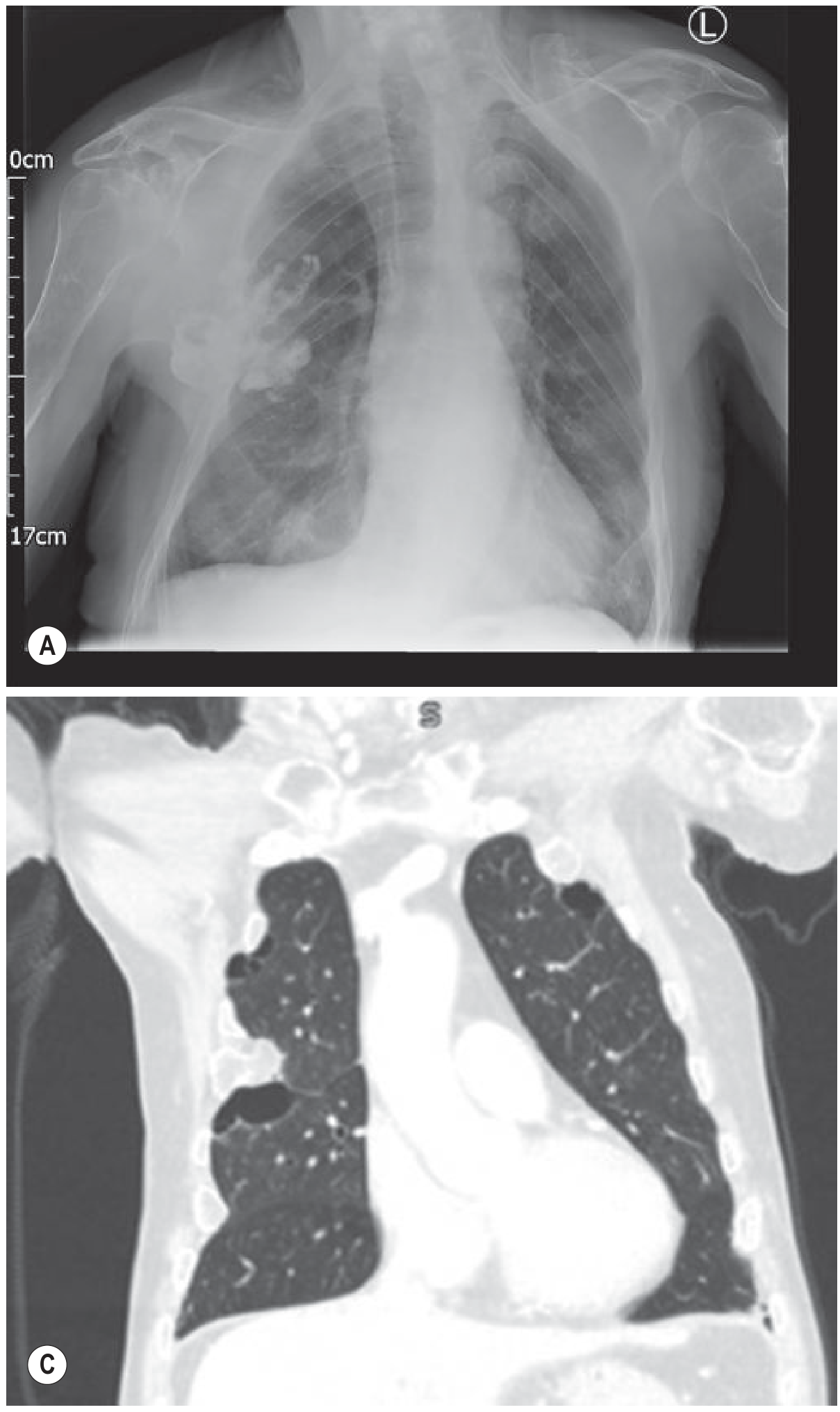
Bilateral pleural effusion: (A) erect CXR showing bilateral lower-zone opacification with concave meniscus higher laterally; (C) coronal CT confirming bilateral effusions with compressed lung
Radiographic progression of free pleural fluid:
- Small (<200 mL): may be undetectable on erect PA; fluid collects under lower lobes (subpulmonary effusion)
- 200-500 mL: posterior then lateral costophrenic angle blunting
- Larger: homogeneous opacification of lower chest with obliterated costophrenic angle and hemidiaphragm; superior margin is concave to the lung and higher laterally (meniscus sign)
- ~1000 mL: effusion reaches level of the 4th anterior rib
- Massive: dense opacification of the hemithorax; may cause mediastinal shift to the contralateral side
Subpulmonary effusion appears as a "high hemidiaphragm" with an unusually lateral peak; left-sided variants show >2 cm separation of the stomach bubble from the lung base.
Supine patients: fluid layers posteriorly, visible as a hazy veil-like opacity over the lower hemithorax with preserved vascular markings (no costophrenic blunting).
- Grainger & Allison's Diagnostic Radiology, pp. 61-63
Ultrasound
- More sensitive than CXR; detects effusions as small as 50 mL
- Simple transudates appear hypoechoic (anechoic) above the diaphragm
- Complex exudates: heterogeneous echogenicity, fibrinous strands, septations, loculations
- Swirling echoes suggest high cellularity (malignant effusion)
- Compressed lung tissue often visible within the effusion
- Strongly preferred to guide thoracentesis - significantly reduces iatrogenic pneumothorax risk
- Rosen's Emergency Medicine, pp. 989-990
CT
- Best for characterizing the pleural space, identifying loculations, pleural thickening/enhancement (suggesting exudate), and underlying parenchymal pathology
Special Types
Hepatic Hydrothorax
- Complicates ~5-10% of patients with cirrhosis
- Caused by small diaphragmatic defects allowing ascitic fluid to pass into the pleural space
- 85% are right-sided; may exist even without detectable ascites
- Nearly all are transudative (unless infected)
- Management: sodium restriction + diuretics → TIPS (70-80% initial response, limited by encephalopathy) → VATS with pleurodesis → indwelling pleural catheters (palliation or bridge to transplant)
- Conventional chest tube drainage is NOT recommended (protein/electrolyte loss, infection, renal failure)
- Murray & Nadel's Textbook of Respiratory Medicine, p. (hepatic disease chapter)
Parapneumonic Effusion / Empyema
- Associated with bacterial pneumonia or lung abscess
- Empyema = frank pus in the pleural space
- Loculated effusion: fluid trapped by adhesions between parietal and visceral pleura
- pH < 7.0 is an indication for chest tube drainage
- Small-bore pigtail catheters (14 Fr) are now accepted first-line over large-bore tubes (28-40 Fr)
- Rosen's Emergency Medicine, p. 989, 991
Hemodynamic Effects
A less-appreciated complication: large right-sided effusions can impair right ventricular filling, producing hemodynamic effects similar to cardiac tamponade. Echocardiographic evidence of this should prompt urgent thoracentesis. Hypoxemia in this setting is partly explained by low cardiac output and low mixed venous PO2 in the setting of venous admixture. - Murray & Nadel's Textbook of Respiratory Medicine, p. 960
Management
When to Perform Thoracentesis
- Diagnostic: most effusions of unknown cause (can defer if obvious CHF and bilateral)
- Urgent therapeutic: massive effusions (>1.5-2 L) causing respiratory/circulatory compromise; empyema for source control
- ED indications: life-threatening pathology (empyema, esophageal rupture) or significant symptomatic relief needed
Contraindications (Relative)
- Coagulopathy / bleeding disorders
- History of pleurodesis
- Chest wall infection
- Pleural adhesions (risk reducible with ultrasound guidance)
Volume Limits
- Remove no more than 1500 mL per single procedure (British Thoracic Society, Grade C) to reduce the risk of re-expansion pulmonary edema
- Re-expansion pulmonary edema (rare): symptoms usually mild; treat with O2 and gentle diuresis
Complications of Thoracentesis
- Iatrogenic pneumothorax (most common - minimized by ultrasound guidance)
- Hemothorax
- Lung laceration
- Infection
- Transient hypoxia (V/Q mismatch)
- Hypotension (especially in volume-depleted patients)
- Re-expansion pulmonary edema
Post-procedure: chest X-ray or bedside ultrasound to rule out pneumothorax. - Rosen's Emergency Medicine, pp. 991-992
Key Summary Table
| Feature | Transudate | Exudate |
|---|---|---|
| Mechanism | Hydrostatic/oncotic pressure | Inflammation, infection, malignancy |
| Protein ratio (PF/serum) | <0.5 | >0.5 |
| LDH ratio (PF/serum) | <0.6 | >0.6 |
| Common causes | CHF, cirrhosis, nephrotic | Pneumonia, TB, malignancy, PE |
| Ultrasound | Hypoechoic, simple | Echogenic, septated, complex |
Recent evidence note: A 2024 systematic review (PMID 39182102) on complicated pleural infections found that surgery and intrapleural fibrinolysis have comparable efficacy - this is relevant for empyema management when simple tube drainage fails.
This is a shared conversation. Sign in to Orris to start your own chat.