
Alzi ers disease.
Reading File
Now I'll search the medical library and PubMed simultaneously for a comprehensive answer on Alzheimer's disease.
Finding Sources
Searching PubMed
"Alzheimer disease"[MeSH Terms] AND treatment
Searching the Web
Alzheimer's disease 2025 2026 new treatments lecanemab donanemab guidelines
Good results. Now let me pull the detailed textbook content from Bradley and Daroff's Neurology (the most comprehensive source) in parallel with paragraph searches.
Reading File
Finding Sources
Finding Sources
Excellent. Now let me pull the detailed clinical content from Harrison's (2025) and Bradley and Daroff's on pathology/clinical features/treatment.
Reading File
Reading File
Excellent - rich content retrieved. Let me get the treatment section from Harrison's and the pathology image.
Reading File
Loading Image
I now have comprehensive content. Let me compile the full response.
Alzheimer's Disease
Overview
Alzheimer's disease (AD) is the most common cause of dementia, accounting for an estimated 60-70% of all dementia cases worldwide. Approximately 55 million people globally are living with dementia, with total U.S. healthcare costs for dementia care reaching $360 billion in 2024 (~$25,000 per patient). AD can begin as early as the third decade of life, but is most prevalent in the elderly population.
- Harrison's Principles of Internal Medicine, 22e
Historical Background
In 1906, German psychiatrist Alois Alzheimer reported a woman in her 50s presenting with paranoia, memory loss, aphasia, and loss of motor task ability. At autopsy, her brain showed diffuse atrophy. Bielschowsky staining revealed the hallmark findings now known as amyloid plaques and neurofibrillary tangles (NFTs).
- Bradley and Daroff's Neurology in Clinical Practice
Pathology
AD is defined by two core pathological findings:
1. Amyloid (Neuritic) Plaques
- Extracellular deposits composed primarily of amyloid-beta (Aβ) peptides, derived from amyloid precursor protein (APP) via proteolytic cleavage
- Aβ deposition follows a sequential pattern: cortex → hippocampus → basal ganglia → thalamus → basal forebrain → brainstem/cerebellum
- Plaques are heterogeneous lesions containing Aβ, tau, and cellular components
2. Neurofibrillary Tangles (NFTs)
- Intraneuronal accumulations of hyperphosphorylated tau protein filaments
- Earliest tau pathology (Braak staging) appears in the locus coeruleus as early as the 3rd or 4th decade, before involving limbic areas or amyloid plaques
- Tau pathology spreads in a prion-like transsynaptic manner through anatomically connected regions
Macroscopic Changes
- Diffuse brain atrophy with reduced brain weight at autopsy
- Preferentially affected: multimodal association areas, hippocampi, cingulate gyrus, locus coeruleus
- Relatively spared: primary motor/somatosensory/auditory/visual cortices, substantia nigra
Here is a histopathology image (Fig. 95.12) showing the classic AD findings:
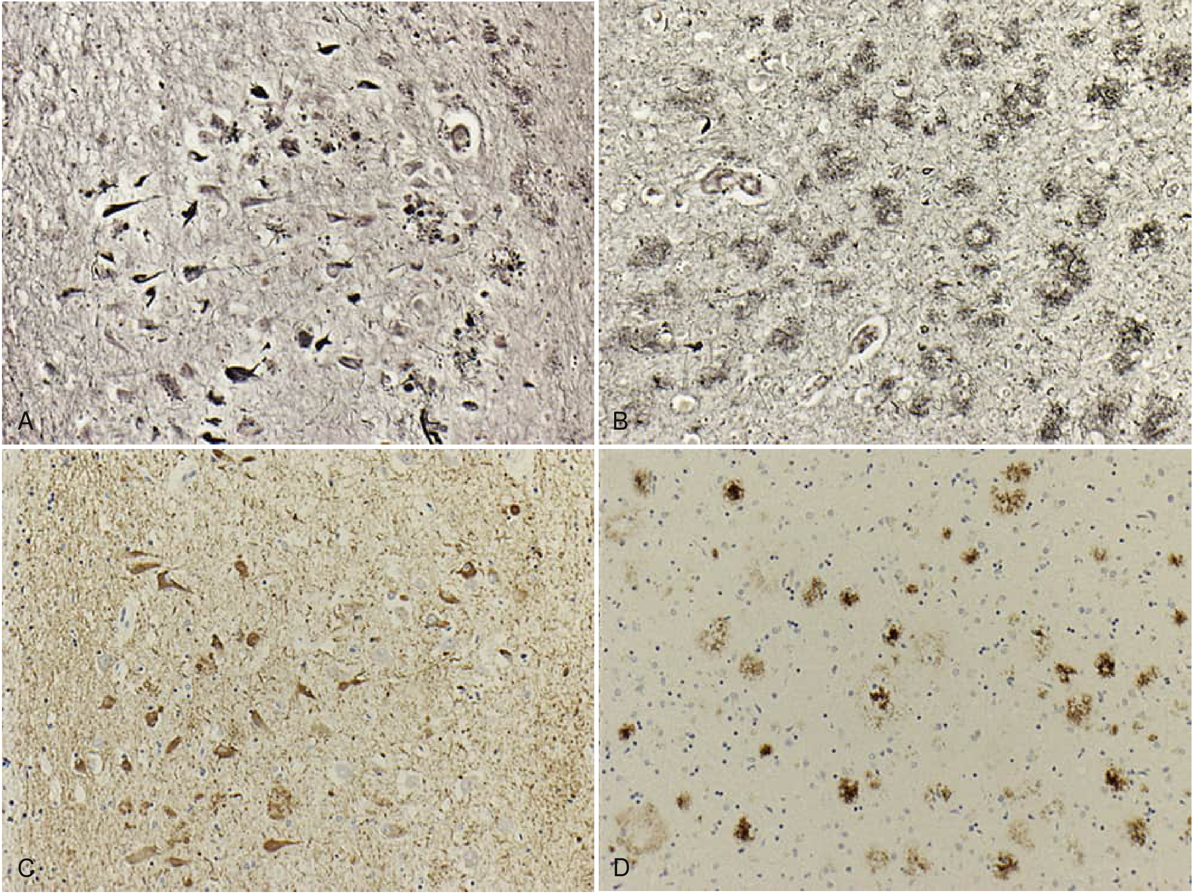
Bielschowsky stain of CA1 hippocampus (A) and temporal cortex (B) showing neurofibrillary tangles and amyloid plaques; Tau stain of CA1 hippocampus (C) demonstrating NFTs; Aβ stain of parietal cortex (D) showing plaques. (Bradley and Daroff's Neurology in Clinical Practice, Courtesy Dr. Joseph Paris.)
Genetics
| Factor | Detail |
|---|---|
| ApoE ε4 allele | Most important genetic risk factor; 1 allele = 2-3x increased risk in women; 2 alleles = 10-15x increased risk in both sexes |
| Early-onset familial AD | Mutations in APP, Presenilin-1 (PSEN1), Presenilin-2 (PSEN2) |
| ApoE function | Transports lipids/cholesterol from astrocytes to neurons; ε4 decreases CSF Aβ42 clearance and promotes Aβ aggregation |
| Trisomy 21 (Down syndrome) | APP gene on chromosome 21 → virtually all develop AD pathology by age 40 |
- Bradley and Daroff's Neurology in Clinical Practice; Thompson & Thompson Genetics and Genomics in Medicine
Clinical Manifestations
Typical Presentation (Amnestic AD)
- Insidious onset of episodic memory impairment
- Progresses to deficits in executive function, language, and visuospatial abilities
- Brain atrophy begins in medial temporal lobes, spreads to inferior temporal, parietal, and frontal cortices
- Depression, social withdrawal, and anxiety may precede cognitive symptoms
Atypical Variants (~20% of cases)
- Posterior cortical atrophy: visual processing dysfunction is the initial complaint
- Logopenic aphasia: word-finding difficulty and difficulty with repetition
- Corticobasal syndrome: asymmetric akinetic-rigid-dystonic features
- Frontal variant: prominent dysexecutive or behavioral changes
Disease Stages
| Stage | Description |
|---|---|
| Subjective cognitive decline (SCD) | Self-perceived memory worsening, not detectable on testing |
| Mild cognitive impairment (MCI) | Noticeable to patient/family, confirmed on testing; ~50% progress to dementia over 4 years (~12%/year) |
| Mild dementia | Memory loss affects daily function; patient aware and often distressed |
| Moderate dementia | Progressive loss of independence; behavioral symptoms emerge |
| Severe dementia | Complete dependency; loss of language, mobility, swallowing |
- Harrison's Principles of Internal Medicine, 22e
Biomarkers & Diagnosis
The AT(N) framework (Alzheimer's Association/NIA 2018 Research Framework) classifies individuals by three biomarker categories:
- A - β-Amyloid deposition (amyloid PET positivity or low CSF Aβ42)
- T - Pathological tau (tau PET positivity or elevated phospho-tau in CSF/plasma)
- (N) - Neurodegeneration (brain atrophy on MRI, FDG-PET hypometabolism, elevated CSF total tau)
Updated 2025 guidelines from the Alzheimer's Association (Rabinovici et al., Alzheimers Dement 2025, PMID 39776249) have updated appropriate use criteria for amyloid and tau PET, emphasizing earlier and more precise use of these imaging tools to guide disease-modifying therapy decisions.
A 2025 Lancet Neurology meta-analysis (Therriault et al., PMID 40818474) confirmed that blood phosphorylated tau (plasma p-tau) is a highly accurate diagnostic biomarker for AD, paving the way for widespread blood-based screening.
Treatment
1. Neurotransmitter-Based (Symptomatic) Therapies
These remain the foundation of treatment for mild-to-severe AD:
| Drug | Class | Dose | Approved Stage |
|---|---|---|---|
| Donepezil | Cholinesterase inhibitor | 10 mg daily | Mild-to-severe |
| Rivastigmine | Cholinesterase inhibitor | 6 mg bid or 9.5 mg/day patch | Mild-to-severe |
| Galantamine | Cholinesterase inhibitor | 24 mg/day (extended release) | Mild-to-moderate |
| Memantine | NMDA antagonist | 10 mg twice daily | Moderate-to-severe |
Mechanism: Cholinesterase inhibitors increase cerebral acetylcholine; memantine blocks overactivated NMDA glutamate receptors.
Efficacy: Average patient on a cholinesterase inhibitor maintains MMSE score for ~1 year vs. 2-3 point decline in placebo-treated patients over the same period.
Common approach: Start a cholinesterase inhibitor at mild stage; add memantine when the patient enters the moderate stage.
Side effects:
-
Cholinesterase inhibitors: GI symptoms (nausea, diarrhea, cramps), vivid dreams, bradycardia, muscle cramps
-
Memantine: constipation, dizziness, headache, somnolence
-
Harrison's Principles of Internal Medicine, 22e
2. Disease-Modifying Therapies (Anti-Amyloid Monoclonal Antibodies)
This is the most significant recent advance in AD treatment:
| Drug | Target | Route | Approval |
|---|---|---|---|
| Lecanemab (Leqembi) | Aβ protofibrils | IV infusion every 2 weeks | FDA approved 2023 (full approval) |
| Donanemab (Kisunla) | Pyroglutamate Aβ in plaques | IV infusion monthly | FDA approved 2024 |
| Aducanumab | Aβ aggregates | IV | Removed from clinical use (ambiguous efficacy) |
Both lecanemab and donanemab received full FDA approval based on Phase 3 RCT evidence showing clinical efficacy (slowing of cognitive decline) in early AD. As of mid-2025, they have been approved in 45+ countries including the USA, UK, EU, China, and Japan.
Key risk: Both drugs carry a risk of amyloid-related imaging abnormalities (ARIA) - either ARIA-E (edema/effusions) or ARIA-H (microhemorrhages/hemosiderosis). ApoE ε4 homozygotes are at substantially higher risk for ARIA and may be excluded from treatment in some protocols.
2026 outlook: Pill-based therapies and subcutaneous formulations of lecanemab are in development/trials.
- Harrison's Principles of Internal Medicine, 22e; Lancet Treatment Review 2025
3. Non-Pharmacological & Supportive Care
- Caregiver education and structured support networks
- Mediterranean diet: A 2025 meta-analysis (Fekete et al., Geroscience 2025, PMID 39797935) confirms a reduced risk of cognitive impairment and AD with Mediterranean diet adherence
- Physical activity and cognitive stimulation
- Management of behavioral and psychological symptoms (agitation, depression, psychosis) with targeted pharmacotherapy when needed
- Advance care planning and legal/financial arrangements early in the disease course
Differential Diagnosis (Key Mimics)
- Lewy body dementia - visual hallucinations, parkinsonism, fluctuating cognition
- Frontotemporal dementia (FTD) - prominent behavioral/personality changes or language syndrome
- Vascular dementia - stepwise progression, focal deficits, vascular risk factors
- LATE (Limbic-predominant Age-related TDP-43 Encephalopathy) - recently characterized; can closely mimic AD clinically (Wolk et al., Alzheimers Dement 2025, PMID 39807681)
- Normal pressure hydrocephalus - triad of dementia, gait apraxia, urinary incontinence
- Hypothyroidism, vitamin B12 deficiency - reversible causes
Key Recent Evidence (2025-2026)
| Study | Finding |
|---|---|
| Therriault et al. Lancet Neurol 2025 (PMID 40818474) | Blood plasma p-tau has high diagnostic accuracy for AD |
| Rabinovici et al. Alzheimers Dement 2025 (PMID 39776249) | Updated PET use criteria for amyloid/tau to guide DMT selection |
| Wolk et al. Alzheimers Dement 2025 (PMID 39807681) | Clinical criteria for LATE - a key mimic of AD |
| Fekete et al. Geroscience 2025 (PMID 39797935) | Mediterranean diet reduces AD/dementia risk (meta-analysis) |
| Fox et al. Lancet 2025 | Lecanemab/donanemab now approved in 45+ countries |
Summary: Alzheimer's disease is a progressive neurodegenerative disorder defined by amyloid plaques and neurofibrillary tangles, presenting with insidious memory loss that advances to global cognitive decline. The field has entered a new era with FDA-approved anti-amyloid therapies (lecanemab, donanemab) that slow disease progression in early stages - a major shift from purely symptomatic treatment. Diagnosis is increasingly biological (AT(N) framework, plasma p-tau), enabling earlier and more precise intervention.
Cushing Syndrome physiology
Reading File
This is a broad physiological topic. Let me search the library headings and paragraphs in parallel, along with PubMed and web search.
Finding Sources
Finding Sources
Searching PubMed
"Cushing syndrome"[MeSH Terms] AND pathophysiology
Searching the Web
Cushing syndrome pathophysiology cortisol HPA axis mechanisms 2025
Good. Now let me pull the detailed physiology content from the best sources - Harrison's (primary endocrine textbook), Campbell Walsh, and search specifically for the physiology/pathophysiology paragraphs.
Finding Sources
Reading File
Excellent. Now let me search for Harrison's dedicated section on Cushing syndrome physiology and get a diagram.
Finding Sources
Finding Sources
Now let me read the Harrison's main CUSHING'S SYNDROME section and Medical Physiology for the core physiology content.
Reading File
Reading File
Excellent. Let me now read the key physiology/clinical features sections from Harrison's and the adrenal anatomy diagram.
Reading File
Loading Image
I have all the key content I need. Let me compile the comprehensive response.
Cushing Syndrome - Physiology & Pathophysiology
1. Normal HPA Axis Physiology (The Foundation)
To understand Cushing syndrome, you must first understand the normal hypothalamic-pituitary-adrenal (HPA) axis - the neuroendocrine negative feedback loop that controls cortisol.
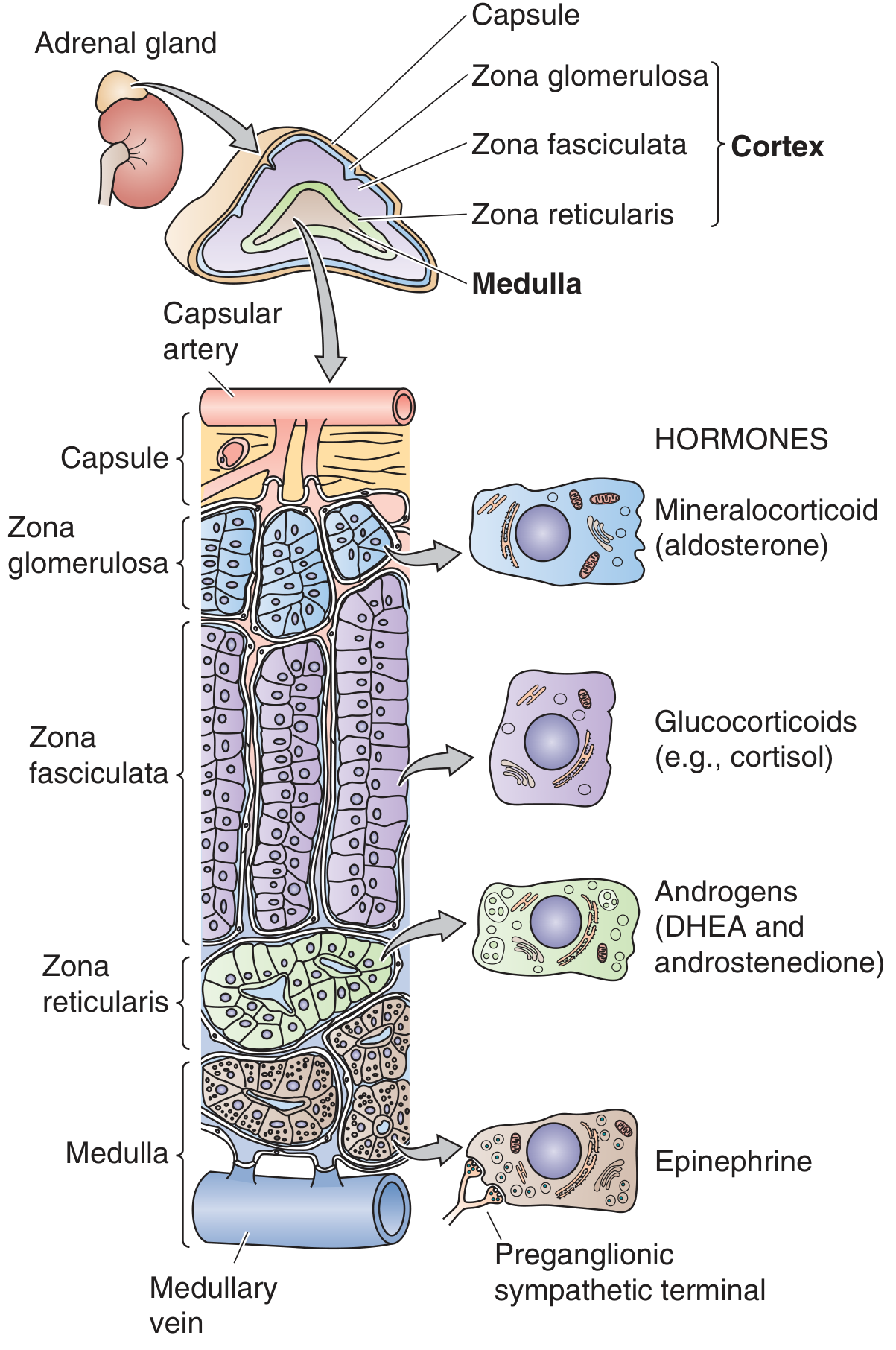
Fig. 50-1 Anatomy of the adrenal gland showing three cortical zones and their respective hormones. Cortisol is produced in the zona fasciculata. (Medical Physiology)
The Axis - Step by Step
Hypothalamus
    │ CRH (Corticotropin-Releasing Hormone)
    ↓
Anterior Pituitary (corticotrope cells)
    │ ACTH (Adrenocorticotropic Hormone) via POMC cleavage
    ↓
Adrenal Cortex (Zona Fasciculata)
    │ Cortisol (~20 mg/day)
    ↓
Target tissues + Negative feedback ──→ back to Hypothalamus & Pituitary
Key points of normal physiology:
-
CRH from the hypothalamus is the most important driver of ACTH release. Vasopressin (ADH) and oxytocin also stimulate it
-
ACTH (cleaved from POMC) stimulates cortisol AND adrenal androgen production, and is also trophic - it maintains adrenocortical cell viability. Without ACTH, all but mineralocorticoid-producing cells undergo apoptosis
-
Cortisol binds receptors in the hypothalamus and pituitary to suppress CRH and ACTH (negative feedback loop)
-
Circadian rhythm: CRH secretion is under control of the suprachiasmatic nucleus. Cortisol peaks in the early morning and reaches a nadir around 11 PM. Loss of this diurnal rhythm is a hallmark of Cushing syndrome
-
Stress (physiological or psychological) is the most powerful modulator of HPA axis activity
-
Campbell Walsh Wein Urology; Medical Physiology
2. Normal Cortisol - Physiological Actions
Cortisol, the primary glucocorticoid in humans, acts on virtually every tissue in the body. Its actions are largely genomic (through glucocorticoid response elements, GREs, altering gene transcription), though non-genomic rapid effects also exist.
| System | Physiological Action of Cortisol |
|---|---|
| Metabolism | Promotes gluconeogenesis in liver; mobilizes amino acids from muscle proteins; increases lipolysis in adipose |
| Immune/Inflammatory | Potent anti-inflammatory and immunosuppressive effects; reduces cytokine production and leukocyte migration |
| Bone | Regulates calcium and bone metabolism |
| CNS | Behavioral effects; mood regulation |
| Cardiovascular | Maintains vascular tone; permissive effect on catecholamines |
| Kidney | At high concentrations, activates mineralocorticoid receptors (sodium retention, potassium loss) |
Medical Physiology; Harrison's Principles of Internal Medicine 22e
3. Definition of Cushing Syndrome
Cushing syndrome is the constellation of clinical features resulting from chronic exposure to excess glucocorticoids, regardless of the cause.
-
Cushing's disease specifically refers to the subset caused by a pituitary corticotrope adenoma (ACTH-secreting)
-
First described by Harvey Cushing in 1912
-
Incidence of endogenous Cushing syndrome: ~1.8-3.2 per million per year (rare)
-
Iatrogenic (exogenous glucocorticoid therapy) is by far the most common cause overall (~1% of the population uses chronic glucocorticoids)
-
Harrison's Principles of Internal Medicine 22e
4. Classification by Cause
ACTH-Dependent (~80-85% of endogenous cases)
| Cause | Detail |
|---|---|
| Cushing's disease (pituitary adenoma) | ~70% of all endogenous cases; corticotrope microadenoma (<10 mm) in 90% of cases; more common in women; ACTH excess drives bilateral adrenal hyperplasia |
| Ectopic ACTH syndrome | ACTH secreted by non-pituitary tumors (small cell lung cancer, bronchial carcinoids, pancreatic tumors); more common in men; often rapid onset with severe hypokalemia |
ACTH-Independent (~10-15% of endogenous cases)
| Cause | Detail |
|---|---|
| Adrenocortical adenoma | Unilateral; autonomous cortisol production; ACTH suppressed by negative feedback; contralateral adrenal atrophies |
| Adrenocortical carcinoma (ACC) | Usually large; may co-secrete multiple steroids |
| Primary bilateral macronodular adrenal hyperplasia (PBMAH) | Aberrant G protein-coupled receptors on adrenal cells (for LH, vasopressin, GIP, serotonin, etc.) → autonomous cortisol production independent of ACTH |
| Primary pigmented nodular adrenal disease (PPNAD) | Part of Carney complex; PRKAR1A germline mutations |
| McCune-Albright syndrome | GNAS activating mutations |
Iatrogenic (Exogenous)
-
Administration of glucocorticoids for anti-inflammatory/immunosuppressive purposes
-
HPA axis is suppressed by exogenous glucocorticoids; abrupt cessation risks adrenal insufficiency
-
Harrison's Principles of Internal Medicine 22e; Campbell Walsh Wein Urology
5. Core Pathophysiology - What Excess Cortisol Does
The pathophysiology of Cushing syndrome is simply the exaggeration of cortisol's normal physiological actions across all organ systems. Here is the mechanistic basis for each clinical feature:
A. Metabolic Effects (Fat & Carbohydrate)
- Central obesity / buffalo hump / moon face: Excess cortisol promotes redistribution of fat from peripheral (limbs) to central (visceral, dorsal cervical, facial) depots. The mechanism involves differential glucocorticoid receptor density and differential sensitivity of fat depots to cortisol-stimulated lipolysis and insulin-stimulated lipogenesis
- Hyperglycemia / diabetes: Cortisol enhances hepatic gluconeogenesis and induces insulin resistance in peripheral tissues (liver, muscle, adipose). Excess cortisol drives overproduction of glucose and impairs glucose uptake
- A 2025 review (DeFronzo & Auchus, Diabetes 2025, PMID 40663715) specifically addresses how hypercortisolism drives glucose dysregulation through multiple mechanisms including hepatic insulin resistance and impaired beta-cell compensation
B. Protein Catabolism (Muscle & Skin)
- Cortisol promotes protein breakdown in muscle, skin, and connective tissue
- In muscle: protein catabolism → proximal myopathy (difficulty rising from chairs, climbing stairs)
- In skin: loss of dermal collagen → thin skin, easy bruising, broad purple striae (stretch marks from dermal tearing as skin cannot accommodate rapid fat deposition)
- In bone: inhibits osteoblast activity and calcium absorption → osteoporosis and vertebral fractures
C. Mineralocorticoid Overflow Effect
- At very high levels, cortisol overwhelms the enzyme 11β-HSD2 (which normally converts active cortisol to inactive cortisone in mineralocorticoid-sensitive cells)
- Excess cortisol then activates mineralocorticoid receptors in the kidney → sodium retention, potassium wasting
- Result: hypertension, hypokalemia, metabolic alkalosis - especially severe in ectopic ACTH syndrome
D. Immune Suppression
- Cortisol suppresses the immune system at multiple levels: reduces cytokine synthesis, inhibits leukocyte trafficking, induces lymphocyte apoptosis
- Clinical result: increased susceptibility to infections, impaired wound healing
E. HPA Axis Disruption
- In ACTH-dependent Cushing's: pituitary tumor cells lose sensitivity to cortisol's normal negative feedback - they keep secreting ACTH despite high cortisol levels
- Loss of circadian rhythm: cortisol remains elevated throughout the day, particularly at the normal 11 PM nadir. This is one of the most sensitive diagnostic indicators
- In ACTH-independent Cushing's: autonomous adrenal cortisol secretion suppresses pituitary ACTH → the normal (contralateral or remaining) adrenal tissue undergoes atrophy
F. Androgenic Effects (in adrenal causes)
- Adrenocortical tumors and ectopic ACTH tumors often co-secrete adrenal androgens (DHEA, androstenedione)
- Result in women: hirsutism, acne, virilization, menstrual irregularity
- Adrenal carcinomas particularly notable for excess androgen co-secretion
G. CNS & Psychiatric Effects
- Glucocorticoid receptors are abundant in the hippocampus and limbic system
- Chronic excess cortisol causes hippocampal atrophy, neuronal damage
- Clinical: depression, anxiety, cognitive impairment, psychosis, insomnia
H. Cardiovascular Effects
- Hypertension from: (1) mineralocorticoid receptor activation, (2) increased angiotensinogen synthesis, (3) enhanced vascular sensitivity to catecholamines
- Insulin resistance and dyslipidemia → accelerated atherosclerosis
- Increased thrombotic risk (cortisol upregulates coagulation factors, reduces fibrinolysis)
6. Signs & Symptoms Summary (From Harrison's Table 398-2)
| System | Features |
|---|---|
| Body fat | Central obesity, rounded face (moon face), buffalo hump |
| Skin | Facial plethora, thin/brittle skin, easy bruising, broad purple striae, acne, hirsutism |
| Bone | Osteopenia, osteoporosis, vertebral fractures; decreased linear growth in children |
| Muscle | Proximal myopathy, weakness |
| Cardiovascular | Hypertension |
| Metabolic | Hyperglycemia/diabetes, dyslipidemia |
| Reproductive | Menstrual irregularity, decreased libido, hypogonadism |
| Neuropsychiatric | Depression, anxiety, cognitive impairment, psychosis |
| Immune | Recurrent/opportunistic infections |
| Eyes | Glaucoma, cataracts |
| Pigmentation | Hyperpigmentation (only in ACTH-dependent forms, from POMC-derived MSH) |
Harrison's Principles of Internal Medicine 22e
7. Key Physiological Principles Used in Diagnosis
The diagnosis of Cushing syndrome exploits three core physiological principles:
- Loss of the cortisol diurnal nadir - Late-night salivary cortisol or midnight plasma cortisol is elevated (normally near zero at 11 PM)
- Loss of feedback sensitivity - The dexamethasone suppression test: exogenous glucocorticoid normally suppresses ACTH/cortisol; in Cushing's, this suppression fails (low-dose DST)
- Excess free cortisol excretion - 24-hour urine free cortisol is elevated because protein-binding capacity is exceeded
Differentiating pituitary from ectopic ACTH: High-dose dexamethasone does suppress a pituitary adenoma (maintains some residual feedback sensitivity) but does NOT suppress ectopic ACTH sources. Inferior petrosal sinus sampling (IPSS) is the gold standard.
8. Genetics (Brief)
- Somatic PRKACA mutations (PKA catalytic subunit): found in ~40% of cortisol-producing adrenal adenomas
- PRKAR1A germline mutations: Carney complex (PPNAD)
- ARMC5 mutations: most common cause of bilateral macronodular adrenal hyperplasia
- GNAS activating mutations: McCune-Albright syndrome
- MEN1, AIP: rare pituitary Cushing's disease associations
Recent Evidence
- Nieman LK et al. Nat Rev Dis Primers 2025 (PMID 39848955) - Comprehensive 2025 primer on Cushing syndrome covering all aspects of pathophysiology through management
- DeFronzo & Auchus, Diabetes 2025 (PMID 40663715) - Deep dive into the mechanisms of glucose dysregulation in hypercortisolism
In summary: Cushing syndrome is a state of chronic glucocorticoid excess that disrupts the normal HPA axis negative feedback loop, either through autonomous cortisol secretion (adrenal) or through ACTH oversecretion that escapes normal suppression (pituitary or ectopic). Every clinical feature - from central obesity and striae to hypertension, myopathy, and osteoporosis - maps directly to the known physiological actions of cortisol acting in excess across its widespread target tissues.
Lesion of Visual Pathway
Finding Sources
Finding Sources
Searching the Web
lesions of visual pathway optic nerve chiasm tract radiation cortex visual field defects anatomy
Let me search the library with different terms to find the dedicated visual pathway sections.
Finding Sources
Finding Sources
Excellent finds. Now let me read the best sources in parallel - Adams & Victor's (dedicated section on chiasm/tract/geniculocalcarine lesions), Localization in Clinical Neurology, and Costanzo Physiology.
Reading File
Reading File
Reading File
Excellent content! Now let me fetch the key visual pathway diagram from Costanzo and read more from Adams & Victor on optic radiation/cortex lesions, plus the Localization in Neurology content.
Loading Image
Reading File
Reading File
I now have comprehensive, high-quality content from multiple authoritative sources. Let me look for the visual field defect diagram and compile the complete response.
Finding Sources
Finding Sources
I have all the material needed for a thorough answer. Here is the comprehensive response:
Lesions of the Visual Pathway
1. The Visual Pathway - Anatomy Overview
The visual pathway runs from the retina → optic nerve → optic chiasm → optic tract → lateral geniculate body (LGB) → optic radiations → primary visual cortex (area 17, calcarine cortex, occipital lobe).
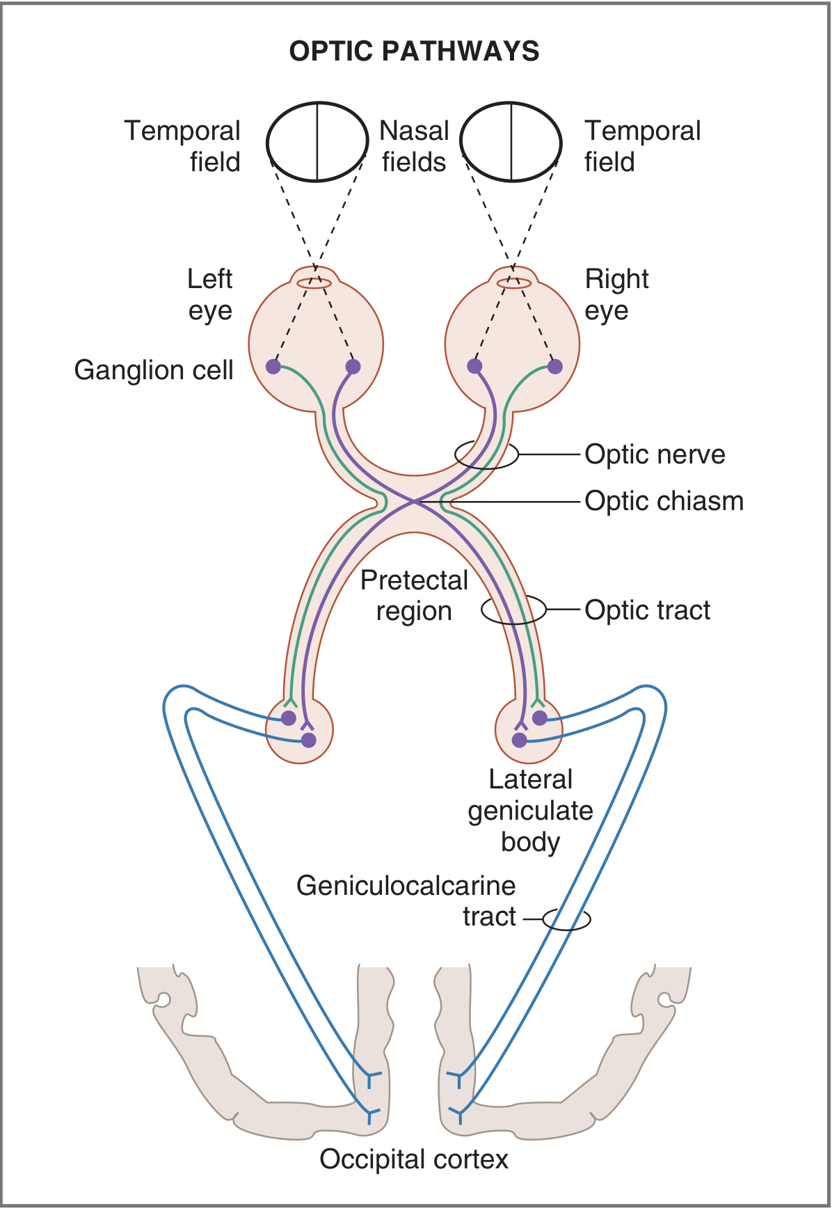
Fig. 3.18 Optic pathways showing the crossing of nasal fibers at the optic chiasm and projection to the occipital cortex. (Costanzo Physiology 7e)
Key Anatomical Principle at the Chiasm
- Temporal visual fields project onto the nasal hemiretina → fibers cross at the chiasm to the contralateral optic tract
- Nasal visual fields project onto the temporal hemiretina → fibers remain uncrossed and ascend ipsilaterally
- The ratio of crossed to uncrossed fibers at the chiasm is approximately 53:47
- Each optic tract therefore carries signals from the contralateral visual hemifield of both eyes
Costanzo Physiology 7e; Localization in Clinical Neurology 8e
2. Lesion-by-Lesion: Visual Field Defects
Each site of damage produces a characteristic and predictable visual field defect. The classic numbered lesions, from front to back:
Lesion 1 - Optic Nerve (Prechiasmal)
Defect: Complete monocular blindness (ipsilateral eye)
- All visual information from that eye is lost because the cut occurs before any fibers cross at the chiasm
- Example: left optic nerve lesion → complete blindness in the left eye
- RAPD (Relative Afferent Pupillary Defect / Marcus Gunn pupil): This is the hallmark of optic nerve disease - the injured eye shows a diminished direct pupillary response to light compared to the consensual response
- Partial optic nerve lesions produce: central scotoma, cecocentral scotoma, arcuate scotoma, or altitudinal defect
Common causes: Optic neuritis (MS), anterior ischemic optic neuropathy (AION), optic nerve compression (tumor, aneurysm), trauma, glaucoma
Lesion 2 - Optic Chiasm (Central)
Defect: Bitemporal hemianopia (heteronymous)
- The crossing nasal fibers from both eyes are disrupted
- Both temporal visual fields are lost (the "blinkers" effect)
- Called heteronymous because the defect is on opposite sides of the vertical meridian in each eye
- Most commonly caused by pituitary adenoma (suprasellar extension)
Other causes of chiasmal compression:
- Craniopharyngioma
- Rathke cleft cyst
- Meningioma of the tuberculum sellae
- Saccular aneurysm (anterior communicating or carotid artery)
- Sarcoidosis
- Metastatic carcinoma
- Hydrocephalus (downward herniation of the third ventricle)
Important variant - Junctional Scotoma (Traquair):
- Lesion at the junction of one optic nerve and the chiasm
- Result: ipsilateral central scotoma (optic nerve involvement) PLUS contralateral superior temporal quadrantanopia (from disruption of Wilbrand's knee - inferior nasal fibers that loop forward into the contralateral optic nerve before crossing)
Pre-fixed vs post-fixed chiasm:
- A posteriorly placed (post-fixed) chiasm → pituitary tumors present with an optic neuropathy pattern rather than bitemporal hemianopia
- An anteriorly placed (pre-fixed) chiasm → pituitary tumors may compress the optic tract instead, producing a homonymous defect
Adams and Victor's Principles of Neurology 12e; Localization in Clinical Neurology 8e
Lesion 3 - Optic Tract (Retrochiasmal - Anterior)
Defect: Contralateral homonymous hemianopia (incongruous)
- Cutting the left optic tract causes loss of the right visual field in both eyes:
- Loss of the temporal visual field of the right eye (crossed fibers)
- Loss of the nasal visual field of the left eye (uncrossed fibers)
- Incongruous = the defect pattern differs between the two eyes (one eye shows more loss than the other, but both on the same side of the vertical meridian)
- Incongruity indicates the lesion is anterior in the retrochiasmal pathway (fibers from the two eyes are not yet fully paired)
- RAPD may be present with optic tract lesions because of asymmetric crossing of pupillary fibers at the chiasm (~53% cross)
Common causes: Craniopharyngioma, temporal lobe herniation, pituitary tumor extending posteriorly
Lesion 4 - Lateral Geniculate Body (LGB)
Defect: Contralateral homonymous hemianopia (may be incongruous or congruous)
- Rare as an isolated lesion
- Lesion of the LGB may spare the pupillary reflex (pupillary fibers leave the optic tract to go to the pretectal area before the LGB)
Lesion 5 - Optic Radiation (Geniculocalcarine Tract)
The optic radiations split into two divisions after leaving the LGB:
A. Temporal Lobe - Meyer's Loop (Inferior Division)
Defect: Contralateral superior homonymous quadrantanopia ("pie in the sky")
- The lower fibers of the optic radiation swing in a wide arc anteriorly and inferiorly over the temporal horn of the lateral ventricle before sweeping posteriorly to the calcarine cortex - this is Meyer's (Archambault's) loop
- These fibers carry information from the inferior retina / superior visual field
- Damage produces a contralateral superior quadrantanopia: upper temporal field of the contralateral eye + upper nasal field of the ipsilateral eye lost
- First described by Harvey Cushing (before his description of the syndrome!)
Common causes: Temporal lobe tumors, temporal lobe epilepsy surgery (anterior temporal lobectomy), temporal lobe hemorrhage/infarct, herpes simplex encephalitis
B. Parietal Lobe (Superior Division)
Defect: Contralateral inferior homonymous quadrantanopia ("pie on the floor")
- The upper fibers pass through the parietal lobe
- Carry information from the superior retina / inferior visual field
- Parietal lobe lesions affect inferior visual field quadrants more than superior
Common causes: MCA territory infarcts, parietal lobe tumors, trauma
Lesion 6 - Primary Visual Cortex (Area 17, Occipital Lobe)
Defect: Contralateral homonymous hemianopia with macular sparing (congruous)
- The calcarine cortex contains a precise retinotopic map of the contralateral visual hemifield
- The macula (central vision, highest acuity) is represented at the occipital pole and has a disproportionately large cortical representation (cortical magnification)
- Macular sparing occurs because:
- The occipital pole has a dual blood supply (both PCA and MCA branches)
- Lesions rarely destroy all neurons representing the macula
- Congruous defect = identical pattern in both eyes → indicates the lesion is posterior in the visual pathway (cortex), where fibers from corresponding retinal points are tightly grouped
- Bilateral occipital lesions → cortical blindness (Anton syndrome - patient may deny blindness)
Altitudinal hemianopia: Confined by a horizontal border (not vertical meridian) - usually caused by bilateral occipital lobe lesions above or below the calcarine sulcus; unilateral altitudinal defect = usually optic neuropathy (NAION)
Special phenomenon - Blindsight: Even with complete occipital destruction, some patients retain unconscious visual processing (reaching accurately toward moving stimuli) attributed to preserved retinocular or geniculoprestriate connections.
Common causes: Posterior cerebral artery (PCA) infarction, occipital lobe tumors, trauma, migraine (transient), arteriovenous malformation
Adams and Victor's Principles of Neurology 12e; Costanzo Physiology 7e; Localization in Clinical Neurology 8e
3. Summary Table
| Site | Lesion | Visual Field Defect | Key Features |
|---|---|---|---|
| Optic nerve | Optic neuritis, AION, glaucoma | Monocular blindness / scotoma | RAPD present; no crossing yet |
| Chiasm (central) | Pituitary adenoma, craniopharyngioma | Bitemporal hemianopia | Heteronymous; "blinkers" |
| Chiasm (junction) | Lateral pituitary / optic nerve-chiasm junction | Ipsilateral central scotoma + contralateral superior temporal quad | Junctional scotoma / Traquair |
| Optic tract | Craniopharyngioma, herniation | Contralateral homonymous hemianopia | Incongruous; RAPD possible |
| LGB | Rare (vascular) | Contralateral homonymous hemianopia | Pupil reflex spared |
| Temporal lobe (Meyer's loop) | Tumour, temporal lobectomy, infarct | Contralateral superior quadrantanopia ("pie in the sky") | Incongruous; temporal lobe signs |
| Parietal lobe | MCA infarct, tumor | Contralateral inferior quadrantanopia ("pie on the floor") | Parietal lobe signs (neglect, etc.) |
| Occipital cortex (calcarine) | PCA infarct, tumor, trauma | Contralateral homonymous hemianopia with macular sparing | Congruous; pupil reflex intact |
| Bilateral occipital | Bilateral PCA infarcts, trauma | Cortical blindness ± Anton syndrome | Normal pupils |
4. Key Distinguishing Points
| Feature | Optic Nerve | Chiasm | Retrochiasmal |
|---|---|---|---|
| Eye involved | 1 eye | Both eyes | Both eyes |
| RAPD | Present | Absent | Usually absent (present in optic tract) |
| Pupil reflex | Impaired | Variable | Intact (post-LGB) |
| Congruity | N/A | N/A | Incongruous (anterior); Congruous (posterior) |
| Macular sparing | No | No | Yes (cortical lesions) |
5. Clinically Important Causes by Location
| Lesion Site | Classic Cause to Know |
|---|---|
| Optic nerve | Multiple sclerosis (optic neuritis - painful monocular visual loss); AION (painless) |
| Optic chiasm | Pituitary macroadenoma - most common; bitemporal hemianopia beginning superiorly |
| Optic tract | Temporal lobe herniation from raised ICP |
| Meyer's loop | Temporal lobectomy for epilepsy → "pie in the sky" quadrantanopia |
| Visual cortex | PCA territory stroke → contralateral hemianopia with macular sparing |
| Bilateral occipital | Cardiac arrest / hypoxia, bilateral PCA stroke → cortical blindness |
A great video to visualize this entire pathway and its lesions:
This is a shared conversation. Sign in to Orris to start your own chat.