
Side determination of testis
testis anatomy epididymis side determination diagram
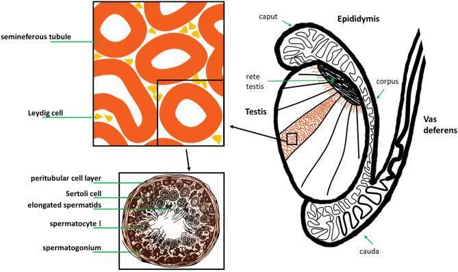
This medical illustration depicts the gross anatomy and histology of the human male reproductive system, specifically the testis and its associated ducts. The main diagram shows a sagittal-like section of the testis, highlighting internal lobules containing seminiferous tubules that converge at the rete testis. The epididymis is shown as a coiled ductal system attached to the posterior testis, divided into the caput (head), corpus (body), and cauda (tail), which then continues as the vas deferens. A magnified histological inset focuses on the interstitial space, showing Leydig cells clustered between cross-sections of seminiferous tubules. A second, high-magnification cross-section of a single seminiferous tubule illustrates the stages of spermatogenesis. Labeled structures include the peritubular cell layer (basal lamina), Sertoli cells for structural support, and germ cells in various stages of maturation: spermatogonia at the periphery, followed by spermatocytes I, and elongated spermatids positioned near the central lumen. This diagram serves as an educational tool for understanding the relationship between macroscopic anatomy and microscopic gametogenesis.
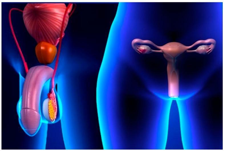
A 3D anatomical diagram illustrating the male and female reproductive systems side-by-side within stylized blue human silhouettes. On the left, the male anatomy includes the penis, a testis shown in cross-section within the scrotum, the epididymis, the vas deferens, the prostate gland, and the urinary bladder superiorly. On the right, the female anatomy depicts the uterus centrally, with the fallopian tubes extending laterally to the ovaries, and the vaginal canal positioned inferiorly. The visual highlights the pelvic location and relative spatial arrangement of primary and secondary sex organs. The image serves as an educational infographic for sexual health and reproductive anatomy, specifically in the context of public health strategies like the SAFE-S (Sexual behavior, Antibiotics, Facial cleanliness, Environmental improvement) strategy for controlling Chlamydia trachomatis and other sexually transmitted infections. The medical specialty is Urology and Gynecology, aimed at a general educational level.
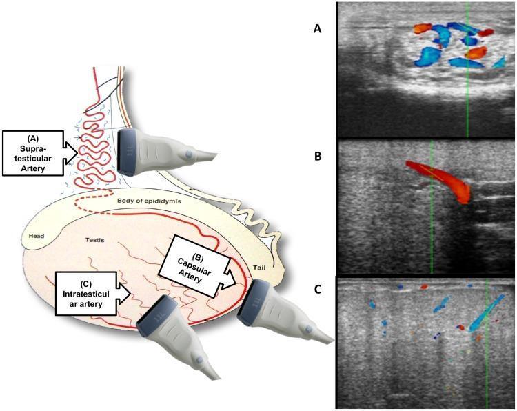
This composite educational graphic details the ultrasonographic assessment of testicular blood flow, combining an anatomical illustration with corresponding color Doppler images. The left panel features a diagram of the male reproductive anatomy, including the testis, epididymis (head, body, and tail), and spermatic cord, indicating three key vascular landmarks and transducer positions. The right panel displays three color Doppler ultrasound frames labeled A, B, and C. Frame A shows the supra-testicular artery (SA) in the spermatic cord, characterized by a complex cluster of tortuous vessels with mixed red and blue signals against a medium-gray B-mode background. Frame B depicts the capsular artery (CA) near the tail of the epididymis, showing a more linear, prominent vessel with a uniform red signal. Frame C illustrates intratesticular arteries (IA) within the testicular parenchyma, appearing as small, dispersed punctate signals within a heterogeneous, speckled echogenic background. This visual serves to teach the mapping and physiological flow characteristics of the testicular arterial supply for diagnostic purposes in reproductive medicine.
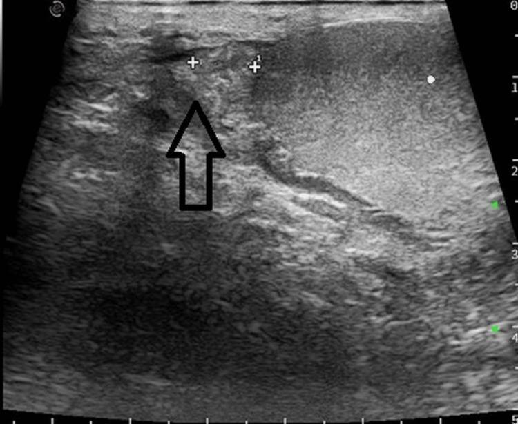
A grayscale diagnostic ultrasound image showing a sagittal view of the right scrotal contents. The image demonstrates the right testis and the right epididymis. The right testis (right side of the image) appears as an oval structure with a homogeneous, mid-level echotexture and a smooth, well-defined tunica albuginea. To the left of the testis, indicated by a black arrow, is the right epididymis. The epididymal head shows normal echogenicity, being slightly hyperechoic or isoechoic relative to the testicular parenchyma, with a characteristic granular echo pattern. Two calipers (+) are placed to measure the dimensions of the epididymal head. No evidence of hydrocele, epididymal enlargement, or abnormal focal lesions is observed in this view, supporting a finding of a normal right epididymis. This diagnostic image serves as a clinical reference for normal male reproductive anatomy and the evaluation of scrotal pathology such as epididymitis or torsion.
Side Determination of the Testis
Key Landmarks
1. Epididymis - Most Reliable Guide
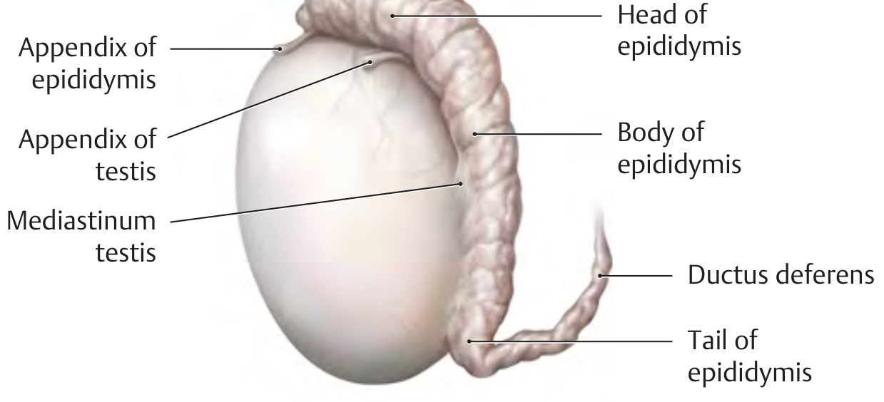
- Head (caput) - attaches to the upper pole of the testis
- Body (corpus) - runs down the posterolateral margin
- Tail (cauda) - at the lower pole, becomes continuous with the ductus deferens
Once you identify which surface of the testis carries the epididymis (posterolateral), you can orient the testis in 3D space. Then the medial surface (bearing the mediastinum testis) and lateral surface become identifiable, and side assignment follows.
2. Mediastinum Testis
- The anterior surface = smooth, free (covered by tunica vaginalis visceral layer)
- The posterior surface = where the epididymis, blood vessels, and ductus deferens enter/exit
3. Ductus Deferens
4. Upper vs Lower Pole
- Upper pole = bears the head of the epididymis (+ appendix testis and appendix epididymis)
- Lower pole = bears the tail of the epididymis, from which the ductus deferens begins
Step-by-Step Method to Determine Side
| Step | Action | Finding |
|---|---|---|
| 1 | Identify the epididymis | It always lies posterolaterally |
| 2 | Locate the head of epididymis | At the upper pole |
| 3 | Locate the tail of epididymis | At the lower pole, continued as vas deferens |
| 4 | Orient the testis so epididymis is facing you posterolaterally | Now anterior surface = smooth convex side |
| 5 | With posterior surface facing you and upper pole up | The side where the epididymis falls indicates the side of the body it came from |
- If the epididymis is to your right when facing the posterior surface (with head up), it is the right testis
- If the epididymis is to your left when facing the posterior surface (with head up), it is the left testis
Additional Clinical Notes
- In the body, the left testis normally hangs lower than the right (due to the longer left spermatic cord/testicular vein draining into the left renal vein at a right angle).
- The tunica vaginalis invests the anterior, medial, and lateral surfaces, but not the posterior surface where the epididymis and vessels attach - this bare posterior area (the "bare area") reinforces which surface is posterior.
- On ultrasound, the epididymis is identified mediastinum-adjacent, posterolateral to the testicular parenchyma, and this is used to label "right" or "left" during scrotal scanning.
Summary Rule (Exam Tip)
The epididymis is the key. It lies posterolaterally. Its head is superior, tail is inferior. With the posterior surface facing you and head upwards, the epididymis points toward the side of origin.