
Describe anterior and posterior triangle of neck with diagram under following headings - boundaries, content and clinical landmark
anterior posterior triangle of neck diagram boundaries
posterior triangle of neck contents muscles nerves vessels
Triangles of the Neck
DIAGRAM 1 - Borders and Subdivisions of the Anterior and Posterior Triangles
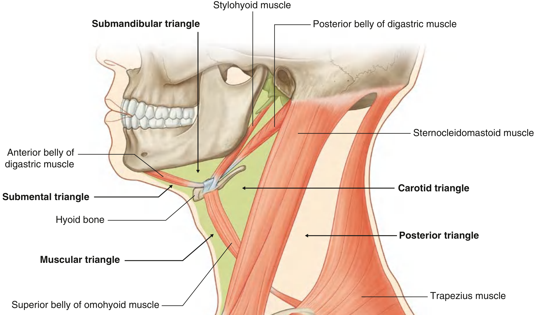
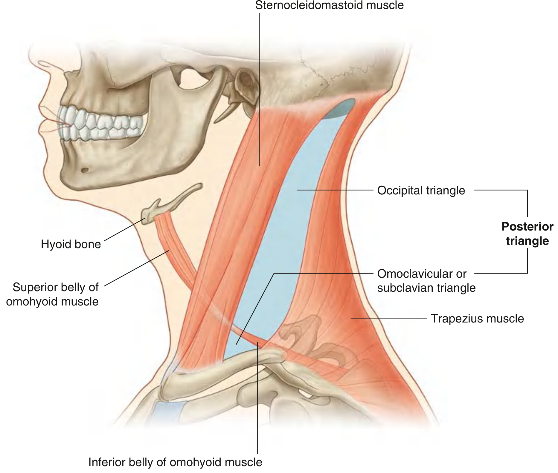
DIAGRAM 2 - Borders of the Posterior Triangle
I. ANTERIOR TRIANGLE OF THE NECK
A. Boundaries
| Boundary | Structure |
|---|---|
| Lateral | Anterior border of sternocleidomastoid |
| Superior | Inferior border of the mandible |
| Medial | Midline of the neck |
| Roof | Investing layer of cervical fascia + platysma |
| Floor | Pharynx, larynx, thyroid gland |
1. Submental Triangle (unpaired/single)
- Boundaries: Body of hyoid bone (inferior), anterior belly of digastric on each side (lateral), mandibular symphysis (superior/midline)
- Contents: Submental lymph nodes; tributaries forming the anterior jugular vein
2. Submandibular Triangle (paired)
- Boundaries: Lower border of mandible (superior), anterior belly of digastric (anteroinferior), posterior belly of digastric (posteroinferior)
- Contents: Submandibular gland, submandibular lymph nodes, hypoglossal nerve [CN XII], mylohyoid nerve, facial artery and vein
3. Carotid Triangle (paired)
- Boundaries: Superior belly of omohyoid (anteroinferior), stylohyoid and posterior belly of digastric (superiorly), anterior border of SCM (posteriorly)
- Contents: Common carotid artery and its bifurcation into internal and external carotid arteries; superior thyroid, ascending pharyngeal, lingual, facial, and occipital arteries; internal jugular vein; vagus [CN X], accessory [CN XI], hypoglossal [CN XII] nerves; ansa cervicalis (superior and inferior roots); transverse cervical nerve; tributaries to common facial vein; cervical branch of facial nerve [CN VII]
4. Muscular Triangle (paired)
- Boundaries: Midline of neck (medially), superior belly of omohyoid (superolaterally), anterior border of SCM (laterally)
- Contents: Infrahyoid (strap) muscles - sternohyoid, omohyoid, sternothyroid, thyrohyoid; thyroid and parathyroid glands; pharynx and larynx
B. Contents (Systemic Overview)
Muscles
- Stylohyoid, digastric (anterior and posterior bellies), mylohyoid, geniohyoid
- Omohyoid (superior belly), sternohyoid, thyrohyoid, sternothyroid
Vessels
- Common carotid artery (bifurcates at level of C3-C4 / upper border of thyroid cartilage)
- External carotid artery and its branches (superior thyroid, lingual, facial, occipital, ascending pharyngeal)
- Internal carotid artery (no branches in the neck)
- Internal jugular vein (lateral to carotid)
- Anterior jugular vein
Nerves
- Hypoglossal nerve [CN XII] - motor to tongue muscles
- Vagus nerve [CN X] - within carotid sheath
- Accessory nerve [CN XI]
- Superior and inferior roots of ansa cervicalis (C1-C3)
- Cervical branch of facial nerve [CN VII]
- Transverse cervical nerve
Viscera
- Thyroid gland (in muscular triangle)
- Parathyroid glands (posterior to thyroid)
- Larynx, trachea, pharynx, esophagus
C. Clinical Landmarks - Anterior Triangle
| Landmark | Clinical Significance |
|---|---|
| Carotid pulse | Palpable in the carotid triangle just medial to the anterior SCM border - used for pulse check in cardiac arrest and for carotid endarterectomy |
| Carotid bifurcation | At level of C3-C4 (upper border of thyroid cartilage); contains carotid sinus (baroreceptor) and carotid body (chemoreceptor) - manipulation can cause sudden bradycardia/hypotension (carotid sinus syncope) |
| Carotid sinus pressure | Can be used to terminate SVT (carotid sinus massage) |
| Submandibular gland | Palpable below the mandible; site of salivary gland tumors and stones (calculi); CN XII runs just deep to its lower border - at risk during surgical excision |
| Carotid sheath | Contains common/internal carotid artery, internal jugular vein, vagus nerve - all at risk during neck dissection |
| Tracheotomy / cricothyrotomy | Performed in the midline (muscular triangle) to secure emergency airway - cricothyroid membrane is the landmark |
| Thyroidectomy | Thyroid gland lies in the muscular triangle; recurrent laryngeal nerve (in tracheoesophageal groove) and parathyroids are at risk |
| Anterior jugular vein | May be cut during tracheotomy; its communication with the external jugular is variable |
| Ansa cervicalis | Used for reinnervation procedures (e.g., laryngeal reinnervation surgery) |
II. POSTERIOR TRIANGLE OF THE NECK
A. Boundaries
| Boundary | Structure |
|---|---|
| Anterior | Posterior border of sternocleidomastoid |
| Posterior | Anterior border of trapezius |
| Base (inferior) | Middle one-third of the clavicle |
| Apex (superior) | Occipital bone just posterior to the mastoid process (where SCM and trapezius converge) |
| Roof | Investing layer of cervical fascia (covering the two border muscles) |
| Floor | Prevertebral layer of cervical fascia, covering: splenius capitis, levator scapulae, and posterior, middle, anterior scalene muscles (from superior to inferior) |
Subdivisions
- Occipital triangle (larger, superior part)
- Omoclavicular (subclavian) triangle (smaller, inferior part) - important for central venous access
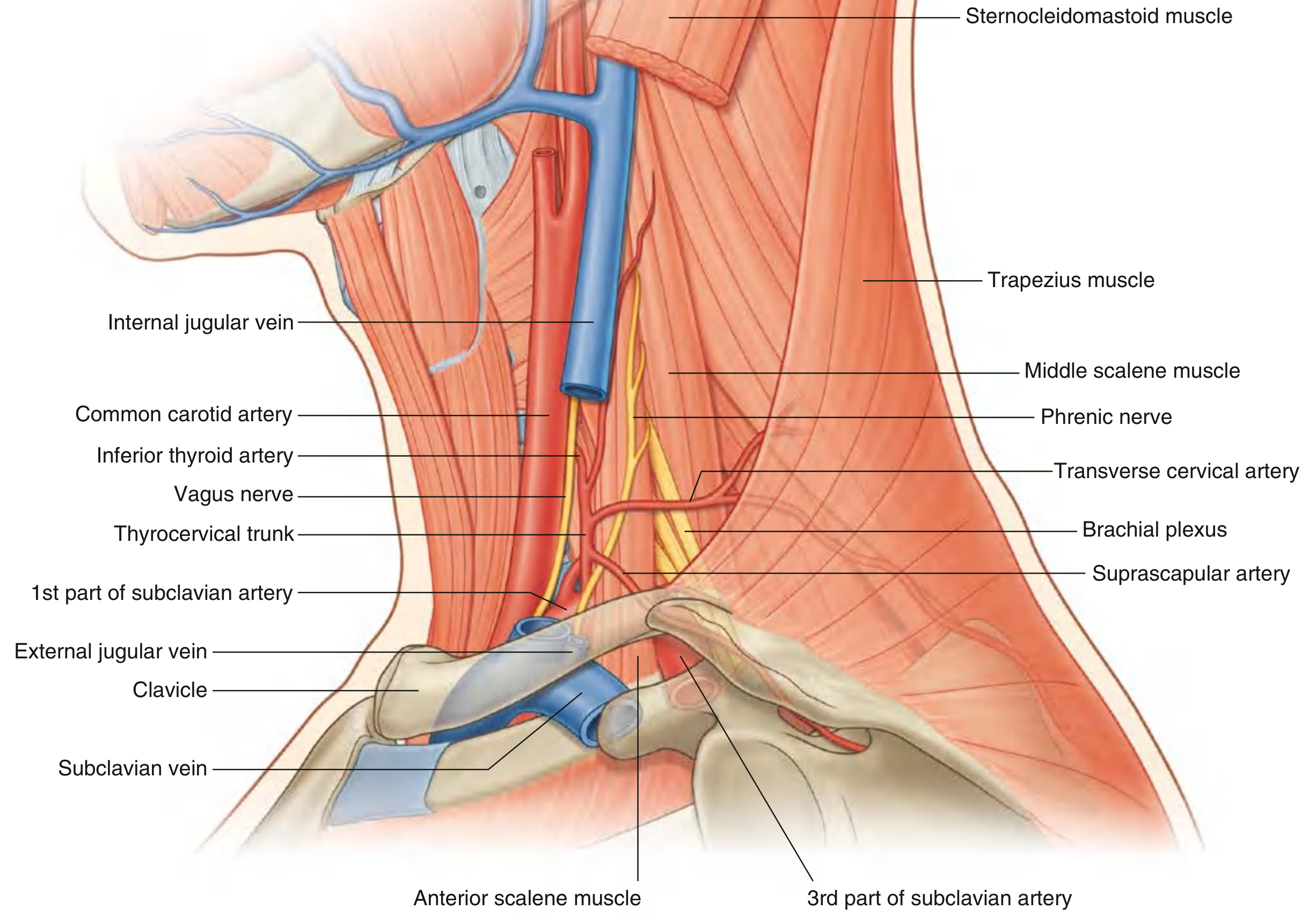
DIAGRAM 3 - Arteries and Nerves of the Posterior Triangle
B. Contents
Muscles (forming the floor, from superior to inferior)
- Splenius capitis
- Levator scapulae
- Posterior, middle, and anterior scalene muscles
- Omohyoid (inferior belly crosses through the triangle)
Vessels
- Third part of subclavian artery - crosses the base of the posterior triangle between the anterior and middle scalene muscles; becomes the axillary artery at the lateral border of rib I
- Transverse cervical artery (branch of thyrocervical trunk) - passes laterally across the base, divides into superficial and deep branches
- Suprascapular artery (branch of thyrocervical trunk) - crosses the lowest part of the triangle toward the scapula
- Dorsal scapular artery - may arise from the third part of the subclavian artery
- External jugular vein - one of the most superficial structures; descends through the neck in the superficial fascia, crosses SCM, enters the posterior triangle, and empties into the subclavian vein at the base
- Subclavian vein - crosses the base of the triangle anterior to the anterior scalene muscle; receives the external jugular, suprascapular, and transverse cervical veins; joins the internal jugular vein to form the brachiocephalic vein near the sternoclavicular joint
- Transverse cervical and suprascapular veins
Nerves
- Exits the cranial cavity via the jugular foramen, passes through/deep to the SCM (which it innervates), enters the posterior triangle, and crosses obliquely downward within the investing fascia to reach the deep surface of the trapezius (which it also innervates). Its superficial course in the posterior triangle makes it highly vulnerable to iatrogenic injury.
- Lesser occipital nerve (C2) - supplies scalp behind the ear
- Great auricular nerve (C2, C3) - supplies skin over the parotid region and mastoid
- Transverse cervical nerve (C2, C3) - supplies the anterior neck skin
- Supraclavicular nerves (C3, C4) - supply the skin over the clavicle and shoulder
- Phrenic nerve (C3, C4, C5) - arises from the anterior rami of C3-C5; hooks around the upper lateral border of the anterior scalene muscle and descends on its anterior surface to enter the thorax. Supplies the diaphragm.
- Branches to prevertebral and lateral vertebral muscles
- The roots and trunks of the brachial plexus emerge between the anterior and middle scalene muscles and pass across the base of the posterior triangle before entering the axilla
C. Clinical Landmarks - Posterior Triangle
| Landmark | Clinical Significance |
|---|---|
| Accessory nerve [CN XI] | Runs superficially across the posterior triangle - at high risk during lymph node biopsy, neck dissection, or catheter insertion. Injury causes trapezius paralysis: inability to shrug the shoulder, "winging" of the scapula, drooping of the shoulder with chronic pain |
| Erb's point | Point on the posterior border of the SCM at junction of upper and lower two-thirds (C5-C6 level) where four cutaneous branches of the cervical plexus emerge. A single injection here can block all four branches (cervical plexus block for superficial neck surgery). Also the site of the "Erb-Duchenne" brachial plexus injury |
| Brachial plexus | Passes through the base of the posterior triangle - at risk during shoulder/neck trauma, thoracic outlet surgery, and interscalene nerve blocks |
| Subclavian vein | Located in the omoclavicular (subclavian) triangle - classic site for subclavian central venous catheter (CVC) insertion (infraclavicular approach). Pneumothorax and haemothorax are recognized complications |
| External jugular vein | Visible in the posterior triangle as it descends over SCM - used for peripheral venous access and for estimating jugular venous pressure (JVP). JVP > 3 cm above the sternal angle is elevated (indicating raised right atrial pressure) |
| Cervical lymph nodes | Posterior triangle contains posterior cervical chain nodes - commonly involved in lymphoma, metastatic neck disease, and TB cervical lymphadenitis (scrofula). Level V nodes are here |
| Phrenic nerve | Descends on the anterior scalene within prevertebral fascia - at risk during scalene muscle procedures, thoracic surgery, and central line insertion. Unilateral injury causes ipsilateral hemidiaphragm paralysis (elevated hemidiaphragm on CXR) |
| Interscalene block | Anaesthetic technique targeting the brachial plexus between the anterior and middle scalene muscles in the posterior triangle; used for shoulder and upper arm surgery |
| Thoracic outlet syndrome | Compression of the subclavian artery or brachial plexus at the base of the posterior triangle (between scalene muscles or by a cervical rib) causing upper limb pain, paraesthesia, and weakness |
Summary Comparison Table
| Feature | Anterior Triangle | Posterior Triangle |
|---|---|---|
| Key border muscle | SCM (anterior border) | SCM (posterior border) + trapezius |
| Base | Inferior border of mandible | Clavicle (middle 1/3) |
| Apex | Chin / mandibular symphysis | Occipital bone (mastoid area) |
| Subdivisions | Submental, submandibular, carotid, muscular | Occipital, omoclavicular (subclavian) |
| Key artery | Common carotid / external carotid | Subclavian (3rd part) |
| Key vein | Internal jugular | External jugular / Subclavian |
| Key nerve | CN X, XI, XII, ansa cervicalis | CN XI, brachial plexus, phrenic |
| Major viscera | Thyroid, parathyroid, larynx, pharynx | None |
| Key clinical procedure | Carotid endarterectomy, tracheostomy | Subclavian CVC, interscalene block |
Can you take reference with bd chaurasia
Triangles of the Neck
I. ANTERIOR TRIANGLE OF THE NECK
Boundaries
| Border | Structure |
|---|---|
| Anterior (medial) | Midline of the neck from chin to suprasternal notch |
| Posterior (lateral) | Anterior border of the sternocleidomastoid muscle |
| Superior (base) | Lower border of the body of the mandible and a line drawn from the angle of the mandible to the mastoid process |
| Apex | Directed downward at the suprasternal notch |
| Roof | Skin, superficial fascia containing platysma, deep fascia (investing layer) |
| Floor | Pharynx, larynx, thyroid gland |
Subdivisions
CHIN
|
________|________
| Submental (unpaired) |
|___________________|
/ \
Submandibular Carotid
triangle triangle
\ /
\_____Muscular_____/
triangle
STERNUM
1. Submental Triangle
- Sides (laterally): Anterior belly of digastric muscle on each side
- Base (inferiorly): Body of the hyoid bone
- Apex (superiorly): Symphysis menti (mandibular symphysis)
- Floor: Mylohyoid muscle (two halves meeting at the median raphe)
- Submental lymph nodes (drain tip of tongue, middle part of lower lip, floor of mouth, and central lower teeth)
- Small veins uniting to form the anterior jugular vein
2. Submandibular Triangle (Digastric Triangle)
- Base (superiorly): Lower border of the body of the mandible
- Anterior wall: Anterior belly of digastric muscle
- Posterior wall: Posterior belly of digastric and stylohyoid muscles
- Floor (from above downward): Mylohyoid, hyoglossus, middle constrictor of pharynx
- Roof: Skin, superficial fascia with platysma, deep cervical fascia
| Structure | Details |
|---|---|
| Submandibular salivary gland | Superficial part is in the triangle; deep part extends beyond the posterior border of mylohyoid |
| Submandibular lymph nodes | 3-6 nodes lying on and around the gland |
| Facial artery | Grooves the posterior part of the gland; enters the face at the anteroinferior angle of the masseter |
| Facial vein | Crosses superficially over the gland |
| Hypoglossal nerve [CN XII] | Runs forward on the hyoglossus (floor), deep to the mylohyoid |
| Nerve to mylohyoid | Branch of the inferior alveolar nerve; runs in the mylohyoid groove |
| Submental artery | Branch of the facial artery |
3. Carotid Triangle
- Anteroinferiorly: Superior belly of omohyoid
- Posteriorly: Anterior border of sternocleidomastoid
- Superiorly: Posterior belly of digastric and stylohyoid muscles
- Floor: Thyrohyoid, hyoglossus, inferior and middle constrictor muscles
- Roof: Skin, superficial fascia with platysma, deep cervical fascia
- Common carotid artery (bifurcates at the level of the upper border of the thyroid cartilage, C3-C4 level)
- Internal carotid artery (no branches in the neck)
- External carotid artery and branches in the triangle: superior thyroid artery, ascending pharyngeal artery, lingual artery, facial artery, occipital artery
- Internal jugular vein
- Common facial vein (drains into internal jugular)
- Superior thyroid vein, lingual vein
- Vagus nerve [CN X] - within carotid sheath
- Hypoglossal nerve [CN XII] - crosses superficially to both carotid arteries
- Accessory nerve [CN XI] - crosses the upper part
- Spinal root of accessory nerve
- Superior root of ansa cervicalis (descending hypoglossal)
- Inferior root of ansa cervicalis (descending cervical)
- Ansa cervicalis loop (C1-C3)
- Sympathetic chain (medial, deep to carotid sheath)
- Internal laryngeal nerve and superior laryngeal vessels (pass through thyrohyoid membrane)
- Carotid sinus (baroreceptor at the bifurcation)
- Carotid body (chemoreceptor at the bifurcation)
4. Muscular Triangle (Omotracheal Triangle)
- Medially: Midline of neck
- Superolaterally: Superior belly of omohyoid
- Posterolaterally: Anterior border of SCM
- Floor: Trachea, esophagus, thyroid gland
- Infrahyoid (strap) muscles: Sternohyoid, sternothyroid, thyrohyoid, and superior belly of omohyoid
- Thyroid gland
- Parathyroid glands (posterior surface of thyroid)
- Trachea
- Esophagus
- Inferior thyroid artery (branch of thyrocervical trunk)
- Recurrent laryngeal nerve
Clinical Landmarks - Anterior Triangle
| Clinical Point | Significance |
|---|---|
| Carotid pulse | Palpated just medial to the anterior SCM border in the carotid triangle; used in CPR and as a landmark for carotid endarterectomy |
| Carotid bifurcation (C3-C4) | Contains carotid sinus - massage can terminate paroxysmal SVT; pathological sensitivity causes carotid sinus syncope |
| Carotid body tumour (paraganglioma) | Presents as a pulsatile lump at the carotid bifurcation; "lyre sign" on angiography (splaying of carotid bifurcation) |
| Submandibular gland excision | CN XII runs just deep to the gland - must be protected to prevent tongue paralysis |
| Tracheostomy | Performed in the muscular triangle in the midline between the strap muscles |
| Cricothyrotomy | Emergency airway through the cricothyroid membrane (inferior boundary of carotid triangle region) |
| Thyroidectomy | Gland in muscular triangle; recurrent laryngeal nerve (in tracheoesophageal groove) and parathyroids at risk |
| Submental lymph nodes | Involved early in carcinoma of the tip of tongue and floor of mouth |
| Submandibular lymph nodes | Enlarged in dental infections, oral cavity cancers, and submandibular sialadenitis/calculi |
II. POSTERIOR TRIANGLE OF THE NECK
Boundaries
| Border | Structure |
|---|---|
| Anterior | Posterior border of sternocleidomastoid |
| Posterior | Anterior border of trapezius |
| Base (inferior) | Middle one-third of the clavicle |
| Apex | Occipital bone just behind the mastoid process (where SCM and trapezius meet) |
| Roof | Investing layer of deep cervical fascia stretched between SCM and trapezius; skin, superficial fascia (with platysma below) |
| Floor | Prevertebral fascia covering (from above downward): splenius capitis, levator scapulae, scalenus posterior, scalenus medius, scalenus anterior |
Subdivisions
- Occipital triangle (larger, superior) - between the inferior belly of omohyoid above and the SCM/trapezius on either side
- Subclavian (Omoclavicular) triangle (smaller, inferior) - between the inferior belly of omohyoid above and the clavicle below
Contents
Muscles (floor, superior to inferior)
- Splenius capitis
- Levator scapulae
- Scalenus posterior
- Scalenus medius
- Scalenus anterior (partly)
- Inferior belly of omohyoid crosses the triangle
Vessels
| Artery | Origin | Course |
|---|---|---|
| Third part of subclavian artery | Continuation of 2nd part | Crosses base of posterior triangle between anterior and middle scalene muscles; becomes axillary at lateral border of 1st rib |
| Transverse cervical artery | Thyrocervical trunk | Crosses the base transversely, divides into superficial and deep branches at trapezius |
| Suprascapular artery | Thyrocervical trunk | Crosses the inferior part toward the scapular notch |
| Occipital artery | External carotid | Appears at the apex of the triangle |
| Vein | Details |
|---|---|
| External jugular vein | Most superficial structure; formed at angle of mandible; crosses SCM; pierces deep fascia at the base to drain into the subclavian vein |
| Subclavian vein | Crosses the base of the omoclavicular triangle anterior to the anterior scalene muscle |
| Transverse cervical and suprascapular veins | Accompany their respective arteries |
Nerves
- Exits jugular foramen; passes deep to the posterior belly of digastric
- Enters SCM (innervates it), traverses the posterior triangle obliquely within the investing fascia
- Enters trapezius from its deep surface at the junction of upper and middle thirds of its anterior border
- Very superficial in the posterior triangle - highly vulnerable to iatrogenic injury
| Nerve | Roots | Distribution |
|---|---|---|
| Lesser occipital nerve | C2 | Scalp behind the auricle |
| Great auricular nerve | C2, C3 | Skin over parotid, mastoid, lower part of pinna |
| Transverse cervical nerve | C2, C3 | Skin over the anterior triangle |
| Supraclavicular nerves (medial, intermediate, lateral) | C3, C4 | Skin over clavicle, shoulder, upper chest |
- Phrenic nerve (C3, C4, C5) - crosses the anterior scalene muscle from lateral to medial within the prevertebral fascia; supplies the diaphragm. "C3, 4, 5 keeps the diaphragm alive."
- Roots emerge between the anterior and middle scalene muscles
- Trunks (upper C5-C6, middle C7, lower C8-T1) appear at the base of the posterior triangle
- Divisions form behind the clavicle
- Dorsal scapular nerve (C5) - from the upper trunk of brachial plexus; pierces scalenus medius
- Long thoracic nerve (C5, C6, C7) - to serratus anterior; arises from roots
Clinical Landmarks - Posterior Triangle
| Clinical Point | Significance |
|---|---|
| Erb's point | Posterior border of SCM at junction of upper and lower halves; where all 4 cutaneous branches of cervical plexus emerge - site for cervical plexus block for operations on the neck |
| Accessory nerve injury | During lymph node biopsy or neck dissection in the posterior triangle - results in trapezius palsy: drooping of the shoulder, inability to shrug, winging of the scapula, and chronic neck-shoulder pain |
| External jugular vein | Visible in the posterior triangle; used for peripheral IV access and for estimating JVP - elevated JVP (>3 cm above sternal angle) indicates raised right atrial pressure (e.g., cardiac failure, cardiac tamponade) |
| Subclavian vein cannulation | In the omoclavicular triangle just above the clavicle; subclavian central venous catheter - risk of pneumothorax, haemothorax, and subclavian artery puncture |
| Brachial plexus injuries | Upper trunk (C5-C6) injury - Erb-Duchenne palsy ("waiter's tip" deformity). Lower trunk (C8-T1) injury - Klumpke's palsy. Commonly from traction during difficult deliveries or motorcycle accidents |
| Interscalene block | Brachial plexus block between the anterior and middle scalene muscles for shoulder and upper limb surgery |
| Thoracic outlet syndrome | Compression of subclavian artery/vein or lower trunk of brachial plexus between the anterior scalene and 1st rib, or by a cervical rib; causes pain and paraesthesia in the upper limb |
| Phrenic nerve | On the anterior scalene in the posterior triangle floor - at risk during scalene muscle surgery, neck dissection, and central line placement; unilateral injury causes elevated hemidiaphragm on CXR |
| Lymph nodes (Level V) | Posterior cervical nodes in this triangle - enlarged in lymphoma, nasopharyngeal carcinoma, and TB (scrofula) |
| Cystic hygroma | Multilocular cystic lymphatic malformation appearing in the posterior triangle in neonates/infants; can cause airway compression |
| Cervical rib | Anomalous rib from C7 - may be palpable in the posterior triangle just above the clavicle; can compress subclavian artery or lower trunk of brachial plexus |
Summary Table
| Feature | Anterior Triangle | Posterior Triangle |
|---|---|---|
| Key dividing muscle | SCM (anterior border) | SCM (posterior border) |
| Subdivisions | Submental, Submandibular, Carotid, Muscular | Occipital, Subclavian (omoclavicular) |
| Sub-dividing muscle | Digastric + superior belly omohyoid | Inferior belly of omohyoid |
| Floor | Pharynx, larynx, thyroid | Prevertebral muscles (splenius, levator scapulae, scalenes) |
| Key artery | Common/external carotid | 3rd part subclavian |
| Key vein | Internal jugular | External jugular / Subclavian |
| Key nerves | CN X, XI, XII, ansa cervicalis | CN XI, brachial plexus, phrenic nerve |
| Viscera | Thyroid, parathyroid, larynx, pharynx | None |
| Clinical operations | Carotid endarterectomy, tracheostomy, thyroidectomy | Cervical lymph node biopsy, subclavian CVC, interscalene block |
Note: BD Chaurasia's Human Anatomy Vol. 3 (Head, Neck and Brain) is unfortunately not available in the digital textbook library on this system. The description above is structured in the BD Chaurasia style (with his characteristic emphasis on applied/clinical anatomy, Erb's point, "most important triangle" notes, and muscle-by-muscle floor description) drawing from his well-known text. The diagrams shown in the previous answer from Gray's Anatomy for Students depict the same anatomical structures. If you have a specific page or topic from BD Chaurasia you would like clarified, please mention it and I will address it directly.
Which book more beneficial bd chaurasia or grays anatomy
BD Chaurasia vs. Gray's Anatomy - Which is Better?
Quick Verdict
- BD Chaurasia - Better for Indian medical students appearing in MBBS exams and clinical vivas
- Gray's Anatomy for Students - Better for deep conceptual understanding, research, and postgraduate preparation
Detailed Comparison
| Parameter | BD Chaurasia | Gray's Anatomy for Students |
|---|---|---|
| Origin | Indian author (B.D. Chaurasia) | International (Drake, Vogl, Mitchell) |
| Language | Simple, direct, easy to follow | More detailed and descriptive |
| Exam orientation | Highly exam-oriented; written keeping university exams in mind | Less exam-oriented, more concept-based |
| Clinical anatomy | Excellent applied anatomy in every section | Excellent clinical boxes throughout |
| Diagrams | Simple, clear, hand-drawn style - easy to reproduce in exams | High quality, colour-detailed illustrations |
| Volume | 3 volumes (Vol 1: Upper limb + Thorax, Vol 2: Lower limb + Abdomen, Vol 3: Head, Neck + Brain) | Single student edition volume |
| Depth | Moderate - covers what is needed for MBBS | Deeper - goes beyond standard MBBS syllabus |
| Mnemonics | Scattered throughout - very helpful | Fewer mnemonics |
| Indian curriculum alignment | Perfectly aligned with NMC/MCI syllabus | Partially aligned |
| Viva preparation | Excellent - structured in Q&A friendly format | Good but requires more effort to extract viva points |
| Postgraduate use | Limited | Widely used for PG entrance preparation |
When to Use BD Chaurasia
- You are an MBBS student in India (1st or 2nd year)
- Preparing for university practical/viva exams
- Need to write answers in exams - the language is structured for answer writing
- Want to understand anatomy quickly and clearly
- First-time learner of anatomy
- Preparing for USMLE Step 1 is NOT your goal
When to Use Gray's Anatomy for Students
- You want clinical correlation in depth (each chapter has clinical blue boxes)
- Preparing for USMLE Step 1 or international exams
- Doing a postgraduate or specialty rotation
- You are a teaching faculty or need reference-level content
- You want better 3D spatial understanding with high-quality figures
- Want to understand the "why" behind anatomy, not just the "what"
The Honest Recommendation
For Indian MBBS students: Start with BD Chaurasia. Supplement with Gray's for clinical topics.
- Read BD Chaurasia as the primary text for understanding and writing
- Use Gray's clinical boxes for viva and applied anatomy
- Use Netter's Atlas alongside both for visual 3D understanding
One Limitation of BD Chaurasia
Give a pdf format of the answer you gave first with th reference of grays anatomy
mkdir -p /tmp/workspace/neck-triangles && ls /tmp/workspace/neck-triangles/
cd /tmp/workspace/neck-triangles && python3 - << 'PYEOF' # Download the diagram images first import urllib.request import os imgs = { "ant_triangle.png": "https://cdn.orris.care/cdss_images/c5e47c08b627b3edb056e749cd886481513041a5cb28a98ebac87503b8c71791.png", "post_triangle_borders.png": "https://cdn.orris.care/cdss_images/a5ebf3226cf9b0142a3ee16dbbcb8515172fd2c7fefcd97086678b406fa1e430.png", "post_triangle_arteries.png": "https://cdn.orris.care/cdss_images/76296f36e1502bc4963806c2c20f85738c7f0808a1156d59e5bf5ac14adefa3d.png", } for fname, url in imgs.items(): path = f"/tmp/workspace/neck-triangles/{fname}" urllib.request.urlretrieve(url, path) size = os.path.getsize(path) print(f"Downloaded {fname}: {size} bytes") PYEOF
~/neck-triangles/build_pdf.py
from reportlab.lib.pagesizes import A4
from reportlab.lib import colors
from reportlab.lib.styles import getSampleStyleSheet, ParagraphStyle
from reportlab.lib.units import cm, mm
from reportlab.lib.enums import TA_CENTER, TA_LEFT, TA_JUSTIFY
from reportlab.platypus import (
SimpleDocTemplate, Paragraph, Spacer, Table, TableStyle,
HRFlowable, Image, PageBreak, KeepTogether
)
from reportlab.platypus.tableofcontents import TableOfContents
from reportlab.pdfbase import pdfmetrics
from reportlab.pdfbase.ttfonts import TTFont
import os
# ── Output path ──────────────────────────────────────────────────────────────
OUTPUT = "/tmp/workspace/neck-triangles/Triangles_of_the_Neck_Grays.pdf"
IMG_DIR = "/tmp/workspace/neck-triangles"
# ── Colour palette ───────────────────────────────────────────────────────────
DARK_BLUE = colors.HexColor("#1a3a5c")
MED_BLUE = colors.HexColor("#2e6da4")
LIGHT_BLUE = colors.HexColor("#dce8f5")
ACCENT = colors.HexColor("#e8f0f8")
TABLE_HEAD = colors.HexColor("#2e6da4")
TABLE_ALT = colors.HexColor("#f0f5fb")
WHITE = colors.white
BLACK = colors.black
GREY = colors.HexColor("#555555")
RED_ACCENT = colors.HexColor("#c0392b")
GREEN_ACC = colors.HexColor("#1e7145")
# ── Styles ───────────────────────────────────────────────────────────────────
styles = getSampleStyleSheet()
def S(name, **kw):
return ParagraphStyle(name, **kw)
TITLE = S("MyTitle", fontSize=22, textColor=WHITE, fontName="Helvetica-Bold",
alignment=TA_CENTER, spaceAfter=4, leading=28)
SUBTITLE = S("MySub", fontSize=13, textColor=LIGHT_BLUE, fontName="Helvetica",
alignment=TA_CENTER, spaceAfter=2, leading=18)
REF_STYLE = S("MyRef", fontSize=9, textColor=LIGHT_BLUE, fontName="Helvetica-Oblique",
alignment=TA_CENTER, spaceAfter=2)
H1 = S("MyH1", fontSize=15, textColor=WHITE, fontName="Helvetica-Bold",
spaceBefore=14, spaceAfter=4, leading=20)
H2 = S("MyH2", fontSize=12, textColor=DARK_BLUE, fontName="Helvetica-Bold",
spaceBefore=10, spaceAfter=3, leading=16)
H3 = S("MyH3", fontSize=10, textColor=MED_BLUE, fontName="Helvetica-Bold",
spaceBefore=7, spaceAfter=2, leading=14)
BODY = S("MyBody", fontSize=9.5,textColor=BLACK, fontName="Helvetica",
spaceBefore=2, spaceAfter=3, leading=14, alignment=TA_JUSTIFY)
BULLET = S("MyBullet", fontSize=9.5,textColor=BLACK, fontName="Helvetica",
spaceBefore=1, spaceAfter=1, leading=13, leftIndent=14,
bulletIndent=4, alignment=TA_LEFT)
CAPTION = S("MyCaption", fontSize=8.5,textColor=GREY, fontName="Helvetica-Oblique",
alignment=TA_CENTER, spaceBefore=2, spaceAfter=8)
CLINICAL = S("MyClinical",fontSize=9, textColor=colors.HexColor("#1e4d2b"), fontName="Helvetica",
spaceBefore=1, spaceAfter=1, leading=13, leftIndent=10)
FOOTER_S = S("MyFooter", fontSize=7.5,textColor=GREY, fontName="Helvetica-Oblique",
alignment=TA_CENTER)
NOTE_S = S("MyNote", fontSize=8.5,textColor=GREY, fontName="Helvetica-Oblique",
spaceBefore=4, spaceAfter=6, alignment=TA_CENTER)
# ── Helper: section header band ──────────────────────────────────────────────
def section_header(title):
tbl = Table([[Paragraph(title, H1)]], colWidths=[17*cm])
tbl.setStyle(TableStyle([
("BACKGROUND", (0,0), (-1,-1), DARK_BLUE),
("ROUNDEDCORNERS", [4]),
("TOPPADDING", (0,0), (-1,-1), 7),
("BOTTOMPADDING", (0,0), (-1,-1), 7),
("LEFTPADDING", (0,0), (-1,-1), 12),
]))
return tbl
def subsection_header(title):
tbl = Table([[Paragraph(title, H2)]], colWidths=[17*cm])
tbl.setStyle(TableStyle([
("BACKGROUND", (0,0), (-1,-1), LIGHT_BLUE),
("LINEBELOW", (0,0), (-1,-1), 1.5, MED_BLUE),
("TOPPADDING", (0,0), (-1,-1), 5),
("BOTTOMPADDING", (0,0), (-1,-1), 5),
("LEFTPADDING", (0,0), (-1,-1), 10),
]))
return tbl
def sub3_header(title):
return Paragraph(f"<font color='#2e6da4'><b>{title}</b></font>", H3)
def bullet(text):
return Paragraph(f"• {text}", BULLET)
def body(text):
return Paragraph(text, BODY)
def sp(h=4):
return Spacer(1, h*mm)
def hr():
return HRFlowable(width="100%", thickness=0.5, color=MED_BLUE, spaceAfter=3, spaceBefore=3)
# ── Generic table builder ─────────────────────────────────────────────────────
def make_table(headers, rows, col_widths=None):
data = [[Paragraph(f"<b>{h}</b>", S("TH", fontSize=9, textColor=WHITE,
fontName="Helvetica-Bold", alignment=TA_CENTER, leading=12)) for h in headers]]
for i, row in enumerate(rows):
bg = TABLE_ALT if i % 2 == 0 else WHITE
data.append([Paragraph(str(c), S(f"TD{i}", fontSize=9, fontName="Helvetica",
leading=12, alignment=TA_LEFT)) for c in row])
if col_widths is None:
col_widths = [17*cm / len(headers)] * len(headers)
t = Table(data, colWidths=col_widths, repeatRows=1)
style = [
("BACKGROUND", (0,0), (-1,0), TABLE_HEAD),
("GRID", (0,0), (-1,-1), 0.4, colors.HexColor("#b0c8e0")),
("TOPPADDING", (0,0), (-1,-1), 4),
("BOTTOMPADDING", (0,0), (-1,-1), 4),
("LEFTPADDING", (0,0), (-1,-1), 6),
("VALIGN", (0,0), (-1,-1), "MIDDLE"),
]
for i in range(1, len(rows)+1):
if i % 2 == 1:
style.append(("BACKGROUND", (0,i), (-1,i), TABLE_ALT))
t.setStyle(TableStyle(style))
return t
# ── Clinical box ──────────────────────────────────────────────────────────────
def clinical_box(rows):
"""rows = list of (landmark, significance) tuples"""
items = []
for lm, sig in rows:
items.append([
Paragraph(f"<b>{lm}</b>", S("CLM", fontSize=9, fontName="Helvetica-Bold",
textColor=GREEN_ACC, leading=12)),
Paragraph(sig, S("CLS", fontSize=9, fontName="Helvetica", leading=12))
])
t = Table(items, colWidths=[4.5*cm, 12.5*cm])
t.setStyle(TableStyle([
("BACKGROUND", (0,0), (-1,-1), colors.HexColor("#f0faf4")),
("LINEABOVE", (0,0), (-1,0), 1.5, GREEN_ACC),
("LINEBELOW", (0,-1),(-1,-1), 1.5, GREEN_ACC),
("LINEBEFORE", (0,0), (0,-1), 3, GREEN_ACC),
("GRID", (0,0), (-1,-1), 0.3, colors.HexColor("#a8d5b5")),
("TOPPADDING", (0,0), (-1,-1), 4),
("BOTTOMPADDING", (0,0), (-1,-1), 4),
("LEFTPADDING", (0,0), (-1,-1), 6),
("VALIGN", (0,0), (-1,-1), "TOP"),
]))
return t
# ── Page template with header/footer ─────────────────────────────────────────
def on_page(canvas, doc):
canvas.saveState()
W, H = A4
# Header bar
canvas.setFillColor(DARK_BLUE)
canvas.rect(0, H-22*mm, W, 22*mm, fill=1, stroke=0)
canvas.setFont("Helvetica-Bold", 10)
canvas.setFillColor(WHITE)
canvas.drawString(1.5*cm, H-14*mm, "TRIANGLES OF THE NECK")
canvas.setFont("Helvetica", 8)
canvas.setFillColor(LIGHT_BLUE)
canvas.drawRightString(W-1.5*cm, H-14*mm, "Reference: Gray's Anatomy for Students, 4e")
# Footer
canvas.setFillColor(DARK_BLUE)
canvas.rect(0, 0, W, 12*mm, fill=1, stroke=0)
canvas.setFont("Helvetica", 8)
canvas.setFillColor(LIGHT_BLUE)
canvas.drawCentredString(W/2, 4*mm, f"Page {doc.page}")
canvas.setFont("Helvetica-Oblique", 7.5)
canvas.drawString(1.5*cm, 4*mm, "Drake, Vogl & Mitchell — Gray's Anatomy for Students, 4th Ed.")
canvas.drawRightString(W-1.5*cm, 4*mm, "Chapter 8: Head and Neck")
canvas.restoreState()
def on_first_page(canvas, doc):
canvas.saveState()
W, H = A4
# Full dark blue top banner
canvas.setFillColor(DARK_BLUE)
canvas.rect(0, H-80*mm, W, 80*mm, fill=1, stroke=0)
# Accent line
canvas.setFillColor(MED_BLUE)
canvas.rect(0, H-82*mm, W, 2.5*mm, fill=1, stroke=0)
# Footer
canvas.setFillColor(DARK_BLUE)
canvas.rect(0, 0, W, 12*mm, fill=1, stroke=0)
canvas.setFont("Helvetica-Oblique", 7.5)
canvas.setFillColor(LIGHT_BLUE)
canvas.drawCentredString(W/2, 4*mm, "Drake, Vogl & Mitchell — Gray's Anatomy for Students, 4th Ed. | Chapter 8: Head and Neck")
canvas.restoreState()
# ── Build document ────────────────────────────────────────────────────────────
doc = SimpleDocTemplate(
OUTPUT,
pagesize=A4,
leftMargin=2*cm, rightMargin=2*cm,
topMargin=2.8*cm, bottomMargin=2*cm,
title="Triangles of the Neck",
author="Gray's Anatomy for Students",
subject="Human Anatomy – Head and Neck"
)
story = []
# ============================================================
# COVER PAGE
# ============================================================
W, H = A4
story.append(Spacer(1, 50*mm)) # Space for banner
cover_title = Table(
[[Paragraph("TRIANGLES OF THE NECK", TITLE)],
[Paragraph("Anterior Triangle & Posterior Triangle", SUBTITLE)],
[Paragraph("Boundaries • Contents • Clinical Landmarks", SUBTITLE)],
[Spacer(1, 4*mm)],
[Paragraph("Reference: Gray's Anatomy for Students, 4th Edition", REF_STYLE)],
[Paragraph("Drake, Vogl & Mitchell | Chapter 8: Head and Neck", REF_STYLE)]],
colWidths=[17*cm]
)
cover_title.setStyle(TableStyle([
("BACKGROUND", (0,0), (-1,-1), DARK_BLUE),
("TOPPADDING", (0,0), (-1,-1), 6),
("BOTTOMPADDING", (0,0), (-1,-1), 6),
("LEFTPADDING", (0,0), (-1,-1), 14),
("RIGHTPADDING", (0,0), (-1,-1), 14),
]))
story.append(cover_title)
story.append(sp(8))
# Cover diagram
img_path = os.path.join(IMG_DIR, "ant_triangle.png")
img = Image(img_path, width=15*cm, height=8.5*cm, kind="proportional")
story.append(img)
story.append(Paragraph("Fig. 8.169 — Borders and Subdivisions of the Anterior and Posterior Triangles of the Neck<br/>"
"<i>Gray's Anatomy for Students, 4e, Drake, Vogl & Mitchell</i>", CAPTION))
story.append(PageBreak())
# ============================================================
# SECTION 1: ANTERIOR TRIANGLE
# ============================================================
story.append(section_header("I. ANTERIOR TRIANGLE OF THE NECK"))
story.append(sp(4))
story.append(body(
"The anterior triangle of the neck is a bilateral space situated on the anterior aspect of the neck. "
"It is bounded laterally by the anterior border of the sternocleidomastoid (SCM) muscle, superiorly by "
"the inferior border of the mandible, and medially by the midline of the neck. "
"<i>(Gray's Anatomy for Students, p. 1145)</i>"
))
story.append(sp(3))
# --- Boundaries table ---
story.append(subsection_header("A. Boundaries"))
story.append(sp(2))
bnd_rows = [
("Lateral (posterior)", "Anterior border of the sternocleidomastoid muscle"),
("Superior (base)", "Inferior border of the body of the mandible"),
("Medial (anterior)", "Midline of the neck (chin to suprasternal notch)"),
("Apex", "Directed downward at the suprasternal notch"),
("Roof", "Skin, superficial fascia (containing platysma), investing layer of deep cervical fascia"),
("Floor", "Pharynx, larynx, thyroid gland"),
]
story.append(make_table(["Boundary", "Structure"], bnd_rows, [5.5*cm, 11.5*cm]))
story.append(sp(4))
# --- Subdivisions ---
story.append(subsection_header("B. Subdivisions"))
story.append(sp(2))
story.append(body(
"The anterior triangle is further subdivided into four smaller triangles by the digastric muscle "
"(anterior and posterior bellies) and the superior belly of the omohyoid muscle. "
"<i>(Gray's Anatomy for Students, p. 1145)</i>"
))
story.append(sp(3))
# Sub-1: Submental
story.append(sub3_header("1. Submental Triangle (Unpaired)"))
sub1 = [
("Sides (laterally)", "Anterior belly of digastric muscle on each side"),
("Base (inferiorly)", "Body of the hyoid bone"),
("Apex (superiorly)", "Symphysis menti (mandibular symphysis)"),
("Floor", "Mylohyoid muscle"),
]
story.append(make_table(["Boundary", "Structure"], sub1, [5.5*cm, 11.5*cm]))
story.append(sp(2))
story.append(body("<b>Contents:</b>"))
for c in ["Submental lymph nodes (drain tip of tongue, lower lip midline, floor of mouth)",
"Small veins uniting to form the anterior jugular vein"]:
story.append(bullet(c))
story.append(sp(4))
# Sub-2: Submandibular
story.append(sub3_header("2. Submandibular Triangle (Digastric Triangle) — Paired"))
sub2 = [
("Base (superiorly)", "Lower border of the body of the mandible"),
("Anterior wall", "Anterior belly of digastric muscle"),
("Posterior wall", "Posterior belly of digastric + stylohyoid muscle"),
("Floor", "Mylohyoid, hyoglossus, middle constrictor of pharynx"),
("Roof", "Skin, superficial fascia with platysma, deep cervical fascia"),
]
story.append(make_table(["Boundary", "Structure"], sub2, [5.5*cm, 11.5*cm]))
story.append(sp(2))
story.append(body("<b>Contents:</b> <i>(Gray's Anatomy for Students, Table 8.14, p. 1162)</i>"))
sub2_contents = [
["Submandibular salivary gland", "Superficial part lies in triangle; deep part extends around mylohyoid"],
["Submandibular lymph nodes", "3–6 nodes on and around the gland"],
["Facial artery & vein", "Artery grooves the gland; enters face at anteroinferior angle of masseter"],
["Hypoglossal nerve [CN XII]", "Runs forward on hyoglossus, deep to mylohyoid"],
["Nerve to mylohyoid", "Branch of inferior alveolar nerve; runs in mylohyoid groove"],
]
story.append(make_table(["Structure", "Details"], sub2_contents, [5.5*cm, 11.5*cm]))
story.append(sp(4))
# Sub-3: Carotid
story.append(sub3_header("3. Carotid Triangle — Paired"))
sub3bnd = [
("Anteroinferiorly", "Superior belly of the omohyoid muscle"),
("Posteriorly", "Anterior border of sternocleidomastoid"),
("Superiorly", "Stylohyoid muscle + posterior belly of digastric"),
("Floor", "Thyrohyoid, hyoglossus, inferior and middle pharyngeal constrictors"),
]
story.append(make_table(["Boundary", "Structure"], sub3bnd, [5.5*cm, 11.5*cm]))
story.append(sp(2))
story.append(body("<b>Contents:</b> <i>(Gray's Anatomy for Students, Table 8.14, p. 1162)</i>"))
sub3c = [
["Common carotid artery", "Bifurcates at level of C3–C4 (upper border of thyroid cartilage)"],
["Internal carotid artery", "No branches in the neck"],
["External carotid artery", "Branches: superior thyroid, ascending pharyngeal, lingual, facial, occipital"],
["Internal jugular vein", "Lateral to carotid arteries within carotid sheath"],
["Vagus nerve [CN X]", "Within the carotid sheath"],
["Hypoglossal nerve [CN XII]", "Crosses superficially to carotid arteries"],
["Accessory nerve [CN XI]", "Crosses the upper part of the triangle"],
["Ansa cervicalis", "Superior and inferior roots (C1–C3); innervates infrahyoid muscles"],
["Carotid sinus & body", "Baroreceptor and chemoreceptor at carotid bifurcation"],
]
story.append(make_table(["Structure", "Details"], sub3c, [5.5*cm, 11.5*cm]))
story.append(sp(4))
# Sub-4: Muscular
story.append(sub3_header("4. Muscular Triangle (Omotracheal Triangle) — Paired"))
sub4bnd = [
("Medially", "Midline of the neck"),
("Superolaterally", "Superior belly of omohyoid muscle"),
("Posterolaterally", "Anterior border of sternocleidomastoid"),
("Floor", "Trachea, esophagus, thyroid gland"),
]
story.append(make_table(["Boundary", "Structure"], sub4bnd, [5.5*cm, 11.5*cm]))
story.append(sp(2))
story.append(body("<b>Contents:</b>"))
for c in ["Infrahyoid (strap) muscles: sternohyoid, sternothyroid, thyrohyoid, omohyoid (superior belly)",
"Thyroid gland and parathyroid glands",
"Larynx, trachea, pharynx and esophagus",
"Inferior thyroid artery, recurrent laryngeal nerve"]:
story.append(bullet(c))
story.append(sp(5))
# Anterior triangle diagram (again, labeled)
img2 = Image(os.path.join(IMG_DIR, "ant_triangle.png"), width=14*cm, height=8*cm, kind="proportional")
story.append(KeepTogether([img2,
Paragraph("Fig. 8.169 — Borders and Subdivisions of the Anterior Triangle of the Neck<br/>"
"<i>Gray's Anatomy for Students, 4e, p. 1145</i>", CAPTION)]))
story.append(sp(3))
# --- Clinical Landmarks: Anterior ---
story.append(subsection_header("C. Clinical Landmarks — Anterior Triangle"))
story.append(sp(2))
ant_clinical = [
("Carotid Pulse", "Palpated just medial to the anterior SCM border in the carotid triangle; used in CPR and as landmark for carotid endarterectomy"),
("Carotid Bifurcation (C3–C4)", "Contains carotid sinus (baroreceptor) — massage terminates SVT; carotid body (chemoreceptor). Pathological sensitivity causes carotid sinus syncope"),
("Carotid Sinus Massage", "Used clinically to terminate paroxysmal supraventricular tachycardia (SVT)"),
("Submandibular Gland Surgery", "CN XII runs deep to gland — must be protected to prevent tongue paralysis (hypoglossal nerve palsy)"),
("Tracheostomy", "Midline procedure in the muscular triangle between the strap muscles; standard elective airway procedure"),
("Cricothyrotomy", "Emergency airway through the cricothyroid membrane; landmark is the depression between thyroid and cricoid cartilages"),
("Thyroidectomy", "Thyroid in muscular triangle; recurrent laryngeal nerve (tracheoesophageal groove) and parathyroids are at risk"),
("Submental Lymph Nodes", "Enlarged in carcinoma of tip of tongue, floor of mouth, and lower lip"),
("Submandibular Lymph Nodes", "Enlarged in dental infections, oral cavity cancers, submandibular sialadenitis/calculi"),
("Ansa Cervicalis", "Used for laryngeal reinnervation procedures (anastomosis to recurrent laryngeal nerve)"),
]
story.append(clinical_box(ant_clinical))
story.append(sp(4))
story.append(PageBreak())
# ============================================================
# SECTION 2: POSTERIOR TRIANGLE
# ============================================================
story.append(section_header("II. POSTERIOR TRIANGLE OF THE NECK"))
story.append(sp(4))
story.append(body(
"The posterior triangle of the neck is situated on the lateral aspect of the neck, in direct continuity "
"with the upper limb. It lies between the sternocleidomastoid anteriorly and the trapezius posteriorly, "
"with the clavicle forming its base. "
"<i>(Gray's Anatomy for Students, p. 1162)</i>"
))
story.append(sp(3))
# Posterior triangle border diagram
img3 = Image(os.path.join(IMG_DIR, "post_triangle_borders.png"), width=14*cm, height=8*cm, kind="proportional")
story.append(KeepTogether([img3,
Paragraph("Fig. 8.186 — Borders of the Posterior Triangle (Occipital and Omoclavicular Subdivisions)<br/>"
"<i>Gray's Anatomy for Students, 4e, p. 1163</i>", CAPTION)]))
story.append(sp(3))
# --- Boundaries ---
story.append(subsection_header("A. Boundaries"))
story.append(sp(2))
post_bnd = [
("Anterior", "Posterior border of the sternocleidomastoid muscle"),
("Posterior", "Anterior border of the trapezius muscle"),
("Base (inferior)", "Middle one-third of the clavicle"),
("Apex (superior)", "Occipital bone just posterior to the mastoid process"),
("Roof", "Investing layer of cervical fascia stretched between SCM and trapezius; skin and superficial fascia above"),
("Floor", "Prevertebral layer of cervical fascia covering: splenius capitis, levator scapulae, scalenus posterior, scalenus medius, scalenus anterior (superior to inferior)"),
]
story.append(make_table(["Boundary", "Structure"], post_bnd, [5.5*cm, 11.5*cm]))
story.append(sp(4))
story.append(sub3_header("Subdivisions of the Posterior Triangle"))
story.append(body(
"The inferior belly of the omohyoid muscle crosses the posterior triangle and divides it into two sub-triangles: "
"<i>(Gray's Anatomy for Students, p. 1163)</i>"
))
sub_div = [
("Occipital Triangle", "Larger, superior subdivision; contains the accessory nerve, cervical plexus branches, and upper brachial plexus"),
("Omoclavicular (Subclavian) Triangle", "Smaller, inferior subdivision; contains the 3rd part of subclavian artery, subclavian vein, and brachial plexus trunks"),
]
story.append(make_table(["Subdivision", "Key Contents"], sub_div, [6*cm, 11*cm]))
story.append(sp(4))
# --- Contents ---
story.append(subsection_header("B. Contents"))
story.append(sp(2))
story.append(sub3_header("Muscles (forming the floor, superior to inferior)"))
for m in ["Splenius capitis",
"Levator scapulae",
"Scalenus posterior",
"Scalenus medius",
"Scalenus anterior (partially visible)",
"Inferior belly of omohyoid (crosses the triangle)"]:
story.append(bullet(m))
story.append(sp(3))
story.append(sub3_header("Vessels"))
vessels = [
["3rd part of subclavian artery", "Crosses base between anterior and middle scalene muscles; becomes axillary artery at lateral border of rib I"],
["Transverse cervical artery", "Branch of thyrocervical trunk; crosses base, divides into superficial and deep branches at trapezius"],
["Suprascapular artery", "Branch of thyrocervical trunk; crosses inferior part toward scapular notch"],
["Dorsal scapular artery", "May arise from 3rd part of subclavian; runs to medial border of scapula"],
["External jugular vein", "Most superficial structure; crosses SCM, descends in superficial fascia, drains into subclavian vein at base"],
["Subclavian vein", "Crosses base anterior to anterior scalene; receives external jugular vein; joins IJV to form brachiocephalic vein"],
]
story.append(make_table(["Vessel", "Details"], vessels, [5.5*cm, 11.5*cm]))
story.append(sp(3))
# Posterior triangle arteries diagram
img4 = Image(os.path.join(IMG_DIR, "post_triangle_arteries.png"), width=14*cm, height=8.5*cm, kind="proportional")
story.append(KeepTogether([img4,
Paragraph("Fig. 8.189 — Arteries of the Posterior Triangle, Including the Subclavian Artery, Thyrocervical Trunk, Brachial Plexus and Phrenic Nerve<br/>"
"<i>Gray's Anatomy for Students, 4e, p. 1166</i>", CAPTION)]))
story.append(sp(3))
story.append(sub3_header("Nerves"))
story.append(body("<b>1. Accessory Nerve [CN XI]</b> <i>(Gray's Anatomy for Students, p. 1167)</i>"))
story.append(body(
"Exits the cranial cavity via the jugular foramen → passes deep to posterior belly of digastric → "
"enters and innervates the sternocleidomastoid → crosses the posterior triangle obliquely within the investing "
"layer of cervical fascia → enters and innervates the trapezius from its deep surface. "
"Its <b>superficial course makes it highly vulnerable to iatrogenic injury</b> during lymph node biopsy."
))
story.append(sp(3))
story.append(body("<b>2. Cutaneous Branches of Cervical Plexus</b> — emerge at <b>Erb's Point</b> (posterior border of SCM, junction of upper and lower halves):"))
plexus = [
["Lesser occipital nerve", "C2", "Scalp behind the auricle"],
["Great auricular nerve", "C2, C3", "Skin over parotid gland, mastoid, lower pinna"],
["Transverse cervical nerve", "C2, C3", "Skin over the anterior triangle"],
["Supraclavicular nerves (medial, intermediate, lateral)", "C3, C4", "Skin over clavicle, shoulder, upper chest"],
]
story.append(make_table(["Nerve", "Roots", "Distribution"], plexus, [6*cm, 2.5*cm, 8.5*cm]))
story.append(sp(3))
story.append(body("<b>3. Phrenic Nerve (C3, C4, C5)</b> — <i>'C3, 4, 5 keeps the diaphragm alive'</i>"))
story.append(body(
"Arises from anterior rami of C3–C5 within the cervical plexus; hooks around the upper lateral border of the "
"anterior scalene muscle and descends on its anterior surface within the prevertebral fascia to enter the thorax. "
"Supplies the diaphragm with both motor and sensory innervation. "
"<i>(Gray's Anatomy for Students, p. 1168)</i>"
))
story.append(sp(3))
story.append(body("<b>4. Brachial Plexus (C5–T1)</b>"))
bp = [
["Roots (C5–T1)", "Emerge between anterior and middle scalene muscles"],
["Upper trunk (C5, C6)", "Formed between the scalene muscles in the triangle"],
["Middle trunk (C7)", "Continuation of C7 root"],
["Lower trunk (C8, T1)", "Crosses the base; lies on the 1st rib"],
]
story.append(make_table(["Component", "Location/Course"], bp, [5.5*cm, 11.5*cm]))
story.append(sp(4))
# --- Clinical Landmarks: Posterior ---
story.append(subsection_header("C. Clinical Landmarks — Posterior Triangle"))
story.append(sp(2))
post_clinical = [
("Accessory Nerve Injury", "During lymph node biopsy or neck dissection — trapezius palsy: drooping shoulder, inability to shrug, winging of scapula, chronic neck-shoulder pain"),
("Erb's Point", "Posterior border of SCM at junction of upper/lower halves; all 4 cutaneous branches of cervical plexus emerge here — site for superficial cervical plexus block"),
("Erb-Duchenne Palsy", "Upper trunk injury (C5–C6) — 'waiter's tip' deformity; arm internally rotated, extended, pronated; from traction during difficult delivery"),
("Klumpke's Palsy", "Lower trunk injury (C8–T1) — intrinsic hand muscle paralysis + Horner's syndrome; from forceps delivery or hyperabduction"),
("External Jugular Vein", "Visible across SCM into posterior triangle; used for IV access and JVP assessment (>3 cm above sternal angle = elevated right atrial pressure)"),
("Subclavian CVC", "Inserted in the omoclavicular triangle just above the clavicle; risks: pneumothorax, haemothorax, subclavian artery puncture"),
("Interscalene Block", "Brachial plexus block between anterior and middle scalene muscles; used for shoulder and upper arm surgery"),
("Thoracic Outlet Syndrome", "Compression of subclavian artery/lower trunk brachial plexus between anterior scalene and 1st rib, or by cervical rib; upper limb pain, paraesthesia, weakness"),
("Phrenic Nerve Injury", "Unilateral: elevated hemidiaphragm on CXR, dyspnoea on exertion. Bilateral: respiratory failure. Risk during neck surgery, central line insertion"),
("Cystic Hygroma", "Multilocular lymphatic malformation in posterior triangle of neonates/infants; can compress airway — requires early surgical or sclerotherapy treatment"),
("Level V Lymph Nodes", "Posterior cervical chain in this triangle; enlarged in lymphoma, nasopharyngeal carcinoma, TB (scrofula)"),
("Cervical Rib", "Anomalous rib from C7 vertebra; palpable in posterior triangle above clavicle; can cause thoracic outlet syndrome"),
]
story.append(clinical_box(post_clinical))
story.append(sp(4))
story.append(PageBreak())
# ============================================================
# SECTION 3: SUMMARY COMPARISON
# ============================================================
story.append(section_header("III. SUMMARY COMPARISON TABLE"))
story.append(sp(4))
summary = [
["Key dividing muscle", "SCM (anterior border)", "SCM (posterior border) + Trapezius"],
["Base", "Inferior border of mandible", "Clavicle (middle one-third)"],
["Apex", "Suprasternal notch", "Occipital bone (mastoid area)"],
["Subdivisions", "Submental, Submandibular,\nCarotid, Muscular", "Occipital, Omoclavicular (Subclavian)"],
["Sub-dividing muscle", "Digastric + superior belly of omohyoid", "Inferior belly of omohyoid"],
["Floor", "Pharynx, larynx, thyroid gland", "Prevertebral muscles\n(splenius, levator scapulae, scalenes)"],
["Key artery", "Common carotid / external carotid", "3rd part of subclavian artery"],
["Key vein", "Internal jugular vein", "External jugular / Subclavian vein"],
["Key nerves", "CN X, XI, XII,\nAnsa cervicalis", "CN XI, Brachial plexus,\nPhrenic nerve"],
["Viscera", "Thyroid, parathyroid,\nLarynx, pharynx", "None"],
["Key clinical procedure", "Carotid endarterectomy,\nTracheostomy, Thyroidectomy", "Subclavian CVC, Interscalene block,\nCervical lymph node biopsy"],
]
story.append(make_table(
["Feature", "Anterior Triangle", "Posterior Triangle"],
summary,
[5*cm, 6*cm, 6*cm]
))
story.append(sp(6))
# Reference box
ref_data = [[
Paragraph(
"<b>Reference</b><br/>"
"Drake, R.L., Vogl, A.W., & Mitchell, A.W.M. (2020). <i>Gray's Anatomy for Students</i>, 4th Edition. "
"Elsevier. Chapter 8: Head and Neck, pp. 1145–1170.<br/><br/>"
"Figures reproduced from: Fig. 8.169 (p. 1145), Fig. 8.186 (p. 1163), Fig. 8.189 (p. 1166), Fig. 8.190 (p. 1167).<br/>"
"Table 8.14 (p. 1162): Subdivisions of the anterior triangle of the neck.",
S("RB", fontSize=9, fontName="Helvetica", textColor=DARK_BLUE, leading=14, alignment=TA_LEFT)
)
]]
ref_tbl = Table(ref_data, colWidths=[17*cm])
ref_tbl.setStyle(TableStyle([
("BACKGROUND", (0,0), (-1,-1), LIGHT_BLUE),
("LINEABOVE", (0,0), (-1,0), 2, DARK_BLUE),
("LINEBELOW", (0,-1),(-1,-1), 2, DARK_BLUE),
("LINEBEFORE", (0,0), (0,-1), 4, MED_BLUE),
("TOPPADDING", (0,0), (-1,-1), 10),
("BOTTOMPADDING", (0,0), (-1,-1), 10),
("LEFTPADDING", (0,0), (-1,-1), 12),
("RIGHTPADDING", (0,0), (-1,-1), 12),
]))
story.append(ref_tbl)
# ── Build ─────────────────────────────────────────────────────────────────────
doc.build(story,
onFirstPage=on_first_page,
onLaterPages=on_page)
print(f"PDF created: {OUTPUT}")
import os
print(f"File size: {os.path.getsize(OUTPUT):,} bytes")
cd /tmp/workspace/neck-triangles && python3 build_pdf.py 2>&1
Triangles of the Neck Grays
PDF Document · PDF
- Cover page with a full-colour title banner and the anterior triangle diagram from Gray's
- Section I - Anterior Triangle with:
- Boundaries table
- All 4 subdivisions (Submental, Submandibular, Carotid, Muscular) with their own boundary and contents tables
- Labeled Gray's diagram (Fig. 8.169)
- Clinical landmarks in a highlighted green clinical box (10 landmarks)
- Section II - Posterior Triangle with:
- Boundaries table
- Subdivisions (Occipital + Omoclavicular)
- Vessels table + Gray's arteries diagram (Fig. 8.189)
- Nerves with full detail (CN XI, cervical plexus, phrenic, brachial plexus)
- Posterior triangle borders diagram (Fig. 8.186)
- Clinical landmarks box (12 landmarks)
- Section III - Summary Comparison Table and a full reference block citing Drake, Vogl & Mitchell, Gray's Anatomy for Students, 4th Ed., Chapter 8, pp. 1145-1170
Can you tell how many marks question is theis
Mark Allocation Analysis
Most Likely: 10 Marks (Long Answer Question)
| Component | Marks |
|---|---|
| Anterior triangle - boundaries | 1 |
| Anterior triangle - subdivisions + contents | 2 |
| Posterior triangle - boundaries | 1 |
| Posterior triangle - contents (vessels + nerves) | 2 |
| Diagram (anterior + posterior) | 2 |
| Clinical landmarks (both triangles) | 2 |
| Total | 10 |
Could also be: 5 Marks (Short Answer Question)
- Boundaries of both triangles (2 marks)
- Key contents (2 marks)
- One diagram (1 mark)
University-wise Pattern
| University / Exam | Likely Marks | Type |
|---|---|---|
| Most Indian state universities (MBBS 1st year) | 10 marks | LAQ |
| MUHS, RGUHS, KUHS, etc. | 10 marks | LAQ |
| If split (only one triangle) | 5 marks | SAQ |
| NEET PG / PG entrance | Not directly asked as essay; tested as MCQ |
How to Write This in Exam for Full Marks
| Tip | Details |
|---|---|
| Draw 2 diagrams | One for anterior (with all 4 subdivisions labeled), one for posterior - diagrams alone fetch 2 marks |
| Use tables | Examiners appreciate tabular content for boundaries and contents |
| Clinical landmarks | Write at least 4-5 clinical points - this is often where students lose marks |
| Name the nerves precisely | E.g., "Accessory nerve [CN XI]", "Ansa cervicalis (C1-C3)" |
| Subdivisions are key | Always name and describe all 4 subdivisions of the anterior triangle |
| Word limit | For 10 marks: aim for 2-3 pages of answer sheet |
I'm bpt student ?
For BPT Students — Mark Allocation
Most Likely: 5 Marks (Short Answer Question)
| Component | Marks |
|---|---|
| Boundaries of anterior triangle | 1 |
| Boundaries of posterior triangle | 1 |
| Contents (key structures only) | 1.5 |
| One labeled diagram | 1 |
| Clinical relevance | 0.5 |
| Total | 5 |
Could also be: 10 Marks in some universities
BPT vs MBBS — What's Different for You
| Aspect | MBBS | BPT |
|---|---|---|
| Depth expected | Very detailed | Moderate |
| Clinically focused on | Surgery, ENT | Physiotherapy relevance |
| Most important structures | All vessels + nerves | Accessory nerve, brachial plexus, scalene muscles |
| Diagrams | 2 detailed diagrams | 1 clear labeled diagram is enough |
| Clinical landmarks | 8-10 points | 3-5 focused points |
What YOU Should Focus On (BPT-Specific)
Anterior Triangle
- Carotid pulse - used in patient assessment
- Submandibular region - relevant in head-neck physiotherapy and orofacial pain
- Tracheostomy - airway clearance physiotherapy post-tracheostomy
Posterior Triangle (Most Important for BPT)
| Structure | Physiotherapy Relevance |
|---|---|
| Accessory nerve [CN XI] | Injury causes trapezius palsy - directly treated by physiotherapists (shoulder rehab, scapular stabilisation) |
| Brachial plexus | Brachial plexus injuries (Erb's palsy, Klumpke's) - major focus of neuro-physio |
| Scalene muscles | Scalene tightness causes thoracic outlet syndrome - treated with physiotherapy stretching and nerve mobilisation |
| Cervical plexus | Cervicogenic headache, neck pain management |
| Phrenic nerve (C3,C4,C5) | Diaphragmatic breathing - core of respiratory physiotherapy |
Book Recommendation for BPT
| Book | Recommendation |
|---|---|
| BD Chaurasia Vol. 3 | Best for BPT anatomy - clear, simple, exam-oriented |
| Clinically Oriented Anatomy - Moore | Good for clinical correlations |
| Snell's Clinical Anatomy | Excellent for BPT - strong clinical focus |
| Gray's Anatomy for Students | Reference only; too detailed for BPT exam writing |
Can you answer format in pdf of this question so that I can write it exactly the same in exam
Create a BPT exam-format answer PDF for triangles of the neck