
Introduction to General and Applied Microbiology: Laboratory Organization and Methods of Identification Explain with images
Reading File
Finding Sources
Finding Sources
Searching Images
microbiology laboratory workflow culture plate Gram stain microscopy diagram
Searching PubMed
microbial identification AND laboratory
Searching the Web
CDC microbiology laboratory identification methods culture microscopy molecular MALDI-TOF
Reading File
Reading File
query_images_db
query_images_db
Introduction to General and Applied Microbiology
Microbiology is the study of microorganisms: bacteria, fungi, viruses, protozoa, algae, and microscopic parasites. It covers their structure, growth, identification, beneficial applications, and role in disease, food, industry, agriculture, and the environment.
- General microbiology studies the basic biology of microorganisms.
- Applied microbiology uses microorganisms or microbiological techniques for practical purposes, such as diagnosis, food production, biotechnology, water testing, and pharmaceutical manufacture.
1. Scope of applied microbiology
| Area | Typical application |
|---|---|
| Medical microbiology | Detection and identification of organisms causing infection |
| Food microbiology | Food fermentation, food safety, spoilage testing |
| Industrial microbiology | Production of antibiotics, enzymes, vitamins, organic acids, alcohol |
| Environmental microbiology | Water-quality testing, sewage treatment, bioremediation |
| Agricultural microbiology | Nitrogen fixation, biofertilizers, plant disease control |
| Pharmaceutical microbiology | Sterility testing, contamination control, vaccine and drug production |
2. Organization of a Microbiology Laboratory
A well-organized laboratory prevents contamination, protects workers, and ensures that results are traceable and reliable.
Functional areas
Specimen receipt
      ↓
Preliminary processing
      ↓
Microscopy and staining
      ↓
Culture and incubation
      ↓
Identification and susceptibility testing
      ↓
Result verification, reporting, storage, and disposal
A. Specimen reception and accessioning
This is the controlled entry point for samples. Staff:
- Check patient or sample identifiers.
- Assess whether the specimen is correctly collected and transported.
- Assign a unique laboratory number.
- Enter details into the register or laboratory information system.
- Prioritize urgent specimens, such as blood cultures or cerebrospinal fluid.
B. Media preparation and sterilization area
This area is used to prepare culture media and supplies.
Common equipment:
- Autoclave
- Hot-air oven
- Water bath
- pH meter
- Balance
- Media bottles and Petri dishes
Sterilization removes or destroys all viable microorganisms, including spores. An autoclave commonly uses saturated steam under pressure.
C. Specimen processing area
Specimens are handled aseptically to prevent contamination of the sample, worker, and environment.
Typical activities include:
- Inoculating culture media
- Preparing smears
- Performing rapid antigen or molecular tests
- Preparing dilutions
- Subculturing colonies
Potentially infectious specimens should be handled in an appropriate biological safety cabinet, following local biosafety procedures.
D. Microscopy and staining area
This section provides rapid preliminary information about microorganisms.
A bright-field microscope commonly uses:
- 10× objective: scanning
- 40× objective: larger organisms, fungi, parasites
- 100× oil-immersion objective: bacteria and yeasts
With a 10× ocular lens and 100× objective, the total magnification is generally 1000×, which is commonly needed to examine bacteria. Bright-field microscopy can resolve many bacteria but not viruses; staining improves contrast. Medical Microbiology, 9e, p. 35.
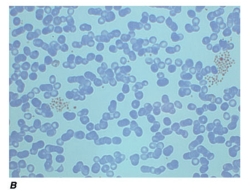
Example: Gram-positive cocci appear purple and may occur in clusters. Microscopy gives early clues about morphology and staining reaction.
E. Culture and incubation area
Culture allows microorganisms to multiply under controlled conditions so they can be isolated and studied.
Key equipment:
- Incubators
- Anaerobic jars or chambers
- Refrigerators and freezers
- Colony counter
- Culture media storage
Culture conditions may vary by:
- Temperature
- Oxygen requirement
- pH
- Nutrient requirements
- Incubation time
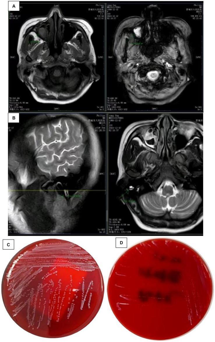
Culture plates permit observation of colony size, color, shape, margin, elevation, hemolysis, and purity.
F. Identification and reporting area
Once a microorganism is isolated, it is identified using a combination of morphology, staining, biochemical reactions, antigen tests, mass spectrometry, and molecular methods.
The report should clearly state:
- Organism detected or isolated
- Level of identification, for example genus or species
- Relevant quantitative result if applicable
- Antimicrobial susceptibility result when required
- Important comments, limitations, or urgent alerts
G. Washing, decontamination, and waste disposal area
All used cultures, contaminated materials, and sharps must be segregated and safely decontaminated. Typical measures include autoclaving infectious waste and using puncture-resistant sharps containers.
3. Laboratory Safety and Good Microbiological Practice
Basic safety rules
- Wear appropriate personal protective equipment: laboratory coat, gloves, and eye protection when needed.
- Never mouth-pipette.
- Disinfect benches before and after work.
- Label all specimens, media, and cultures clearly.
- Avoid aerosol formation during vortexing, centrifugation, and pipetting.
- Wash hands before leaving the laboratory.
- Dispose of cultures and sharps in designated containers.
- Report spills, injuries, and exposure incidents immediately.
- Do not eat, drink, or apply cosmetics in the laboratory.
- Use aseptic technique at every stage.
Aseptic technique
Aseptic technique is the set of procedures used to prevent unwanted microorganisms from entering a culture, specimen, reagent, or work environment. It includes sterile instruments, minimal exposure of media, correct labeling, and proper disinfection.
4. Methods of Identification of Microorganisms
Microbial identification is usually stepwise. No single test is sufficient for every organism.
Specimen
  ↓
Direct examination
  ↓
Culture and isolation
  ↓
Colony characteristics
  ↓
Microscopy and staining
  ↓
Biochemical / antigen / molecular / MALDI-TOF methods
  ↓
Confirmation and reporting
A. Direct microscopic examination
This is often the first laboratory method.
1. Wet mount
A drop of specimen is placed on a slide and examined directly.
Uses:
- Motility of bacteria
- Fungal elements
- Protozoal trophozoites
- Yeast cells
2. Gram stain
The Gram stain is one of the most important differential stains in bacteriology.
| Result | Appearance | Interpretation |
|---|---|---|
| Gram-positive bacteria | Purple or blue | Thick peptidoglycan wall retains crystal violet |
| Gram-negative bacteria | Pink or red | Thin peptidoglycan layer does not retain crystal violet after decolorization |
Main Gram-stain steps:
- Crystal violet
- Iodine
- Decolorizer
- Safranin counterstain
It also reveals:
- Shape: cocci, bacilli, coccobacilli, spiral forms
- Arrangement: clusters, chains, pairs, tetrads
- Presence of pus cells or epithelial cells
- Approximate bacterial load
- Mixed versus pure populations
3. Acid-fast stain
Used mainly for organisms with waxy, lipid-rich cell walls, especially Mycobacterium species.
- Acid-fast bacilli retain the primary red stain.
- Non-acid-fast cells take the counterstain.
4. Special stains
Examples include:
- Endospore stain
- Capsule stain
- Lactophenol cotton blue stain for fungi
- India ink preparation for encapsulated yeasts
- Fluorescent stains
B. Culture methods
Culture is the growth of microorganisms on artificial media.
Types of culture media
| Type | Purpose | Example |
|---|---|---|
| Basal or simple media | Supports non-fastidious organisms | Nutrient agar |
| Enriched media | Supports fastidious organisms | Blood agar, chocolate agar |
| Selective media | Inhibits some organisms while allowing others to grow | MacConkey agar |
| Differential media | Distinguishes organisms by visible reactions | MacConkey agar for lactose fermentation |
| Enrichment broth | Increases small numbers of a target organism | Selenite F broth |
| Transport media | Preserves organisms during transport | Stuart or Amies medium |
| Anaerobic media | Supports anaerobic organisms | Thioglycollate medium |
Colony morphology
After incubation, colonies are examined for:
- Size
- Shape
- Margin
- Elevation
- Surface texture
- Pigment
- Opacity
- Hemolysis
- Odor
- Growth on selective media
These features provide a presumptive identification, not always a final one.
C. Biochemical identification
Biochemical tests identify organisms by detecting enzymes or metabolic activities.
| Test | What it detects | Example use |
|---|---|---|
| Catalase | Breakdown of hydrogen peroxide | Distinguishes staphylococci from streptococci |
| Coagulase | Clotting activity | Helps identify Staphylococcus aureus |
| Oxidase | Cytochrome oxidase | Useful for Pseudomonas, Neisseria, Vibrio |
| Indole | Tryptophan metabolism | Often positive in Escherichia coli |
| Urease | Urea breakdown | Often positive in Proteus |
| Citrate utilization | Ability to use citrate | Part of enteric bacterial identification |
| Carbohydrate fermentation | Acid or gas production | Differentiates enteric organisms |
| Triple sugar iron agar | Sugar fermentation, gas, H₂S | Screening enteric Gram-negative bacilli |
Automated biochemical systems can test many reactions simultaneously.
D. Immunological methods
These methods detect either microbial antigens or antibodies produced by the host.
Examples:
- Latex agglutination
- Enzyme-linked immunosorbent assay
- Rapid antigen detection tests
- Immunofluorescence
- Lateral-flow immunochromatographic tests
They are particularly useful when rapid testing is needed or when culture is slow or difficult.
E. Molecular methods
Molecular techniques detect nucleic acids, such as DNA or RNA.
Examples
- Polymerase chain reaction (PCR)
- Real-time PCR
- Multiplex PCR
- Nucleic-acid amplification tests
- DNA sequencing
- 16S rRNA gene sequencing for bacteria
- Whole-genome sequencing
Advantages
- Rapid
- Highly sensitive and specific
- May identify organisms that are difficult or slow to culture
- Can detect resistance genes in selected settings
Limitations
- May detect nonviable organisms
- Can be affected by contamination
- Requires technical expertise and quality controls
- A positive molecular result does not always prove active disease
F. MALDI-TOF mass spectrometry
Matrix-assisted laser desorption/ionization time-of-flight mass spectrometry, usually shortened to MALDI-TOF MS, identifies cultured organisms by analyzing their protein spectral pattern and comparing it with a reference database.
It is widely used for identification of bacteria, fungi, and mycobacteria grown in culture. Tietz Textbook of Laboratory Medicine, 7th Edition, “Background” section. The CDC laboratory protocol illustrates its use for mold identification.
Benefits:
- Fast identification after colony growth
- High throughput
- Lower reagent use than multiple conventional biochemical tests
- Useful for many routine organisms
Limitations:
- Identification depends on the quality and coverage of the reference database.
- Some closely related species need molecular confirmation.
- It generally identifies an organism after it has been grown, rather than replacing culture in every situation.
5. Quality Control in the Microbiology Laboratory
Quality control ensures that laboratory results are accurate, reproducible, and clinically meaningful.
Important elements include:
- Checking media sterility and performance
- Monitoring incubator temperatures
- Verifying stains with known control organisms
- Calibrating equipment
- Using positive and negative controls in molecular tests
- Maintaining reference strains
- Documenting corrective action when problems occur
- Participating in external quality-assessment programs
6. Key Takeaway
A microbiology laboratory identifies microorganisms through a logical sequence:
Correct specimen → safe handling → direct examination → culture → identification → quality-controlled reporting
Traditional microscopy and culture remain foundational. Biochemical tests, immunological assays, molecular methods, and MALDI-TOF MS add speed and precision. The best method depends on the organism, specimen type, purpose of testing, laboratory resources, and need for rapid results.
This is a shared conversation. Sign in to Orris to start your own chat.