
Sickle cell anemia
sickle cell anemia blood smear red blood cells
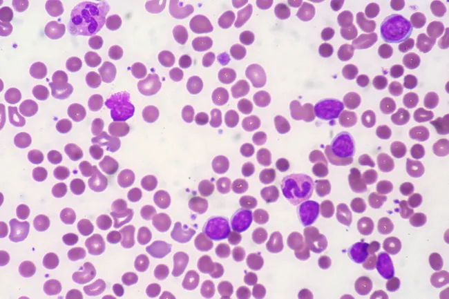
Peripheral blood smear from a patient with known chronic lymphocytic leukemia (CLL) showing autoimmune hemolytic anemia (AIHA). Brightfield microscopy of Wright-Giemsa stained smear at 1000x (oil immersion) reveals two dominant cell populations: numerous small mature lymphocytes typical of CLL and red blood cells showing spherocytic deformation. A smudge cell is evident just left of center, a classical clue for CLL. Red cells display reduced central pallor consistent with spherocytes, indicating intravascular or extravascular hemolysis driven by autoantibodies. The background shows mild polychromasia and rare nucleated RBCs, consistent with a robust reticulocyte response; the corrected reticulocyte count in this case is markedly elevated (>7%). Platelets are not significantly abnormal. These features collectively support autoimmune destruction of erythrocytes in the setting of CLL. Diagnostic significance lies in recognizing AIHA as a common autoimmune complication in CLL, with a clinical picture of sudden Hb decline and compensatory erythropoiesis. Differential considerations include drug-induced hemolysis, other causes of hemolytic anemia, or marrow infiltration by CLL. Clinical correlation includes therapy implications (steroids, rituximab) and close monitoring of hemolysis markers. This image is a valuable teaching resource for hematology morphology, AIHA in CLL, and differential diagnosis in anemia.
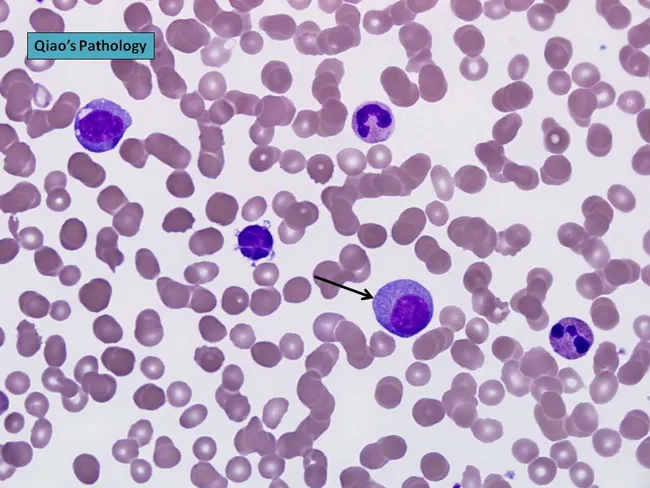
Peripheral blood smear prepared with Wright-Giemsa stain and examined under brightfield illumination using a 100× oil immersion objective reveals a single large plasma cell among a field of mature erythrocytes. The plasma cell displays features typical of clonal plasma cells: basophilic cytoplasm, an eccentrically placed round nucleus, and a conspicuous perinuclear hof; background shows rouleaux formation of red cells. This image exemplifies hematologic involvement by a plasma cell dyscrasia. In plasma cell leukemia, circulating plasma cells constitute more than 20% of leukocytes, a hallmark distinguishing it from conventional multiple myeloma, although both conditions share monoclonal plasma cell proliferation. The presence of circulating plasma cells is associated with younger patient age, higher incidence of anemia, thrombocytopenia, organomegaly, and possible lymphadenopathy; however lytic bone lesions may be less common. Immunophenotypic patterns often include lack of CD56 expression relative to myeloma. Clinically, this morphological finding warrants confirmation with complete blood count, peripheral smear review, quantitative plasma cell percentage, serum protein electrophoresis, and immunofixation to identify monoclonal immunoglobulin isotype (IgD/IgE or light-chain predominance). Early detection of circulating plasma cells supports prognosis assessment and guides therapy decisions in plasma cell dyscrasia management and monitoring, including risk stratification and treatment response evaluation for practice in clinics.
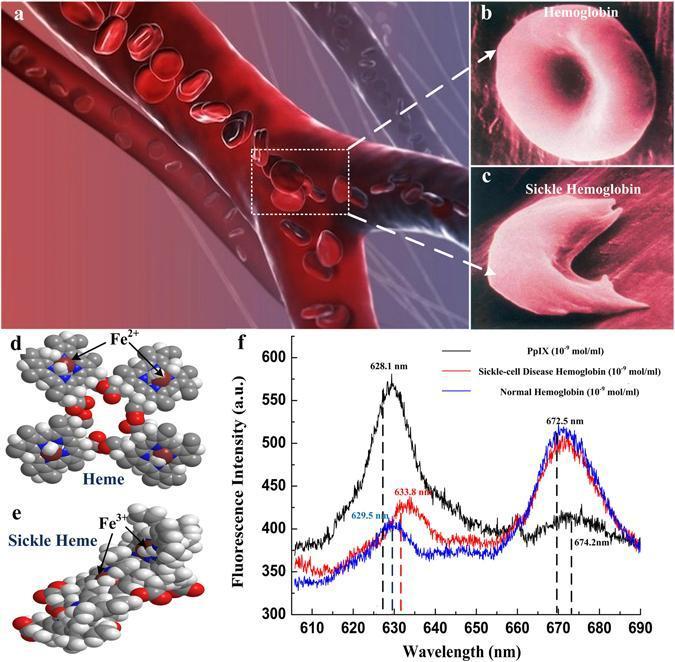
A multi-panel medical illustration and data graphic comparing normal hemoglobin and sickle cell disease (SCD) hemoglobin. (a) A 3D schematic of a bifurcating blood vessel showing the movement of red blood cells. (b, c) Magnified views contrasting the flexible, biconcave disc morphology of normal hemoglobin with the rigid, elongated, crescent or sickle shape characteristic of SCD hemoglobin, illustrating the mechanism for vascular occlusion. (d, e) Ball-and-stick molecular models detailing Heme (with Fe2+) and Sickle Heme (associated with Fe3+), emphasizing structural differences at the molecular level. (f) A comparative fluorescence spectrum plot showing Protoporphyrin (black), Sickle-cell Disease Hemoglobin (red), and Normal Hemoglobin (blue) across wavelengths of 610–690 nm. The graph highlights diagnostic spectral shifts: Protoporphyrin peaks at ~628.1 nm, while hemoglobin variants show distinctive primary peaks at ~672.5 nm and secondary peaks showing shifts (629.5 nm for normal vs. 633.8 nm for sickle). This composite image serves to correlate clinical hematology, cellular morphology, and molecular spectroscopy in the study of Sickle Cell Anemia.
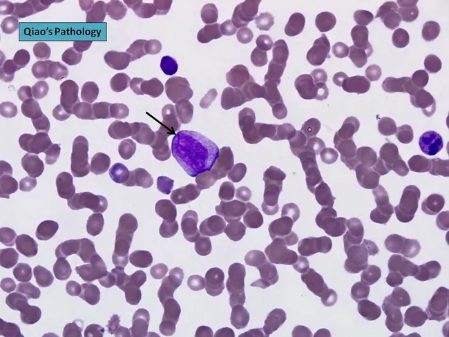
Comprehensive description: Brightfield light microscopy image of a peripheral blood smear stained with Wright-Giemsa, captured at high magnification to visualize circulating plasma cells. The central plasmablast shows an enlarged, irregular nucleus with a prominent nucleolus; the cytoplasm is moderately basophilic. This cell appears larger than surrounding erythrocytes and is consistent with malignant plasma cells typical of plasma cell leukemia. The background demonstrates rouleaux formation of red blood cells, a common feature in paraproteinemias due to increased serum proteins. The smear illustrates plasmacytosis that, when exceeding 20% of leukocytes, supports a diagnosis of plasma cell leukemia. In PCL, neoplastic plasma cells may be CD56 negative and often secrete monoclonal immunoglobulin with IgD or IgE isotypes or light-chain restriction; cytogenetic abnormalities are frequently observed. Morphology alone cannot definitively distinguish PCL from aggressive myeloma; immunophenotyping and cytogenetic studies are essential for precise classification. This image is valuable for hematology education, cytology review, and discussions of malignant plasma cell disorders, including primary versus secondary PCL, and their prognostic significance. Potential clinical utilities include morphologic confirmation in suspected PCL, training in recognition of plasmablasts, and correlation with CBC abnormalities and clinical features such as anemia, thrombocytopenia, lymphadenopathy, and organomegaly.
"sickle cell disease" AND treatment
Sickle cell anemia pediatric ppt
Who is the target audience for this presentation? · How detailed should the content be? · Approximately how many slides?
sickle cell anemia blood smear peripheral blood
Peripheral blood smear from a patient with known chronic lymphocytic leukemia (CLL) showing autoimmune hemolytic anemia (AIHA). Brightfield microscopy of Wright-Giemsa stained smear at 1000x (oil immersion) reveals two dominant cell populations: numerous small mature lymphocytes typical of CLL and red blood cells showing spherocytic deformation. A smudge cell is evident just left of center, a classical clue for CLL. Red cells display reduced central pallor consistent with spherocytes, indicating intravascular or extravascular hemolysis driven by autoantibodies. The background shows mild polychromasia and rare nucleated RBCs, consistent with a robust reticulocyte response; the corrected reticulocyte count in this case is markedly elevated (>7%). Platelets are not significantly abnormal. These features collectively support autoimmune destruction of erythrocytes in the setting of CLL. Diagnostic significance lies in recognizing AIHA as a common autoimmune complication in CLL, with a clinical picture of sudden Hb decline and compensatory erythropoiesis. Differential considerations include drug-induced hemolysis, other causes of hemolytic anemia, or marrow infiltration by CLL. Clinical correlation includes therapy implications (steroids, rituximab) and close monitoring of hemolysis markers. This image is a valuable teaching resource for hematology morphology, AIHA in CLL, and differential diagnosis in anemia.
Peripheral blood smear prepared with Wright-Giemsa stain and examined under brightfield illumination using a 100× oil immersion objective reveals a single large plasma cell among a field of mature erythrocytes. The plasma cell displays features typical of clonal plasma cells: basophilic cytoplasm, an eccentrically placed round nucleus, and a conspicuous perinuclear hof; background shows rouleaux formation of red cells. This image exemplifies hematologic involvement by a plasma cell dyscrasia. In plasma cell leukemia, circulating plasma cells constitute more than 20% of leukocytes, a hallmark distinguishing it from conventional multiple myeloma, although both conditions share monoclonal plasma cell proliferation. The presence of circulating plasma cells is associated with younger patient age, higher incidence of anemia, thrombocytopenia, organomegaly, and possible lymphadenopathy; however lytic bone lesions may be less common. Immunophenotypic patterns often include lack of CD56 expression relative to myeloma. Clinically, this morphological finding warrants confirmation with complete blood count, peripheral smear review, quantitative plasma cell percentage, serum protein electrophoresis, and immunofixation to identify monoclonal immunoglobulin isotype (IgD/IgE or light-chain predominance). Early detection of circulating plasma cells supports prognosis assessment and guides therapy decisions in plasma cell dyscrasia management and monitoring, including risk stratification and treatment response evaluation for practice in clinics.
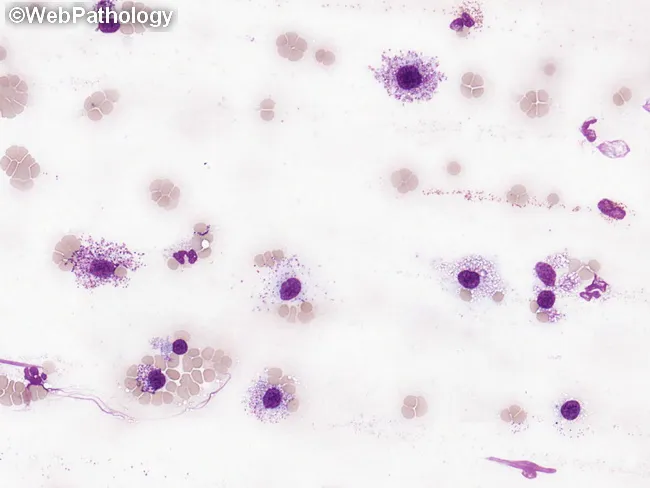
This peripheral blood smear was prepared and stained with Wright-Giemsa and examined by bright-field light microscopy to assess circulating hematopoietic cells in suspected systemic mastocytosis (SM). The smear shows rare, but conspicuous, mast cells with round to polygonal shapes and abundant cytoplasmic granules that impart a deep purple, metachromatic appearance. Nuclei are generally round to ovoid and may be slightly irregular or bilobed in some cells; granules occupy most of the cytoplasm and may obscure nuclear detail. The surrounding leukocytes include eosinophils and neutrophils, and the erythrocyte background is normocytic with occasional anisocytosis. In SM, circulating mast cells are typically infrequent; when they constitute a substantial fraction of leukocytes (≥10%), the finding is highly suggestive of mast cell leukemia (MCL). The image exemplifies an increased mast cell burden consistent with aggressive SM or MCL, often accompanied by cytopenias such as anemia or thrombocytopenia and potential associated hematologic neoplasms (e.g., CMML, MDS/MPN). This morphologic snapshot supports integration with phenotypic assays (CD117/c-KIT), flow cytometry, and molecular testing for KIT mutations, to establish diagnosis, prognostication, and therapeutic planning. Relevant keywords: systemic mastocytosis, mast cell leukemia, circulating mast cells, metachromatic granules, Wright-Giemsa, toluidine blue, KIT D816V, CD117, SM-AHN. This description emphasizes morphology guiding diagnosis appropriately.
sickle cell vaso-occlusion pathophysiology diagram
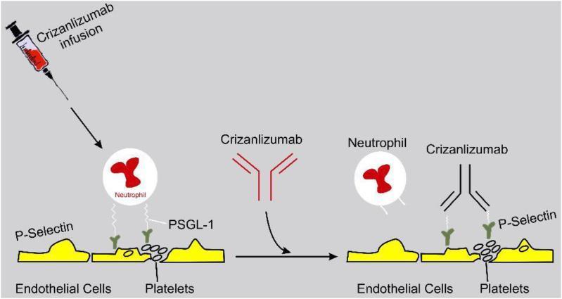
This pathophysiology diagram illustrates the mechanism of action of Crizanlizumab in managing vaso-occlusion, particularly relevant to sickle cell disease. The illustration is divided into two comparative stages: pretreatment (left) and post-treatment (right). On the left, a neutrophil is shown adhering to the vascular endothelium and activated platelets. This adhesion is mediated by the interaction between P-selectin, expressed on the surface of endothelial cells and platelets, and P-selectin glycoprotein ligand-1 (PSGL-1) located on the neutrophil. In the center, a syringe depicts the infusion of Crizanlizumab, a humanized monoclonal antibody. On the right, the diagram demonstrates the therapeutic effect: Crizanlizumab molecules bind directly to P-selectin on the endothelial cells and platelets. This binding competitively inhibits the P-selectin/PSGL-1 interaction, resulting in the detachment of the neutrophil from the vessel wall and preventing further leukocyte adhesion. The diagram highlights the drug's role in reducing multicellular adhesion and subsequent vaso-occlusive crises.
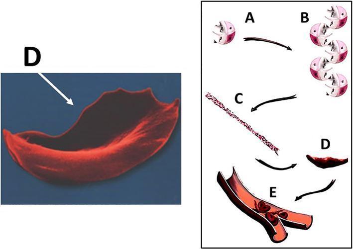
This educational graphic illustrates the molecular and cellular pathophysiology of sickle cell disease (SCD) leading to vaso-occlusion. The diagram is divided into a microscopic anatomical illustration and a high-magnification scanning electron micrograph. Steps A through D depict the hemoglobin S (HbS) polymerization process: (A) individual deoxygenated hemoglobin tetramers, (B) aggregation of hemoglobin molecules, (C) formation of long, rigid hemoglobin polymers or strands, and (D) the resulting transformation of a flexible biconcave red blood cell into a rigid, crescent or sickle-shaped erythrocyte. A corresponding scanning electron micrograph (labeled D) highlights the characteristic elongated, spicular morphology of a sickled cell. Section E shows a cross-section of a small bifurcating blood vessel where these rigid, abnormally shaped cells are aggregating, causing mechanical obstruction (vaso-occlusion) of the lumen. This process demonstrates the mechanism behind tissue ischemia and subsequent complications such as osteonecrosis in SCD patients. The content is suitable for medical students and clinicians studying hematology and musculoskeletal pathology.
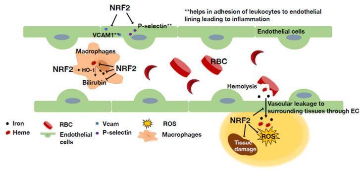
This pathophysiology diagram illustrates the protective roles of NRF2 in the context of sickle cell disease (SCD) within the vascular microenvironment. The illustration shows a blood vessel lined with green endothelial cells containing normal biconcave and crescent-shaped (sickled) red blood cells (RBCs). Key mechanisms depicted include: 1) Endothelial Protection: NRF2 inhibits the expression of adhesion molecules VCAM1 and P-selectin, which normally promote leukocyte adhesion and inflammation. 2) Heme Clearance: Within macrophages, NRF2 upregulates Heme Oxygenase-1 (HO-1), facilitating the breakdown of toxic free heme (from hemolysis) into bilirubin and iron. 3) Vascular Integrity: NRF2 is shown mitigating tissue damage and reactive oxygen species (ROS) accumulation in the surrounding tissue, specifically addressing vascular leakage through the endothelial basement membrane. The diagram highlights how NRF2 serves as a critical regulator across multiple cell types (macrophages, endothelial cells, and peripheral tissues) to reduce vaso-occlusion, inflammation, and oxidative stress induced by chronic hemolysis in SCD.
sickle cell disease complications organ damage spleen
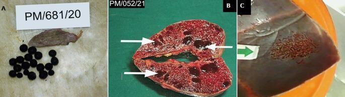
This composite of gross autopsy photographs displays pathological findings in Sickle Cell Disease (SCD) patients. Panel A shows an autosplenectomy specimen, characterized by a severely shrunken, fibrotic splenic remnant (approximately 5x3x1cm) alongside numerous dark, ovoid pigment gallstones (bilirubinate stones), which are common complications of chronic hemolysis. Panel B presents a sagittal cut section of a moderately enlarged spleen containing multiple, well-circumscribed, dark brown to black regenerative nodules (white arrows). These represent sites of extramedullary hematopoiesis, where the body attempts to compensate for chronic anemia. Panel C illustrates a gross specimen of the skull vault, where a green arrow points to a localized region of cortical bone thinning and irregular bone resorption. This reflects bone marrow expansion and compensatory remodeling of the calvarium typical in SCD. Collectively, these images demonstrate multi-organ manifestations of chronic hematologic stress, including splenic atrophy, biliary complications, and skeletal changes, pertinent to medical education in pathology and hematology.
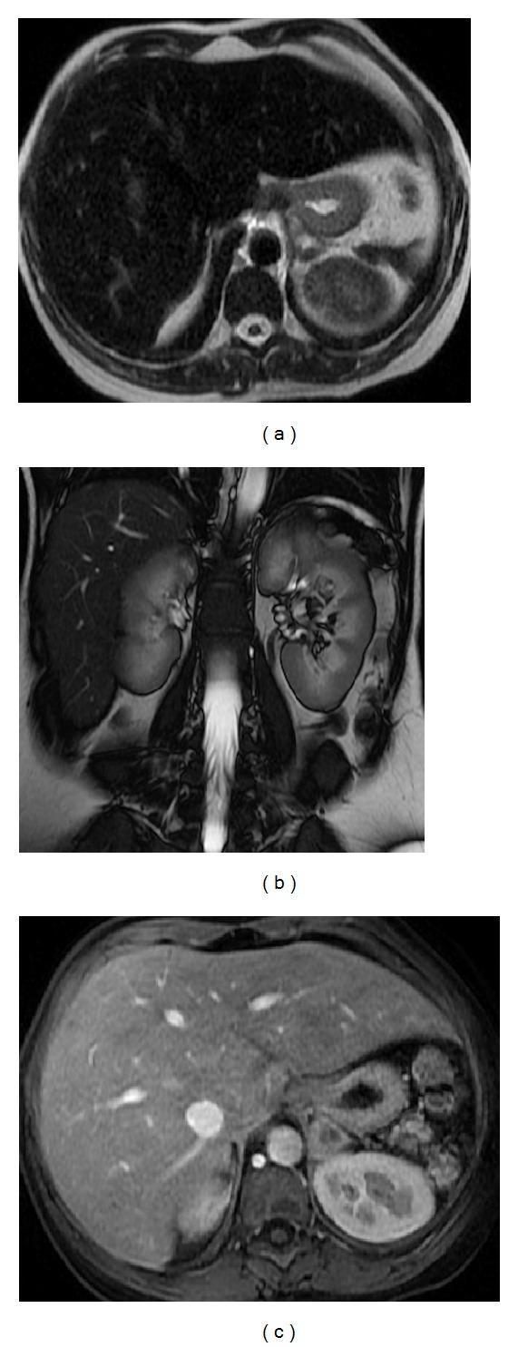
This diagnostic MRI series demonstrates findings consistent with autosplenectomy and secondary iron deposition in a patient with homozygous sickle cell disease. The image set includes: (a) an axial T2-weighted Single Shot Fast Spin Echo (SSFSE) image, (b) a coronal Steady-State Free Precession (SSFP) image, and (c) a post-contrast axial 3D-GRE T1-weighted image with fat suppression during the arterial phase. A key visual feature is the severe atrophy of the spleen, appearing as a small, retracted remnant in the left upper quadrant. Both the liver and the remnant spleen demonstrate diffusely diminished signal intensity (appearing dark/hypointense) across T1 and T2 sequences. This signal void is characteristic of significant iron deposition (hemosiderosis) resulting from chronic blood transfusions. The post-contrast arterial phase image (c) highlights the vascular structures with fat suppression, further emphasizing the hepatic parenchymal signal loss compared to normal tissues. This series illustrates the end-stage splenic complications of sickle cell disease and the multi-organ impact of iron overload.
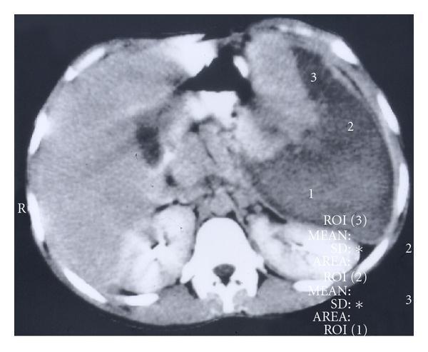
This abdominal axial CT scan demonstrates massive splenomegaly with extensive splenic infarction in a pediatric patient with sickle cell anemia. The spleen is significantly enlarged, occupying a substantial portion of the left upper quadrant and displacing adjacent structures. The splenic parenchyma exhibits a heterogeneous, mottled appearance characterized by large, wedge-shaped and irregular regions of low attenuation (hypodensity), representing infarcted tissue that has lost its blood supply. These infarcted areas are marked with numbered Regions of Interest (ROI 1, 2, and 3) and involve more than 50% of the organ's volume, meeting the criteria for massive splenic infarction. The remaining hyperdense areas represent viable, enhancing splenic tissue. Visible surrounding landmarks include the liver on the right, the kidneys posteriorly, and the vertebral column. The image illustrates a critical complication of sickle cell disease where vaso-occlusive crises lead to major organ ischemia rather than the typical progressive atrophy and autosplenectomy.
mkdir -p /tmp/workspace/sickle-cell-ppt && ls /tmp/workspace/sickle-cell-ppt/
~/sickle-cell-ppt/build_pptx.py
import json, subprocess, base64
from io import BytesIO
from pptx import Presentation
from pptx.util import Inches, Pt, Emu
from pptx.dml.color import RGBColor
from pptx.enum.text import PP_ALIGN, MSO_ANCHOR
from pptx.util import Inches, Pt

# ── Color palette ────────────────────────────────────────────────
DARK_RED    = RGBColor(0xA0, 0x00, 0x00)   # deep crimson
MED_RED     = RGBColor(0xC0, 0x20, 0x20)
LIGHT_RED   = RGBColor(0xF5, 0xD5, 0xD5)
DARK_GRAY   = RGBColor(0x2C, 0x2C, 0x2C)
MED_GRAY    = RGBColor(0x55, 0x55, 0x55)
WHITE       = RGBColor(0xFF, 0xFF, 0xFF)
GOLD        = RGBColor(0xD4, 0xA0, 0x17)
LIGHT_GOLD  = RGBColor(0xFF, 0xF3, 0xCC)
TEAL        = RGBColor(0x00, 0x6B, 0x6B)
LIGHT_TEAL  = RGBColor(0xCC, 0xEE, 0xEE)
BLUE        = RGBColor(0x1A, 0x4E, 0x8C)
LIGHT_BLUE  = RGBColor(0xD6, 0xE4, 0xF7)
ORANGE      = RGBColor(0xC8, 0x60, 0x00)
LIGHT_BG    = RGBColor(0xFA, 0xFA, 0xFC)

prs = Presentation()
prs.slide_width  = Inches(13.333)
prs.slide_height = Inches(7.5)
blank = prs.slide_layouts[6]   # fully blank

# ── Helper utilities ─────────────────────────────────────────────
def add_rect(slide, x, y, w, h, fill_color, border_color=None, border_width=0):
    from pptx.util import Pt
    shape = slide.shapes.add_shape(1, Inches(x), Inches(y), Inches(w), Inches(h))
    shape.fill.solid()
    shape.fill.fore_color.rgb = fill_color
    if border_color:
        shape.line.color.rgb = border_color
        shape.line.width = Pt(border_width)
    else:
        shape.line.fill.background()
    return shape

def add_textbox(slide, x, y, w, h, text, font_size=18, bold=False, italic=False,
                color=DARK_GRAY, align=PP_ALIGN.LEFT, word_wrap=True,
                font_name="Calibri", v_anchor=None):
    tb = slide.shapes.add_textbox(Inches(x), Inches(y), Inches(w), Inches(h))
    tf = tb.text_frame
    tf.word_wrap = word_wrap
    if v_anchor:
        tf.vertical_anchor = v_anchor
    tf.margin_left = Pt(2)
    tf.margin_right = Pt(2)
    tf.margin_top = Pt(2)
    tf.margin_bottom = Pt(2)
    p = tf.paragraphs[0]
    p.alignment = align
    r = p.add_run()
    r.text = text
    r.font.name = font_name
    r.font.size = Pt(font_size)
    r.font.bold = bold
    r.font.italic = italic
    r.font.color.rgb = color
    return tb, tf

def add_bullet_slide(slide, title, bullets, accent=DARK_RED):
    """Standard content slide with colored header bar + bullets."""
    # Header bar
    add_rect(slide, 0, 0, 13.333, 1.1, accent)
    add_textbox(slide, 0.3, 0.1, 12.5, 0.9, title,
                font_size=28, bold=True, color=WHITE, align=PP_ALIGN.LEFT,
                v_anchor=MSO_ANCHOR.MIDDLE)
    # Content background
    add_rect(slide, 0, 1.1, 13.333, 6.4, LIGHT_BG)
    # Bullets
    tb = slide.shapes.add_textbox(Inches(0.4), Inches(1.25), Inches(12.5), Inches(5.9))
    tf = tb.text_frame
    tf.word_wrap = True
    for i, (level, text) in enumerate(bullets):
        p = tf.paragraphs[0] if i == 0 else tf.add_paragraph()
        p.level = level
        indent = "    " * level
        bullet_char = "•" if level == 0 else "◦"
        r = p.add_run()
        r.text = f"{indent}{bullet_char}  {text}"
        r.font.name = "Calibri"
        r.font.size = Pt(17 - level * 1.5)
        r.font.color.rgb = DARK_GRAY if level > 0 else DARK_GRAY
        r.font.bold = (level == 0)
    return slide

def add_two_column_slide(slide, title, left_items, right_items,
                          left_header="", right_header="", accent=DARK_RED):
    add_rect(slide, 0, 0, 13.333, 1.1, accent)
    add_textbox(slide, 0.3, 0.1, 12.5, 0.9, title,
                font_size=28, bold=True, color=WHITE, v_anchor=MSO_ANCHOR.MIDDLE)
    add_rect(slide, 0, 1.1, 13.333, 6.4, LIGHT_BG)
    # Divider
    add_rect(slide, 6.55, 1.2, 0.05, 6.1, MED_GRAY)
    for col, (header, items) in enumerate([(left_header, left_items),
                                            (right_header, right_items)]):
        x = 0.3 if col == 0 else 6.7
        if header:
            add_textbox(slide, x, 1.15, 6.0, 0.45, header,
                        font_size=16, bold=True, color=accent)
        tb = slide.shapes.add_textbox(Inches(x), Inches(1.65), Inches(6.1), Inches(5.5))
        tf = tb.text_frame
        tf.word_wrap = True
        for i, item in enumerate(items):
            p = tf.paragraphs[0] if i == 0 else tf.add_paragraph()
            r = p.add_run()
            r.text = f"•  {item}"
            r.font.name = "Calibri"
            r.font.size = Pt(15.5)
            r.font.color.rgb = DARK_GRAY

def add_image_to_slide(slide, url, x, y, w, h):
    try:
        result = json.loads(subprocess.check_output(
            ["python", "/tmp/skills/shared/scripts/fetch_images.py", url],
            timeout=30
        ))
        if result and result[0].get("base64"):
            b64 = result[0]["base64"]
            if "," in b64:
                b64 = b64.split(",", 1)[1]
            raw = base64.b64decode(b64)
            slide.shapes.add_picture(BytesIO(raw), Inches(x), Inches(y),
                                      Inches(w), Inches(h))
            return True
    except Exception as e:
        print(f"Image fetch failed for {url}: {e}")
    return False

def add_table_slide(slide, title, headers, rows, accent=DARK_RED):
    add_rect(slide, 0, 0, 13.333, 1.0, accent)
    add_textbox(slide, 0.3, 0.05, 12.5, 0.9, title,
                font_size=26, bold=True, color=WHITE, v_anchor=MSO_ANCHOR.MIDDLE)
    add_rect(slide, 0, 1.0, 13.333, 6.5, LIGHT_BG)
    from pptx.util import Inches as I
    cols = len(headers)
    col_w = [I(13.0 / cols)] * cols
    tbl = slide.shapes.add_table(len(rows)+1, cols,
                                   I(0.17), I(1.1),
                                   I(13.0), I(0.45*(len(rows)+1))).table
    tbl.columns[0].width = col_w[0]
    # Header row
    for j, h in enumerate(headers):
        cell = tbl.cell(0, j)
        cell.fill.solid()
        cell.fill.fore_color.rgb = accent
        p = cell.text_frame.paragraphs[0]
        p.alignment = PP_ALIGN.CENTER
        r = p.add_run()
        r.text = h
        r.font.bold = True
        r.font.size = Pt(13)
        r.font.color.rgb = WHITE
        r.font.name = "Calibri"
    for i, row in enumerate(rows):
        bg = LIGHT_RED if i % 2 == 0 else WHITE
        for j, val in enumerate(row):
            cell = tbl.cell(i+1, j)
            cell.fill.solid()
            cell.fill.fore_color.rgb = bg
            p = cell.text_frame.paragraphs[0]
            r = p.add_run()
            r.text = str(val)
            r.font.size = Pt(12)
            r.font.name = "Calibri"
            r.font.color.rgb = DARK_GRAY

# ════════════════════════════════════════════════════════════════
# SLIDE DEFINITIONS
# ════════════════════════════════════════════════════════════════

# ── SLIDE 1: Title slide ─────────────────────────────────────────
slide = prs.slides.add_slide(blank)
add_rect(slide, 0, 0, 13.333, 7.5, DARK_RED)
add_rect(slide, 0, 2.8, 13.333, 2.4, MED_RED)
add_textbox(slide, 0.6, 0.6, 12.0, 1.8,
            "SICKLE CELL ANEMIA",
            font_size=52, bold=True, color=WHITE, align=PP_ALIGN.CENTER)
add_textbox(slide, 0.6, 2.4, 12.0, 0.5,
            "A Comprehensive Pediatric Perspective",
            font_size=22, italic=True, color=LIGHT_RED, align=PP_ALIGN.CENTER)
add_textbox(slide, 0.6, 3.05, 12.0, 0.5,
            "For Medical Students  |  Pediatric Hematology",
            font_size=18, color=WHITE, align=PP_ALIGN.CENTER)
add_textbox(slide, 0.6, 3.6, 12.0, 0.5,
            "Sources: Robbins & Kumar Pathology • Goldman-Cecil Medicine • Harriet Lane Handbook",
            font_size=13, italic=True, color=LIGHT_RED, align=PP_ALIGN.CENTER)
add_textbox(slide, 0.6, 6.8, 12.0, 0.5,
            "2026",
            font_size=16, color=WHITE, align=PP_ALIGN.CENTER)

# ── SLIDE 2: Outline ─────────────────────────────────────────────
slide = prs.slides.add_slide(blank)
add_rect(slide, 0, 0, 13.333, 1.1, DARK_RED)
add_textbox(slide, 0.3, 0.1, 12.5, 0.9, "Presentation Outline",
            font_size=28, bold=True, color=WHITE, v_anchor=MSO_ANCHOR.MIDDLE)
add_rect(slide, 0, 1.1, 13.333, 6.4, LIGHT_BG)
topics = [
    ("1. Introduction & Epidemiology", DARK_RED),
    ("2. Genetics & Molecular Basis", BLUE),
    ("3. Pathophysiology", TEAL),
    ("4. Sickle Cell Subtypes", ORANGE),
    ("5. Clinical Features in Children", DARK_RED),
    ("6. Acute Complications", BLUE),
    ("7. Chronic Complications", TEAL),
    ("8. Diagnosis & Newborn Screening", ORANGE),
    ("9. Management — General Principles", DARK_RED),
    ("10. Disease-Modifying Therapies", BLUE),
    ("11. Curative Approaches", TEAL),
    ("12. Prognosis & Key Takeaways", ORANGE),
]
for i, (topic, color) in enumerate(topics):
    col = i % 2
    row = i // 2
    x = 0.4 + col * 6.5
    y = 1.25 + row * 1.0
    add_rect(slide, x, y, 6.2, 0.82, color)
    add_textbox(slide, x+0.1, y+0.05, 5.9, 0.72, topic,
                font_size=15, bold=True, color=WHITE, v_anchor=MSO_ANCHOR.MIDDLE)

# ── SLIDE 3: Introduction ─────────────────────────────────────────
slide = prs.slides.add_slide(blank)
add_bullet_slide(slide, "Introduction", [
    (0, "Most common familial hemolytic anemia worldwide"),
    (0, "Autosomal recessive disorder of hemoglobin structure"),
    (0, "Caused by a single point mutation in the β-globin gene"),
    (1, "Glutamic acid → Valine substitution at position 6 (Glu6Val)"),
    (1, "Results in production of sickle hemoglobin (HbS)"),
    (0, "Under deoxygenated conditions, HbS polymerizes → sickle-shaped RBCs"),
    (0, "Two major consequences: Hemolytic anemia + Vascular occlusion"),
    (0, "Symptoms typically appear at 5–6 months of age (as HbF declines)"),
    (0, "Life expectancy: median 5th decade; significant morbidity throughout childhood"),
], accent=DARK_RED)

# ── SLIDE 4: Epidemiology ─────────────────────────────────────────
slide = prs.slides.add_slide(blank)
add_bullet_slide(slide, "Epidemiology", [
    (0, "Global burden: 300,000–400,000 affected births annually"),
    (1, ">75% occur in Sub-Saharan Africa"),
    (0, "Prevalence is driven by malaria selective pressure"),
    (1, "HbS heterozygotes are protected against Plasmodium falciparum malaria"),
    (1, "Endemic: equatorial Africa, India, Mediterranean, Middle East"),
    (0, "United States prevalence:"),
    (1, "~8% of African Americans are heterozygous HbS carriers (sickle cell trait)"),
    (1, "~1 in 600 African Americans have sickle cell anemia (HbSS)"),
    (1, "~1 in 1,250 are HbS/HbC compound heterozygotes"),
    (0, "Worldwide, HbS gene is also concentrated in Europe (London, Paris, Manchester) due to migration"),
    (0, "Newborn screening programs now identify most affected children early in life"),
], accent=DARK_RED)

# ── SLIDE 5: Genetics ─────────────────────────────────────────────
slide = prs.slides.add_slide(blank)
add_rect(slide, 0, 0, 13.333, 1.1, BLUE)
add_textbox(slide, 0.3, 0.1, 12.5, 0.9, "Genetics of Sickle Cell Anemia",
            font_size=28, bold=True, color=WHITE, v_anchor=MSO_ANCHOR.MIDDLE)
add_rect(slide, 0, 1.1, 13.333, 6.4, LIGHT_BG)
# Left column: genetics
tb = slide.shapes.add_textbox(Inches(0.3), Inches(1.2), Inches(7.5), Inches(6.0))
tf = tb.text_frame
tf.word_wrap = True
genetics_points = [
    "Gene: HBB (β-globin gene on chromosome 11)",
    "Mutation: A→T transversion at codon 6 (GAG→GTG)",
    "Amino acid change: Glutamic acid → Valine (Glu6Val)",
    "Inheritance: Autosomal recessive",
    "HbSS (homozygous): Sickle cell anemia — most severe",
    "HbAS (heterozygous): Sickle cell trait — usually asymptomatic",
    "Each offspring of 2 carrier parents:",
    "   25% chance of HbSS (affected)",
    "   50% chance of HbAS (carrier)",
    "   25% chance of HbAA (unaffected)",
    "HbF (fetal hemoglobin) is protective — inhibits HbS polymerization",
    "Symptoms manifest at ~5–6 months when HbF transitions to HbA",
]
for i, pt in enumerate(genetics_points):
    p = tf.paragraphs[0] if i == 0 else tf.add_paragraph()
    r = p.add_run()
    r.text = f"•  {pt}" if not pt.startswith(" ") else pt
    r.font.name = "Calibri"
    r.font.size = Pt(15)
    r.font.color.rgb = DARK_GRAY
# Right pedigree box
add_rect(slide, 8.0, 1.3, 5.0, 5.5, LIGHT_BLUE)
add_textbox(slide, 8.1, 1.4, 4.8, 0.5, "Inheritance Pattern",
            font_size=16, bold=True, color=BLUE, align=PP_ALIGN.CENTER)
pedigree_text = [
    "Parents (carriers): HbAS × HbAS",
    "",
    "Offspring probabilities:",
    "  HbAA  →  25%  Normal",
    "  HbAS  →  50%  Carrier (Sickle Trait)",
    "  HbSS  →  25%  Sickle Cell Anemia",
    "",
    "Key concept: Co-inheritance of α-thalassemia",
    "reduces HbS concentration → less sickling",
    "",
    "HbF levels inversely correlate",
    "with disease severity",
]
tb2 = slide.shapes.add_textbox(Inches(8.1), Inches(2.0), Inches(4.7), Inches(4.5))
tf2 = tb2.text_frame
tf2.word_wrap = True
for i, pt in enumerate(pedigree_text):
    p = tf2.paragraphs[0] if i == 0 else tf2.add_paragraph()
    r = p.add_run()
    r.text = pt
    r.font.name = "Calibri"
    r.font.size = Pt(14)
    r.font.color.rgb = DARK_GRAY

# ── SLIDE 6: Normal vs HbS Hemoglobin ────────────────────────────
slide = prs.slides.add_slide(blank)
add_rect(slide, 0, 0, 13.333, 1.1, BLUE)
add_textbox(slide, 0.3, 0.1, 12.5, 0.9, "Normal HbA vs. Sickle HbS",
            font_size=28, bold=True, color=WHITE, v_anchor=MSO_ANCHOR.MIDDLE)
add_rect(slide, 0, 1.1, 13.333, 6.4, LIGHT_BG)
add_rect(slide, 0.2, 1.2, 6.2, 5.9, LIGHT_BLUE)
add_rect(slide, 6.9, 1.2, 6.2, 5.9, LIGHT_RED)
add_textbox(slide, 0.3, 1.3, 6.0, 0.5, "Normal Hemoglobin (HbA)",
            font_size=17, bold=True, color=BLUE, align=PP_ALIGN.CENTER)
add_textbox(slide, 7.0, 1.3, 6.0, 0.5, "Sickle Hemoglobin (HbS)",
            font_size=17, bold=True, color=DARK_RED, align=PP_ALIGN.CENTER)
normal_pts = [
    "α₂β₂ tetramer structure",
    "β-globin: Glutamic acid at position 6",
    "Charged, hydrophilic residue",
    "Remains soluble when deoxygenated",
    "Biconcave disc shape — flexible",
    "Life span: ~120 days",
    "Flows freely through capillaries",
    "HbA₂ (α₂δ₂) = ~3%  |  HbF = ~1%",
]
sickle_pts = [
    "α₂β_S² tetramer structure",
    "β-globin: Valine at position 6 (Glu6Val)",
    "Non-polar, hydrophobic residue",
    "Polymerizes when deoxygenated → long fibers",
    "Elongated, rigid sickle/crescent shape",
    "Life span: ~20 days (1/6 of normal!)",
    "Obstructs microvascular flow",
    "HbS replaces HbA completely in HbSS",
]
for col, pts in enumerate([normal_pts, sickle_pts]):
    color = BLUE if col == 0 else DARK_RED
    x = 0.35 if col == 0 else 7.05
    tb = slide.shapes.add_textbox(Inches(x), Inches(1.9), Inches(5.9), Inches(5.0))
    tf = tb.text_frame
    tf.word_wrap = True
    for i, pt in enumerate(pts):
        p = tf.paragraphs[0] if i == 0 else tf.add_paragraph()
        r = p.add_run()
        r.text = f"•  {pt}"
        r.font.name = "Calibri"
        r.font.size = Pt(15.5)
        r.font.color.rgb = DARK_GRAY

# ── SLIDE 7: Pathophysiology I ───────────────────────────────────
slide = prs.slides.add_slide(blank)
add_rect(slide, 0, 0, 13.333, 1.1, TEAL)
add_textbox(slide, 0.3, 0.1, 12.5, 0.9, "Pathophysiology — HbS Polymerization",
            font_size=28, bold=True, color=WHITE, v_anchor=MSO_ANCHOR.MIDDLE)
add_rect(slide, 0, 1.1, 13.333, 6.4, LIGHT_BG)
add_rect(slide, 0.2, 1.2, 8.0, 6.1, LIGHT_TEAL)
steps = [
    ("Step 1: Deoxygenation", "In capillary beds, O₂ is released. HbS becomes deoxygenated."),
    ("Step 2: Polymerization", "Valine-6 hydrophobic contacts allow deoxyHbS to self-assemble into long, rigid polymers."),
    ("Step 3: Cell Distortion", "Polymers distort the RBC membrane → elongated, rigid sickle/crescent shape."),
    ("Step 4: Initial Reversibility", "On return to lungs, reoxygenation disaggregates polymers — sickling initially reversible."),
    ("Step 5: Irreversible Sickling", "Repeated cycles → Ca²⁺ influx, K⁺/H₂O loss, membrane skeleton damage → irreversibly sickled cells (ISC)."),
    ("Step 6: Two Consequences", "① Hemolytic anemia (RBC lifespan ↓ to ~20 days)  ② Vaso-occlusion → ischemia"),
]
y_start = 1.3
for title, detail in steps:
    add_textbox(slide, 0.35, y_start, 7.7, 0.28, title,
                font_size=14, bold=True, color=TEAL)
    add_textbox(slide, 0.45, y_start + 0.3, 7.5, 0.5, detail,
                font_size=13, color=DARK_GRAY)
    y_start += 0.92
# Right sidebar
add_rect(slide, 8.4, 1.3, 4.7, 5.8, LIGHT_BLUE)
add_textbox(slide, 8.5, 1.4, 4.5, 0.4, "Factors Worsening Sickling",
            font_size=15, bold=True, color=BLUE)
factors = [
    "Low oxygen tension (hypoxia)",
    "Acidosis (low pH)",
    "Dehydration (↑ intracellular Hb conc.)",
    "Low temperature (hypothermia)",
    "Infection / inflammation",
    "Prolonged transit in microvasculature",
    "High HbS concentration",
    "Fever",
    "Strenuous exercise",
]
tb = slide.shapes.add_textbox(Inches(8.5), Inches(1.9), Inches(4.5), Inches(5.0))
tf = tb.text_frame
tf.word_wrap = True
for i, f in enumerate(factors):
    p = tf.paragraphs[0] if i == 0 else tf.add_paragraph()
    r = p.add_run()
    r.text = f"⚠  {f}"
    r.font.name = "Calibri"
    r.font.size = Pt(14)
    r.font.color.rgb = DARK_GRAY

# ── SLIDE 8: Pathophysiology II ──────────────────────────────────
slide = prs.slides.add_slide(blank)
add_rect(slide, 0, 0, 13.333, 1.1, TEAL)
add_textbox(slide, 0.3, 0.1, 12.5, 0.9, "Pathophysiology — Vaso-occlusion & Hemolysis",
            font_size=28, bold=True, color=WHITE, v_anchor=MSO_ANCHOR.MIDDLE)
add_rect(slide, 0, 1.1, 13.333, 6.4, LIGHT_BG)
add_two_column_slide(slide, "",
    left_items=[
        "Sickled RBCs are rigid — cannot deform through capillaries",
        "Increased RBC adhesion to vascular endothelium",
        "Endothelial activation → upregulation of adhesion molecules (VCAM-1, P-selectin)",
        "Leukocyte adhesion compounds obstruction",
        "Vascular inflammation → thrombosis, ischemia, infarction",
        "Tissues at highest risk: spleen, bone marrow, brain, lungs",
        "Intravascular hemolysis releases free Hb → depletes plasma haptoglobin",
        "Free Hb scavenges nitric oxide (NO) → impaired vasodilation",
        "Loss of NO → platelet activation, vasoconstriction, endothelial activation",
    ],
    right_items=[
        "Extravascular hemolysis: macrophages phagocytose sickled RBCs",
        "↑ Unconjugated bilirubin → jaundice, pigment gallstones",
        "Compensatory erythroid hyperplasia in bone marrow",
        "Marrow expansion → bone changes (frontal bossing, crew-cut skull X-ray)",
        "Extramedullary hematopoiesis: liver, spleen",
        "Chronic anemia → hypoxia-induced fatty changes in heart/liver/kidneys",
        "Mean RBC lifespan only ~20 days (normal: ~120 days)",
        "Baseline Hb: 6–9 g/dL in HbSS",
    ],
    left_header="VASO-OCCLUSION MECHANISM",
    right_header="HEMOLYSIS & SYSTEMIC EFFECTS",
    accent=TEAL
)

# ── SLIDE 9: Pathophysiology image slide ─────────────────────────
slide = prs.slides.add_slide(blank)
add_rect(slide, 0, 0, 13.333, 1.1, TEAL)
add_textbox(slide, 0.3, 0.1, 12.5, 0.9, "HbS Polymerization — Visual Overview",
            font_size=28, bold=True, color=WHITE, v_anchor=MSO_ANCHOR.MIDDLE)
add_rect(slide, 0, 1.1, 13.333, 6.4, LIGHT_BG)
added = add_image_to_slide(slide,
    "https://cdn.orris.care/cdss_images/pmc_clinical_VQA_f8dbf9a0401d7490eb41f3739219b79ded37bcca788b00b1e8efca32aa66b099.jpg",
    0.3, 1.2, 7.5, 6.0)
add_textbox(slide, 8.0, 1.3, 5.1, 5.8,
    "Key steps shown:\n\nA–B: Deoxygenated HbS tetramers begin aggregating\n\nC: Polymer fibers form long rigid strands\n\nD: Sickle-shaped RBC — rigid, crescent morphology\n\nE: Vascular occlusion at bifurcation\n\nThe polymer formation is the central pathogenic event in SCD.\n\nNote: Normal biconcave RBC can deform to 3 µm; sickled cells cannot deform below ~4 µm → get trapped in capillaries.",
    font_size=14, color=DARK_GRAY, word_wrap=True)

# ── SLIDE 10: SCD Subtypes ────────────────────────────────────────
slide = prs.slides.add_slide(blank)
add_table_slide(slide, "Sickle Cell Disease — Subtypes & Severity",
    headers=["Genotype", "Name", "Severity", "HbS%", "Hb (g/dL)", "Key Features"],
    rows=[
        ["HbSS", "Sickle Cell Anemia", "Most severe", "~100%", "6–9", "Classic disease; most complications"],
        ["HbSβ⁰-thal", "Sickle-β⁰-thalassemia", "Severe (like HbSS)", "~80–90%", "6–9", "No normal HbA; similar to HbSS"],
        ["HbSC", "Hemoglobin SC disease", "Mild–moderate", "~50%", "9–12", "Retinopathy, AVN, splenomegaly common"],
        ["HbSβ⁺-thal", "Sickle-β⁺-thalassemia", "Mild–moderate", "~60–70%", "8–12", "Some HbA present; variable severity"],
        ["HbAS", "Sickle Cell Trait", "Usually asymptomatic", "~40%", "Normal", "Protective against malaria; can have renal/exertional complications"],
    ],
    accent=ORANGE
)

# ── SLIDE 11: Clinical Features — Pediatric Overview ─────────────
slide = prs.slides.add_slide(blank)
add_bullet_slide(slide, "Clinical Features in Children — Overview", [
    (0, "Symptoms typically begin at 5–6 months of age as HbF transitions to HbA"),
    (0, "The clinical picture in children is dominated by three main problems:"),
    (1, "① Vaso-occlusive pain crises — most common reason for hospitalization"),
    (1, "② Hemolytic anemia — chronic fatigue, pallor, jaundice"),
    (1, "③ Infection susceptibility — due to functional asplenia"),
    (0, "Children with SCD experience pain on ~50% of days"),
    (0, "Phenotypic variability is striking — some have severe disease, others relatively mild"),
    (0, "Co-inheritance of HbF (hereditary persistence) and α-thalassemia are protective"),
    (0, "Key pediatric milestones affected:"),
    (1, "Dactylitis (hand-foot syndrome) — often first presentation in infants <2 years"),
    (1, "Splenic sequestration — common in toddlers; can be life-threatening"),
    (1, "Stroke — 10% of children by age 20 without screening/prevention"),
    (1, "Aplastic crisis (parvovirus B19) — profound anemia, reticulocytopenia"),
], accent=DARK_RED)

# ── SLIDE 12: Dactylitis & Early Presentations ───────────────────
slide = prs.slides.add_slide(blank)
add_bullet_slide(slide, "Dactylitis — First Presentation in Infants", [
    (0, "Also called 'hand-foot syndrome'"),
    (0, "Typically occurs in infants and toddlers (<2 years old)"),
    (0, "Often the first clinical manifestation of sickle cell disease"),
    (0, "Caused by vaso-occlusion in the small bones of hands and feet"),
    (0, "Presentation:"),
    (1, "Painful, symmetrical swelling of fingers and/or toes"),
    (1, "Warmth and tenderness of dorsum of hands/feet"),
    (1, "Low-grade fever may be present"),
    (1, "Child becomes irritable and refuses to use affected limbs"),
    (0, "Management:"),
    (1, "Oral analgesics (acetaminophen ± ibuprofen for mild-moderate pain)"),
    (1, "Oral opioids for moderate-severe pain"),
    (1, "Hydration and warmth"),
    (1, "Admission if fever, severe pain, or concern for osteomyelitis"),
    (0, "Usually self-limiting; resolves within 1–2 weeks"),
], accent=DARK_RED)

# ── SLIDE 13: Vaso-occlusive Crisis ──────────────────────────────
slide = prs.slides.add_slide(blank)
add_two_column_slide(slide,
    "Vaso-occlusive Pain Crisis (VOC)",
    left_items=[
        "Most common acute complication of SCD",
        "Hallmark of disease — unpredictable frequency",
        "Precipitants: infection, dehydration, cold, stress, hypoxia, exertion",
        "Often no identifiable trigger",
        "Infants: dactylitis (hands/feet)",
        "Older children/adults: limbs, back, trunk, chest",
        "Physical exam usually normal; may have swelling/tenderness",
        "No single diagnostic test exists — based on clinical history",
        "~30% of adults have pain on >95% of days",
    ],
    right_items=[
        "ER/Inpatient management (Harriet Lane):",
        "IV opioids: morphine, hydromorphone (fentanyl if renal/hepatic dysfunction)",
        "Patient-controlled analgesia (PCA) with breakthrough dosing",
        "Ketamine if poor opioid response",
        "IV fluids for dehydration",
        "Incentive spirometry (reduces ACS risk)",
        "Monitor SpO₂ continuously",
        "Home management (mild-moderate):",
        "NSAIDs (ibuprofen) + acetaminophen",
        "Oral opioids (oxycodone, morphine, hydrocodone)",
        "Adequate hydration, warmth, rest",
    ],
    left_header="CLINICAL FEATURES",
    right_header="MANAGEMENT",
    accent=DARK_RED
)

# ── SLIDE 14: Peripheral Blood Smear ─────────────────────────────
slide = prs.slides.add_slide(blank)
add_rect(slide, 0, 0, 13.333, 1.1, DARK_RED)
add_textbox(slide, 0.3, 0.1, 12.5, 0.9, "Peripheral Blood Smear in Sickle Cell Anemia",
            font_size=28, bold=True, color=WHITE, v_anchor=MSO_ANCHOR.MIDDLE)
add_rect(slide, 0, 1.1, 13.333, 6.4, LIGHT_BG)
added = add_image_to_slide(slide,
    "https://cdn.orris.care/cdss_images/2dfe6fd01f79a953140287b75bcd46526b5243e0b7a1b580e0fe82a79e1ca6a0.png",
    0.3, 1.2, 7.5, 5.9)
tb = slide.shapes.add_textbox(Inches(8.0), Inches(1.3), Inches(5.1), Inches(5.8))
tf = tb.text_frame
tf.word_wrap = True
smear_findings = [
    ("Findings on PBS:", True),
    ("• Irreversibly sickled cells (ISC)", False),
    ("   — Elongated, boat/crescent shape", False),
    ("• Target cells", False),
    ("• Anisocytosis (variation in size)", False),
    ("• Poikilocytosis (variation in shape)", False),
    ("• Polychromasia (reticulocytes)", False),
    ("• Howell-Jolly bodies (splenic dysfunction)", False),
    ("• Nucleated RBCs in severe anemia", False),
    ("", False),
    ("Laboratory findings:", True),
    ("• Hb: 6–9 g/dL (HbSS)", False),
    ("• Reticulocyte count: ↑↑ (5–15%)", False),
    ("• ↑ Unconjugated bilirubin", False),
    ("• ↑ LDH, ↓ Haptoglobin (hemolysis)", False),
    ("• MCV: normal to slightly elevated", False),
    ("• WBC: often elevated (leukocytosis)", False),
]
for i, (text, bold) in enumerate(smear_findings):
    p = tf.paragraphs[0] if i == 0 else tf.add_paragraph()
    r = p.add_run()
    r.text = text
    r.font.name = "Calibri"
    r.font.size = Pt(14)
    r.font.bold = bold
    r.font.color.rgb = DARK_RED if bold else DARK_GRAY

# ── SLIDE 15: Acute Chest Syndrome ───────────────────────────────
slide = prs.slides.add_slide(blank)
add_two_column_slide(slide,
    "Acute Chest Syndrome (ACS)",
    left_items=[
        "Leading cause of death and hospitalization in SCD",
        "Defined by: NEW pulmonary infiltrate on CXR + at least one of:",
        "  — Fever  |  Cough  |  Chest pain  |  Tachypnea  |  Hypoxia",
        "Pathophysiology: vaso-occlusion in pulmonary vasculature, fat embolism, infection",
        "Often begins 24–72 hours after onset of pain crisis",
        "Common precipitants: pneumonia (Chlamydia, Mycoplasma, RSV in children)",
        "Can progress rapidly → respiratory failure, death",
        "Most common in children aged 2–4 years",
    ],
    right_items=[
        "Admit ALL patients",
        "IV cephalosporin + oral macrolide antibiotic",
        "Supplemental O₂ → target SpO₂ >95%",
        "Analgesia + IV fluids (as for VOC)",
        "Incentive spirometry",
        "Simple transfusion for moderate illness",
        "Exchange transfusion for rapidly deteriorating or severe ACS",
        "High-dose dexamethasone: controversial",
        "Bronchodilators if bronchospasm",
        "ICU for respiratory failure",
        "Prevention: hydroxyurea ↓ ACS frequency by ~50%",
    ],
    left_header="CLINICAL FEATURES",
    right_header="MANAGEMENT",
    accent=BLUE
)

# ── SLIDE 16: Stroke in Children ─────────────────────────────────
slide = prs.slides.add_slide(blank)
add_bullet_slide(slide, "Stroke — A Devastating Pediatric Complication", [
    (0, "Stroke occurs in ~10% of SCD children by age 20 years (up to 25% by age 40)"),
    (0, "Most common neurologic complication in children with SCD"),
    (0, "Types:"),
    (1, "Acute ischemic stroke: cerebral vasculopathy, large vessel occlusion"),
    (1, "Hemorrhagic stroke: also increased risk in SCD"),
    (1, "Silent cerebral infarction: no clinical symptoms; present in ~35% on MRI"),
    (0, "Presentation: hemiparesis, facial droop, dysphasia, LOC, behavioral change"),
    (0, "Screening: Transcranial Doppler (TCD) — annual screening from age 2"),
    (1, "TCD velocity >200 cm/sec = HIGH RISK → initiate chronic transfusion program"),
    (0, "Acute treatment:"),
    (1, "Emergency exchange transfusion (goal HbS <30%)"),
    (0, "Prevention (secondary):"),
    (1, "Chronic monthly RBC transfusion program — most effective"),
    (1, "Exchange transfusion preferred to avoid iron overload"),
    (1, "Hydroxyurea alternative if transfusion not feasible"),
    (1, "Hematopoietic stem cell transplantation for eligible patients"),
], accent=BLUE)

# ── SLIDE 17: Splenic Sequestration ──────────────────────────────
slide = prs.slides.add_slide(blank)
add_bullet_slide(slide, "Splenic Sequestration Crisis", [
    (0, "Most common in young children (before auto-splenectomy occurs)"),
    (0, "Sudden, massive trapping of blood in the spleen"),
    (0, "Presentation:"),
    (1, "Rapidly enlarging, tender spleen"),
    (1, "Hemoglobin drop of ≥2 g/dL below baseline"),
    (1, "Signs of hypovolemic shock: pallor, tachycardia, lethargy, hypotension"),
    (0, "Can be life-threatening — requires URGENT intervention"),
    (0, "Management:"),
    (1, "Admit immediately"),
    (1, "IV fluid resuscitation"),
    (1, "Simple transfusion with HbS-negative blood (goal Hct <30% to avoid hyperviscosity)"),
    (1, "Serial abdominal examinations"),
    (0, "Recurrence rate is high — splenectomy considered after second episode"),
    (0, "Parent education: teach spleen palpation at home"),
    (0, "Auto-splenectomy: progressive splenic infarction occurs over years → functional asplenia by ~5 years in HbSS"),
    (1, "Results in susceptibility to encapsulated organisms"),
], accent=DARK_RED)

# ── SLIDE 18: Aplastic Crisis ─────────────────────────────────────
slide = prs.slides.add_slide(blank)
add_bullet_slide(slide, "Aplastic Crisis", [
    (0, "Temporary cessation of erythropoiesis → profound worsening of anemia"),
    (0, "Most common cause: Parvovirus B19 (erythrovirus)"),
    (1, "Virus directly infects erythroid progenitors in bone marrow"),
    (1, "In normal individuals: self-limited (5–7 day marrow suppression barely noticed)"),
    (1, "In SCD: catastrophic — lifespan of RBCs already only 20 days"),
    (0, "Clinical features:"),
    (1, "Acute viral prodrome (fever, facial rash — 'slapped cheek' syndrome in younger children)"),
    (1, "Severe pallor, fatigue, dyspnea"),
    (1, "Hemoglobin: far below baseline (may be <4 g/dL)"),
    (1, "KEY: Very low reticulocyte count (reticulocytopenia) despite severe anemia"),
    (0, "Diagnosis:"),
    (1, "Parvovirus B19 serology and PCR"),
    (1, "Type and screen"),
    (0, "Management:"),
    (1, "Simple RBC transfusion if severe anemia"),
    (1, "Isolate patient (parvovirus B19 contagious to immunocompromised and pregnant contacts)"),
    (1, "IV fluids; supportive care"),
    (1, "Usually self-resolving within 1–2 weeks as marrow recovers"),
], accent=ORANGE)

# ── SLIDE 19: Infection & Functional Asplenia ────────────────────
slide = prs.slides.add_slide(blank)
add_two_column_slide(slide,
    "Infection Risk & Functional Asplenia",
    left_items=[
        "Auto-splenectomy in HbSS by ~5 years of age",
        "Progressive splenic infarction → loss of splenic filtering",
        "Functional asplenia → inability to clear encapsulated bacteria",
        "Encapsulated organisms: highest risk:",
        "  — Streptococcus pneumoniae (most important!)",
        "  — Haemophilus influenzae type b",
        "  — Neisseria meningitidis",
        "Septicemia with S. pneumoniae was historically #1 cause of death in SCD children",
        "Salmonella osteomyelitis: characteristic (vs. Staph aureus in healthy children)",
    ],
    right_items=[
        "PREVENTION (critical):",
        "Penicillin V prophylaxis: start at diagnosis (usually 2 months)",
        "  — Continues until at least age 5 years",
        "  — Some centers continue indefinitely",
        "Vaccinations:",
        "  — Pneumococcal (PCV13 + PPSV23)",
        "  — Meningococcal (MenACWY + MenB)",
        "  — Haemophilus influenzae type b",
        "  — Annual influenza vaccine",
        "Fever management (Harriet Lane):",
        "Fever ≥38.3°C: immediate evaluation",
        "Blood cultures + empiric IV ceftriaxone",
        "Add vancomycin if meningitis suspected",
    ],
    left_header="RISK FACTORS",
    right_header="PREVENTION & TREATMENT",
    accent=ORANGE
)

# ── SLIDE 20: Chronic Complications Table ────────────────────────
slide = prs.slides.add_slide(blank)
add_table_slide(slide, "Multiorgan Chronic Complications of SCD",
    headers=["System", "Acute Complications", "Chronic Complications"],
    rows=[
        ["Neurologic", "Ischemic stroke, hemorrhagic stroke, venous sinus thrombosis", "Silent cerebral infarction, moyamoya disease, cognitive dysfunction"],
        ["Cardiorespiratory", "Acute chest syndrome", "Pulmonary hypertension, asthma, sleep-disordered breathing, myocardial impairment"],
        ["Renal", "Acute renal failure, hematuria, UTI", "Enuresis, proteinuria, chronic renal failure, hyposthenuria"],
        ["Urologic", "Priapism", "Erectile dysfunction"],
        ["Orthopedic", "Osteomyelitis", "Avascular necrosis (femoral/humeral head)"],
        ["Ophthalmologic", "Retinal artery occlusion, vitreous hemorrhage", "Sickle retinopathy, retinal detachment"],
        ["Splenic/Hepatic", "Splenic sequestration, acute hepatic sequestration", "Auto-splenectomy, sickle hepatopathy, gallstones"],
        ["Hematologic", "Aplastic crisis (Parvovirus B19)", "Chronic hemolytic anemia, iron overload (from transfusions)"],
        ["Skeletal", "Bone infarction (pain crisis)", "Frontal bossing, crew-cut skull X-ray, growth retardation"],
    ],
    accent=TEAL
)

# ── SLIDE 21: Priapism ────────────────────────────────────────────
slide = prs.slides.add_slide(blank)
add_bullet_slide(slide, "Priapism in Pediatric SCD", [
    (0, "Painful, prolonged erection not related to sexual stimulation"),
    (0, "Caused by vaso-occlusion in penile vasculature → ischemic priapism"),
    (0, "Affects up to 30% of males with SCD; can begin in childhood"),
    (0, "Two patterns:"),
    (1, "Stuttering priapism: recurrent brief episodes (<3 hours) — warning sign"),
    (1, "Fulminant priapism: sustained (>4–6 hours) — emergency"),
    (0, "Complications of untreated priapism:"),
    (1, "Fibrosis of corpus cavernosum → permanent erectile dysfunction"),
    (0, "Management:"),
    (1, "Conservative: pain relief, IV fluids, warm baths, gentle exercise"),
    (1, "Oral α-adrenergic agents (etilefrine, pseudoephedrine) for early episodes"),
    (1, "If >1 hour: urologic consultation"),
    (1, "Penile aspiration ± intracavernosal α-agonist injection (phenylephrine)"),
    (1, "PDE5 inhibitors for stuttering priapism prevention (under urologic guidance)"),
    (1, "Antiandrogens for recurrent episodes"),
    (0, "Transfusion: may be indicated for persistent priapism unresponsive to above"),
    (0, "Education: Boys should be taught to report episodes >1 hour immediately"),
], accent=TEAL)

# ── SLIDE 22: Avascular Necrosis ──────────────────────────────────
slide = prs.slides.add_slide(blank)
add_bullet_slide(slide, "Avascular Necrosis (AVN) & Orthopedic Complications", [
    (0, "AVN (aseptic necrosis) results from vaso-occlusion of bone end arteries"),
    (0, "Most common sites: femoral head (hip), humeral head (shoulder)"),
    (0, "Prevalence: ~10% of SCD patients; increases with age"),
    (0, "HbSC and HbSβ⁺-thal have higher rates of AVN despite milder overall disease"),
    (0, "Clinical features:"),
    (1, "Insidious onset of hip/shoulder pain, reduced range of motion"),
    (1, "Pain worsens with weight-bearing"),
    (0, "Diagnosis: MRI (most sensitive early), X-ray (subchondral collapse in later stages)"),
    (0, "X-ray shows 'crescent sign' → collapse of articular surface"),
    (0, "Management:"),
    (1, "Conservative: NSAIDs, activity modification, physiotherapy, crutches"),
    (1, "Core decompression for early-stage AVN"),
    (1, "Total hip/shoulder arthroplasty for advanced disease"),
    (0, "Other orthopedic complications:"),
    (1, "Osteomyelitis: Salmonella spp. most common organism (unlike healthy children)"),
    (1, "Bone marrow hyperplasia → frontal bossing, 'crew-cut' skull radiograph"),
], accent=TEAL)

# ── SLIDE 23: Diagnosis & Newborn Screening ───────────────────────
slide = prs.slides.add_slide(blank)
add_two_column_slide(slide,
    "Diagnosis & Newborn Screening",
    left_items=[
        "NEWBORN SCREENING (gold standard):",
        "Heel-prick blood spot at birth",
        "Hemoglobin electrophoresis (HPLC)",
        "Result pattern 'FS' = HbF + HbS (no HbA) → HbSS",
        "Result 'FSC' = HbF + HbS + HbC → HbSC",
        "Result 'FSA' = HbF + HbS + HbA → HbSβ⁺-thal or sickle trait",
        "False negatives if high HbF present (neonates) — repeat at 3–6 months",
        "Sickle prep / Sickledex: rapid tests; positive in all sickle hemoglobinopathies",
        "Confirm ALL positive screens with HPLC or Hb electrophoresis",
    ],
    right_items=[
        "ADDITIONAL DIAGNOSTIC TESTING:",
        "CBC with differential (baseline Hb, reticulocyte count)",
        "Peripheral blood smear: sickled cells, target cells, Howell-Jolly bodies",
        "Hemoglobin electrophoresis: definitive genotyping",
        "HPLC: quantifies HbS, HbA, HbF, HbC",
        "Genetic testing (ARMS-PCR): confirms β-globin gene mutation",
        "Liver function tests, bilirubin (baseline)",
        "Renal function: BUN, creatinine, urinalysis",
        "Transcranial Doppler (TCD): annual from age 2 for stroke risk",
        "Brain MRI if neurological symptoms",
        "Echocardiography: screen for pulmonary hypertension",
    ],
    left_header="NEWBORN SCREENING",
    right_header="CONFIRMATORY TESTING",
    accent=ORANGE
)

# ── SLIDE 24: General Management Principles ──────────────────────
slide = prs.slides.add_slide(blank)
add_bullet_slide(slide, "General Management Principles", [
    (0, "SCD is a chronic, life-long disease requiring multidisciplinary care"),
    (0, "Hematology team central; involve pediatrician, neurologist, pulmonologist, nephrologist as needed"),
    (0, "Nutritional support:"),
    (1, "Folic acid supplementation (increased erythropoiesis depletes folate)"),
    (1, "High fluid intake to prevent dehydration and sickling"),
    (0, "Patient and family education:"),
    (1, "Recognize acute complications (fever, pain, acute anemia signs)"),
    (1, "Teach spleen palpation for early detection of sequestration"),
    (1, "Avoid precipitants: extreme cold, dehydration, high altitude, strenuous exercise"),
    (1, "When to present to ER (red flag symptoms)"),
    (0, "Regular outpatient monitoring:"),
    (1, "Annual TCD from age 2; ophthalmology from age 10; echocardiography"),
    (1, "Urine albumin:creatinine annually from age 10"),
    (0, "Psychosocial support for child and family — chronic disease impacts quality of life"),
    (0, "Transition from pediatric to adult care: critical period — structured transition programs"),
    (0, "Penicillin V prophylaxis from diagnosis until at least age 5"),
], accent=DARK_RED)

# ── SLIDE 25: Hydroxyurea ─────────────────────────────────────────
slide = prs.slides.add_slide(blank)
add_two_column_slide(slide,
    "Hydroxyurea — First-Line Disease-Modifying Therapy",
    left_items=[
        "Mechanism of action:",
        "  ① ↑ Fetal hemoglobin (HbF) production → less HbS polymerization",
        "  ② ↓ Leukocyte and platelet counts → less vascular adhesion",
        "  ③ Releases nitric oxide (NO) → vasodilation",
        "  ④ ↓ Cell adhesion molecules",
        "Indications:",
        "  All patients with HbSS aged ≥9 months (recent NHLBI guidelines)",
        "  Recurrent VOC, ACS, severe anemia, priapism, stroke prevention",
        "Dose: 15–35 mg/kg/day; titrate to maximum tolerated dose",
        "Monitoring: CBC every 4–8 weeks (myelosuppression)",
    ],
    right_items=[
        "Proven benefits (RCT evidence):",
        "  ↓ Pain crises by ~44%",
        "  ↓ ACS episodes by ~50%",
        "  ↓ Hospitalizations",
        "  ↑ Hemoglobin",
        "  ↓ Transfusion requirements",
        "Side effects:",
        "  Myelosuppression (neutropenia — hold if ANC <2000)",
        "  Teratogenic (not in pregnancy)",
        "  GI side effects (nausea)",
        "Duration: Indefinite — lifelong therapy recommended",
        "Key teaching point: HU is SAFE and EFFECTIVE from 9 months of age",
        "Underused in clinical practice due to provider/patient hesitancy",
    ],
    left_header="MECHANISM & INDICATIONS",
    right_header="BENEFITS & SAFETY",
    accent=BLUE
)

# ── SLIDE 26: Transfusion Therapy ────────────────────────────────
slide = prs.slides.add_slide(blank)
add_bullet_slide(slide, "Transfusion Therapy in Pediatric SCD", [
    (0, "Simple transfusion: raises Hb and dilutes HbS percentage"),
    (1, "Indications: severe anemia (Hb <5 g/dL or ≥2 below baseline), aplastic crisis, splenic sequestration"),
    (1, "Goal: Hct <30% to avoid hyperviscosity syndrome"),
    (0, "Exchange transfusion (partial or complete): replaces HbS with HbA"),
    (1, "Indications: stroke, ACS with rapid deterioration, multiorgan failure"),
    (1, "Goal HbS: <30% for stroke; <50% for ACS"),
    (0, "Chronic transfusion program: monthly exchange transfusions"),
    (1, "Primary prevention of stroke: TCD velocity >200 cm/sec"),
    (1, "Secondary prevention of stroke: prior stroke history"),
    (0, "Use ONLY leukoreduced, phenotypically matched blood (ABO, Rh, Kell, Duffy, Kidd)"),
    (1, "Reduces alloimmunization (complication affecting 20–50% of chronically transfused patients)"),
    (0, "Complications of chronic transfusion:"),
    (1, "Iron overload → requires iron chelation therapy (deferoxamine, deferasirox)"),
    (1, "Alloimmunization → delayed hemolytic transfusion reactions"),
    (1, "Hyperviscosity if over-transfused"),
], accent=BLUE)

# ── SLIDE 27: New Therapies ───────────────────────────────────────
slide = prs.slides.add_slide(blank)
add_bullet_slide(slide, "Emerging & Newer Therapies", [
    (0, "L-glutamine (Endari): FDA-approved 2017; reduces oxidative stress in RBCs"),
    (1, "↓ VOC and hospitalizations; approved for children ≥5 years"),
    (0, "Voxelotor (Oxbryta): FDA-approved 2019; binds HbS → reduces polymerization"),
    (1, "↑ Hemoglobin, ↓ hemolysis markers; approved ≥4 years"),
    (0, "Crizanlizumab (Adakveo): FDA-approved 2019; anti-P-selectin monoclonal antibody"),
    (1, "Blocks P-selectin on endothelium/platelets → prevents cellular adhesion"),
    (1, "↓ Frequency of VOC; IV infusion every 4 weeks; approved ≥16 years"),
    (0, "Mitapivat: Pyruvate kinase activator → ↑ RBC ATP → reduces sickling"),
    (1, "Recent systematic review (2025): shows efficacy and safety promise"),
    (0, "CRISPR/Cas9 gene editing (Casgevy — exa-cel; FDA approved 2023):"),
    (1, "Reactivates HbF by disrupting BCL11A enhancer → silences HbS production"),
    (1, "Clinical trials show sustained HbF elevation, resolution of VOC for >1 year"),
    (1, "Potentially curative — one-time treatment"),
    (0, "Lentiviral gene therapy (Zynteglo — betibeglogene):"),
    (1, "Adds anti-sickling β-globin gene to patient's own HSCs"),
    (1, "Sustained reduction in hemolysis and VOC in clinical trials"),
], accent=TEAL)

# ── SLIDE 28: Curative — HSCT ─────────────────────────────────────
slide = prs.slides.add_slide(blank)
add_bullet_slide(slide, "Curative Approaches — Hematopoietic Stem Cell Transplantation", [
    (0, "Only established curative therapy for SCD"),
    (0, "Best results in young children with HLA-matched sibling donors"),
    (0, "Outcomes with HLA-matched sibling HSCT:"),
    (1, "Overall survival: >95%"),
    (1, "Event-free survival: ~85–90%"),
    (1, "Best performed before significant organ damage accumulates"),
    (0, "Limitations:"),
    (1, "Only 10–20% of patients have an HLA-matched sibling available"),
    (1, "Risk of graft failure, graft-versus-host disease (GVHD)"),
    (1, "Risk of infertility from conditioning chemotherapy"),
    (0, "Alternative donor HSCT (haploidentical, unrelated):"),
    (1, "Growing evidence of efficacy"),
    (1, "Higher morbidity and mortality than matched sibling transplant"),
    (1, "Reduced-intensity conditioning improving outcomes in adults"),
    (0, "Indications in children:"),
    (1, "Stroke (primary or secondary prevention)"),
    (1, "Recurrent ACS despite hydroxyurea"),
    (1, "Severe/frequent VOC unresponsive to hydroxyurea"),
    (0, "Decision-making: risk of transplant vs. risk of ongoing SCD complications"),
], accent=TEAL)

# ── SLIDE 29: Specific Red Flag Symptoms ─────────────────────────
slide = prs.slides.add_slide(blank)
add_rect(slide, 0, 0, 13.333, 1.1, DARK_RED)
add_textbox(slide, 0.3, 0.1, 12.5, 0.9, "Red Flag Symptoms — When to Admit",
            font_size=28, bold=True, color=WHITE, v_anchor=MSO_ANCHOR.MIDDLE)
add_rect(slide, 0, 1.1, 13.333, 6.4, LIGHT_BG)
red_flags = [
    ("🌡 FEVER", "≥38.3°C / 101°F in a child with SCD → immediate evaluation\n→ Blood cultures + empiric IV ceftriaxone"),
    ("🫁 CHEST PAIN / DYSPNEA", "New pulmonary infiltrate + any respiratory symptom\n→ Acute chest syndrome — admit, IV antibiotics + O₂"),
    ("🧠 NEUROLOGICAL SYMPTOMS", "Hemiparesis, facial droop, dysphasia, altered LOC\n→ Stroke — emergency exchange transfusion"),
    ("💉 SEVERE ANEMIA", "Hb ≥2 g/dL below baseline + enlarged spleen\n→ Splenic sequestration — urgent transfusion"),
    ("😴 EXTREME PALLOR / LETHARGY", "Low Hb + reticulocytopenia after viral illness\n→ Aplastic crisis (parvovirus B19)"),
    ("🔴 PRIAPISM", "Erection >1 hour in a boy with SCD\n→ Urologic emergency — aspiration may be needed"),
    ("😖 UNCONTROLLED PAIN", "Pain unresponsive to home analgesics\n→ IV opioids, PCA, monitor for ACS"),
    ("⚠ OTHER RED FLAGS", "Atypical pain, severe headache, severe jaundice, severe lethargy"),
]
cols = 2
for i, (flag, detail) in enumerate(red_flags):
    col = i % cols
    row = i // cols
    x = 0.25 + col * 6.5
    y = 1.2 + row * 1.5
    add_rect(slide, x, y, 6.3, 1.4, LIGHT_RED)
    add_textbox(slide, x+0.1, y+0.05, 6.1, 0.35, flag,
                font_size=14, bold=True, color=DARK_RED)
    add_textbox(slide, x+0.1, y+0.45, 6.1, 0.9, detail,
                font_size=12, color=DARK_GRAY, word_wrap=True)

# ── SLIDE 30: Prognosis ───────────────────────────────────────────
slide = prs.slides.add_slide(blank)
add_bullet_slide(slide, "Prognosis & Natural History", [
    (0, "Historical (pre-treatment era): many children died of sepsis in first 5 years of life"),
    (0, "Modern era (with newborn screening, prophylactic penicillin, hydroxyurea): markedly improved outcomes"),
    (0, "Current median life expectancy in high-income countries: 5th–6th decade"),
    (1, "Still significantly reduced compared to general population"),
    (0, "Leading causes of death in children:"),
    (1, "Sepsis (now reduced with vaccinations and penicillin prophylaxis)"),
    (1, "Stroke"),
    (1, "Acute chest syndrome"),
    (0, "Leading causes of death in adults:"),
    (1, "Acute chest syndrome"),
    (1, "Organ failure (renal, cardiac, pulmonary)"),
    (0, "Phenotypic variability is striking — cannot predict severity at birth"),
    (0, "Modifying factors that improve prognosis:"),
    (1, "High HbF levels (genetic or induced by hydroxyurea)"),
    (1, "Co-inheritance of α-thalassemia (reduces intracellular Hb concentration)"),
    (1, "Early initiation of hydroxyurea"),
    (1, "Access to comprehensive sickle cell center care"),
    (0, "Gene therapy (CRISPR/Cas9) may transform prognosis toward cure"),
], accent=ORANGE)

# ── SLIDE 31: Key Takeaways ───────────────────────────────────────
slide = prs.slides.add_slide(blank)
add_rect(slide, 0, 0, 13.333, 1.1, DARK_RED)
add_textbox(slide, 0.3, 0.1, 12.5, 0.9, "Key Takeaways for Medical Students",
            font_size=28, bold=True, color=WHITE, v_anchor=MSO_ANCHOR.MIDDLE)
add_rect(slide, 0, 1.1, 13.333, 6.4, LIGHT_BG)
takeaways = [
    ("1. GENETICS", "Autosomal recessive; Glu6Val mutation in β-globin (GAG→GTG). HbSS = most severe."),
    ("2. PATHOPHYSIOLOGY", "Deoxygenation → HbS polymerization → rigid sickle cells → hemolysis + vaso-occlusion."),
    ("3. FIRST SYMPTOMS", "Dactylitis at 5–6 months of age as HbF declines. Beware fever — functional asplenia."),
    ("4. ACUTE COMPLICATIONS", "Vaso-occlusive crisis, ACS (most deadly), stroke (TCD screen annually), splenic sequestration, aplastic crisis (parvovirus B19)."),
    ("5. NEWBORN SCREENING", "HPLC/Hb electrophoresis at birth. Early diagnosis enables prophylactic penicillin and monitoring."),
    ("6. HYDROXYUREA", "First-line DMT from 9 months. Induces HbF. Reduces VOC by 44%, ACS by 50%. Underused!"),
    ("7. CURATIVE THERAPY", "HLA-matched sibling HSCT. CRISPR/Cas9 gene therapy (FDA-approved 2023) is the future."),
    ("8. SPLEEN", "Auto-splenectomy by age 5 in HbSS. Penicillin prophylaxis + pneumococcal/Hib/meningococcal vaccines are life-saving."),
]
for i, (heading, body) in enumerate(takeaways):
    col = i % 2
    row = i // 2
    x = 0.25 + col * 6.5
    y = 1.2 + row * 1.3
    add_rect(slide, x, y, 6.3, 1.22, LIGHT_TEAL if i % 4 < 2 else LIGHT_RED)
    add_textbox(slide, x+0.1, y+0.05, 6.1, 0.35, heading,
                font_size=13, bold=True, color=TEAL if i % 4 < 2 else DARK_RED)
    add_textbox(slide, x+0.1, y+0.42, 6.1, 0.78, body,
                font_size=12, color=DARK_GRAY, word_wrap=True)

# ── SLIDE 32: References ──────────────────────────────────────────
slide = prs.slides.add_slide(blank)
add_rect(slide, 0, 0, 13.333, 1.1, MED_GRAY)
add_textbox(slide, 0.3, 0.1, 12.5, 0.9, "References & Further Reading",
            font_size=28, bold=True, color=WHITE, v_anchor=MSO_ANCHOR.MIDDLE)
add_rect(slide, 0, 1.1, 13.333, 6.4, LIGHT_BG)
refs = [
    "1.  Kumar V, Abbas A, Aster J. Robbins & Kumar Basic Pathology, 11th Ed. Elsevier, 2023. (Hemoglobinopathies, Ch. 10)",
    "2.  Goldman L, Schafer AI. Goldman-Cecil Medicine, 27th Ed. Elsevier, 2024. (Sickle Cell Disease, Ch. 149)",
    "3.  The Johns Hopkins Hospital. The Harriet Lane Handbook, 23rd Ed. Elsevier, 2021. (Hematology, Ch. 14)",
    "4.  NHLBI Evidence-Based Management of Sickle Cell Disease: Expert Panel Report, 2014.",
    "5.  Yawn BP, et al. Management of Sickle Cell Disease — Summary of the 2014 Evidence-Based Report by Expert Panel Members. JAMA. 2014;312(10):1033–1048.",
    "6.  Ahmed R, et al. CRISPR/Cas9 System as a Promising Therapy in Thalassemia and Sickle Cell Disease: A Systematic Review. Mol Biotechnol. 2026 Jan. [PMID: 39794549]",
    "7.  Chaudhary SR, et al. Efficacy and safety of Mitapivat in sickle cell disease: a systematic review. Expert Opin Pharmacother. 2025 Jul. [PMID: 40621739]",
    "8.  Ware RE, et al. Hydroxycarbamide versus chronic transfusion for maintenance of transcranial doppler flow velocities in children with sickle cell anaemia—TCD With Transfusions Changing to Hydroxyurea (TWiTCH): a multicentre, open-label, phase 3, non-inferiority trial. Lancet. 2016.",
    "9.  Frangoul H, et al. CRISPR-Cas9 Gene Editing for Sickle Cell Disease and β-Thalassemia. N Engl J Med. 2021;384:252–260.",
]
tb = slide.shapes.add_textbox(Inches(0.4), Inches(1.2), Inches(12.5), Inches(6.0))
tf = tb.text_frame
tf.word_wrap = True
for i, ref in enumerate(refs):
    p = tf.paragraphs[0] if i == 0 else tf.add_paragraph()
    r = p.add_run()
    r.text = ref
    r.font.name = "Calibri"
    r.font.size = Pt(13)
    r.font.color.rgb = DARK_GRAY

# ── Save ──────────────────────────────────────────────────────────
out_path = "/tmp/workspace/sickle-cell-ppt/Sickle_Cell_Anemia_Pediatric.pptx"
from io import BytesIO
buf = BytesIO()
prs.save(buf)
with open(out_path, "wb") as f:
    f.write(buf.getvalue())
print(f"Saved: {out_path}")
print(f"Total slides: {len(prs.slides)}")
cd /tmp/workspace/sickle-cell-ppt && python build_pptx.py 2>&1
requests directly to embed the images.python3 -c " import requests, base64 from io import BytesIO urls = [ 'https://cdn.orris.care/cdss_images/pmc_clinical_VQA_f8dbf9a0401d7490eb41f3739219b79ded37bcca788b00b1e8efca32aa66b099.jpg', 'https://cdn.orris.care/cdss_images/2dfe6fd01f79a953140287b75bcd46526b5243e0b7a1b580e0fe82a79e1ca6a0.png' ] for u in urls: r = requests.get(u, timeout=15) print(u.split('/')[-1][:30], 'status:', r.status_code, 'bytes:', len(r.content)) "
add_image_to_slide function to use requests and rebuild.~/sickle-cell-ppt/build_pptx.py
Successfully replaced 1 instance(s) of the string in '/tmp/workspace/sickle-cell-ppt/build_pptx.py'
cd /tmp/workspace/sickle-cell-ppt && python build_pptx.py 2>&1
Where is ppt
Sickle Cell Anemia Pediatric
Presentation · PPTX
| # | Topic |
|---|---|
| 1 | Title slide |
| 2 | Outline |
| 3–4 | Introduction & Epidemiology |
| 5–6 | Genetics & Normal vs HbS Hemoglobin |
| 7–9 | Pathophysiology (polymerization, vaso-occlusion, image slide) |
| 10 | SCD Subtypes table (HbSS, HbSC, HbSβ-thal) |
| 11–12 | Pediatric clinical features & Dactylitis |
| 13 | Vaso-occlusive crisis (management included) |
| 14 | Peripheral blood smear with embedded image |
| 15 | Acute chest syndrome |
| 16 | Stroke & TCD screening |
| 17 | Splenic sequestration |
| 18 | Aplastic crisis (Parvovirus B19) |
| 19 | Infection & functional asplenia |
| 20 | Multiorgan chronic complications table |
| 21–22 | Priapism & Avascular necrosis |
| 23 | Diagnosis & newborn screening |
| 24 | General management principles |
| 25 | Hydroxyurea (mechanism, dosing, evidence) |
| 26 | Transfusion therapy |
| 27 | Newer therapies (CRISPR, crizanlizumab, voxelotor) |
| 28 | HSCT — curative approach |
| 29 | Red flag symptoms — when to admit |
| 30 | Prognosis & natural history |
| 31 | Key takeaways for medical students |
| 32 | References |