I now have comprehensive content from multiple authoritative sources. Let me compile the complete MSN-level notes:
Parkinson's Disease - MSN Notes
Sources: Harrison's Principles of Internal Medicine 22E (2025), Textbook of Family Medicine 9e, Bradley and Daroff's Neurology in Clinical Practice, Neuroanatomy through Clinical Cases 3rd Ed, Schwartz's Principles of Surgery 11th Ed
1. OVERVIEW & EPIDEMIOLOGY
- Second most common progressive neurodegenerative disorder after Alzheimer's disease
- Affects ~1% of the population over age 60; 4-5% over age 85
- Uncommon before age 40; peak onset 55-65 years
- Higher incidence in men than women
- Up to 15% of patients have a first- or second-degree relative with PD (genetic predisposition)
- First described by James Parkinson in 1817 ("An Essay on the Shaking Palsy")
2. PATHOLOGY & PATHOPHYSIOLOGY
2a. Neuropathology
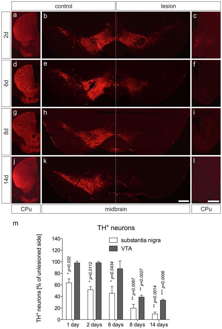
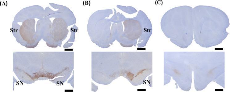
- Hallmark: Loss of pigmented dopaminergic neurons in the substantia nigra pars compacta (SNc)
- Resulting dopamine depletion in the striatum (putamen and caudate nucleus)
- Formation of Lewy bodies - eosinophilic intracytoplasmic inclusions in residual neurons, composed primarily of alpha-synuclein (α-syn)
- Neuronal loss also occurs in: locus coeruleus, dorsal raphe nucleus, nucleus basalis of Meynert, enteric nervous system, dorsal vagal nucleus
- Braak's staging: proposes pathology begins in the olfactory bulb and enteric nervous system before ascending to the SNc ("body-first" vs "brain-first" forms)
2b. Basal Ganglia Circuit Disruption
In normal function, dopamine from SNc modulates the direct and indirect pathways of the basal ganglia:
- Direct pathway (D1 receptors): facilitates movement - reduced activity in PD
- Indirect pathway (D2 receptors): inhibits movement - disinhibited in PD
In PD:
- Dopamine loss → increased firing of STN (subthalamic nucleus) and GPi (globus pallidus interna)
- Excessive inhibition of the thalamus
- Reduced activation of cortical motor systems
- = Poverty of movement (parkinsonism)
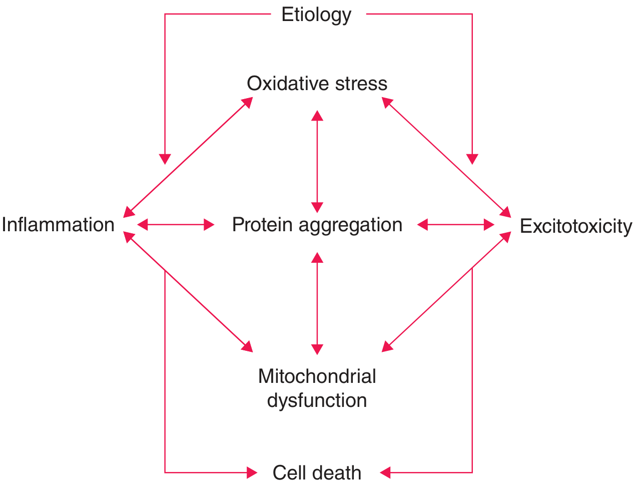
Harrison's Principles of Internal Medicine 22E - Pathogenetic factors in PD leading to cell death
2c. Pathogenesis - Key Mechanisms
| Mechanism | Description |
|---|
| Oxidative stress | Excess free radicals from dopamine metabolism damage neurons |
| Protein aggregation | α-synuclein misfolds, forms toxic oligomers and Lewy bodies |
| Mitochondrial dysfunction | Complex I inhibition (notably by MPTP exposure); impairs energy production |
| Excitotoxicity | Excess glutamate via STN over-activation damages neurons |
| Neuroinflammation | Microglial activation, inflammatory cytokines amplify neurodegeneration |
3. ETIOLOGY & RISK FACTORS
Genetic Causes (~10-15% of cases)
| Gene | Mutation Type | Inheritance |
|---|
| SNCA (alpha-synuclein) | Point mutations, duplications, triplications | AD |
| LRRK2 (leucine-rich repeat kinase 2) | Most common genetic cause in adults | AD |
| Parkin (PARK2) | Most common cause of early-onset PD | AR |
| PINK1 | Early-onset PD | AR |
| DJ-1 | Rare early-onset PD | AR |
| GBA (glucocerebrosidase) | Risk factor, especially in Ashkenazi Jews | - |
Environmental / Other Risk Factors
- MPTP exposure (heroin contaminant) - induces parkinsonism via mitochondrial Complex I inhibition
- Pesticide/herbicide exposure (rotenone, paraquat)
- Head trauma (repeated)
- Well water consumption, rural living
- Protective factors: smoking, caffeine use (epidemiological associations)
4. CLINICAL FEATURES
4a. CARDINAL MOTOR Features (TRAP)
| Feature | Description |
|---|
| T - Tremor | Resting tremor ("pill-rolling"), 4-6 Hz; typically asymmetric at onset; presenting symptom in ~70% of patients; virtually pathognomonic when asymmetric resting tremor is present |
| R - Rigidity | Increased resistance throughout range of motion; "cogwheel rigidity" (ratchety quality on passive movement); "lead-pipe" rigidity |
| A - Akinesia/Bradykinesia | Slowness of movement; difficulty initiating; progressive; often described by patient as "weakness" though strength testing is normal; micrographia is a key feature |
| P - Postural Instability | Late finding; impaired righting reflexes; "pull test" positive; major cause of falls |
4b. Other Motor Features
- Masked facies (hypomimia): reduced facial expression
- Micrographia: progressively smaller handwriting
- Shuffling, narrow-based gait with reduced arm swing
- Festination: involuntary acceleration of gait
- Freezing of gait ("feet stuck to floor") - especially doorways, turning
- Dysarthria: soft, monotone speech (hypophonia)
- Dysphagia: in ~35% subjectively; up to 82% on objective testing; all phases (oral, pharyngeal, esophageal) affected
- En bloc turning (turning in many steps rather than pivoting)
4c. Non-Motor Features (often PRECEDE motor symptoms)
Autonomic Dysfunction:
- Orthostatic hypotension (dizziness, syncope)
- Constipation (most common GI complaint; may precede motor symptoms by years)
- Urinary dysfunction (urgency, nocturia)
- Sexual dysfunction
- Excessive sweating
Neuropsychiatric:
- Depression and anxiety (very common; up to 40-50%)
- Dementia/Cognitive decline - in advanced disease; "Parkinson's Disease Dementia (PDD)"
- Psychosis/Hallucinations - often medication-induced (visual hallucinations)
- Impulse control disorders - associated with dopamine agonist use
Sleep Disorders:
- REM Sleep Behavior Disorder (RBD) - acting out dreams; may precede PD by years/decades
- Excessive daytime sleepiness
- Insomnia
Sensory:
- Anosmia/Hyposmia - often an early/premotor feature
- Pain and sensory disturbances
Clinical Pearl: Constipation, anosmia, and REM sleep behavior disorder can precede motor symptoms by years to decades - these are premotor biomarkers of PD.
5. DIAGNOSIS
5a. Clinical Diagnosis (MDS Criteria)
Core feature (required): Parkinsonism = Bradykinesia + Rigidity and/or Tremor
UK Brain Bank Criteria (historically used, >90% pathologic accuracy):
- Parkinsonism (bradykinesia + rigidity)
- Asymmetric onset
- Resting tremor present
- Good response to levodopa
Levels of certainty:
- Clinically Established PD: core features + supportive criteria + no exclusion/red flags
- Clinically Probable PD: core features + fewer supportive criteria
5b. Supportive Features (increase diagnostic confidence)
- Unequivocal beneficial response to dopaminergic therapy
- Presence of levodopa-induced dyskinesia
- Rest tremor (asymmetric)
- Anosmia or cardiac sympathetic denervation on MIBG scintigraphy
5c. Red Flags / Atypical Features (suggest alternative diagnosis)
- Early falls
- Early severe dysautonomia
- Early dementia/cognitive impairment
- Hallucinations not induced by drugs
- Upward gaze palsy (→ PSP)
- Cerebellar signs (→ MSA)
- Poor or no levodopa response
- Bilateral symmetric onset
- Exposure to dopamine-blocking drugs
5d. Investigations
- Brain MRI: rule out vascular parkinsonism, subdural hematoma, NPH, MSA ("hot cross bun" sign in pons)
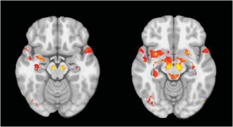
- DaTscan (SPECT): dopamine transporter imaging - reduced asymmetric uptake in posterior putamen; confirms dopaminergic degeneration
- FDG-PET: shows hypometabolism in posterior parietal and occipital regions
- Skin biopsy: α-synuclein deposition (emerging biomarker)
- Seed Amplification Assays (SAA): detect α-synuclein in CSF
- MIBG cardiac scintigraphy: cardiac postganglionic sympathetic denervation
- Polysomnography: RBD without atonia
Routine bloodwork, CT/MRI may be normal. No single test confirms PD; it remains a clinical diagnosis.
6. DIFFERENTIAL DIAGNOSIS
| Condition | Distinguishing Features |
|---|
| Essential Tremor | Action/postural tremor (not resting); symmetric; no rigidity/bradykinesia; responds to alcohol |
| MSA (Multiple System Atrophy) | Early autonomic failure, cerebellar signs, poor levodopa response |
| PSP (Progressive Supranuclear Palsy) | Early falls, vertical gaze palsy, axial rigidity, no tremor |
| DLB (Dementia with Lewy Bodies) | Dementia precedes or concurrent with parkinsonism; visual hallucinations early; fluctuating cognition |
| Corticobasal Syndrome | Asymmetric limb dystonia, alien limb phenomenon, apraxia |
| Vascular Parkinsonism | Lower body predominance ("lower half parkinsonism"), vascular risk factors, MRI white matter changes |
| Drug-Induced Parkinsonism | Exposure to dopamine antagonists (antipsychotics, metoclopramide); symmetric; resolves on drug withdrawal |
| Wilson's Disease | Age <40, liver disease, Kayser-Fleischer rings (important to exclude!) |
| Normal Pressure Hydrocephalus | Triad: gait apraxia, dementia, urinary incontinence |
7. PHARMACOLOGICAL MANAGEMENT
Drug therapy should be initiated when symptoms cause functional impairment. Current therapies do not slow disease progression.
7a. Levodopa/Carbidopa (Sinemet) - Gold Standard
- Mechanism: Levodopa = dopamine precursor; crosses BBB → converted to dopamine in brain. Carbidopa = peripheral dopa-decarboxylase inhibitor (prevents peripheral conversion, reduces dose needed and side effects)
- Carbidopa saturated at 70-100 mg/day
- Starting dose: Carbidopa/levodopa 25 mg/100 mg, ½ tablet TID; increase by ½ tablet every 4-7 days
- Most effective for bradykinesia and rigidity; also benefits tremor
- Long-term complications:
- Motor fluctuations: "wearing off" (symptom return before next dose), "on-off" phenomenon
- Dyskinesias: involuntary choreiform movements (peak-dose or diphasic)
- Confusion, hallucinations (especially in elderly)
- Nausea, orthostatic hypotension
7b. Dopamine Agonists
| Drug | Route | Notes |
|---|
| Pramipexole (Mirapex) | Oral | Non-ergot; D2/D3 agonist |
| Ropinirole (Requip) | Oral | Non-ergot; D2/D3 agonist |
| Rotigotine (Neupro) | Transdermal patch | 24-hr coverage |
| Cabergoline | Oral | Ergot (cardiac valve risk) |
| Bromocriptine | Oral | Ergot; less used now |
| Apomorphine | SC injection | Rescue therapy for off episodes |
- Used as monotherapy in early PD (delay levodopa and reduce dyskinesia risk) or adjunct to levodopa
- Side effects: Nausea, somnolence, hallucinations, orthostatic hypotension, impulse control disorders (gambling, hypersexuality - important counseling point)
7c. MAO-B Inhibitors (Monoamine Oxidase B)
| Drug | Dose | Notes |
|---|
| Selegiline (Eldepryl) | 5 mg BID | May have neuroprotective effect (controversial) |
| Rasagiline (Azilect) | 1 mg OD | Once daily; used as adjunct or monotherapy in early PD |
| Safinamide | 50-100 mg OD | Adjunct to levodopa |
- Inhibit dopamine breakdown → extend dopamine effect
- Selegiline + tyramine-rich foods = hypertensive crisis risk (lesser risk than non-selective MAO inhibitors)
7d. COMT Inhibitors (Catechol-O-Methyltransferase)
| Drug | Dose | Notes |
|---|
| Entacapone (Comtan) | 200 mg with each levodopa dose | No liver monitoring needed |
| Tolcapone (Tasmar) | 100-200 mg TID | Requires LFT monitoring (hepatotoxicity risk) |
| Stalevo | - | Carbidopa + Levodopa + Entacapone (combination pill) |
- Extend the duration of levodopa action; reduce "wearing off"
- Side effects: diarrhea, urine discoloration (orange-brown), dyskinesia
7e. Anticholinergics
| Drug | Dose | Notes |
|---|
| Trihexyphenidyl (Artane) | 1-2 mg TID | Primarily for tremor |
| Benztropine (Cogentin) | 0.5-6 mg/day | Tremor and rigidity |
- Used primarily for tremor-predominant PD in younger patients
- Avoid in elderly (memory impairment, confusion, hallucinations, urinary retention, constipation)
7f. Amantadine
- Mechanism: NMDA receptor antagonist + weak dopaminergic/anticholinergic effects
- Reduces levodopa-induced dyskinesias (unique among PD drugs)
- Useful in early and late PD
- Side effects: livedo reticularis, edema, confusion, hallucinations
7g. Managing Motor Complications
| Problem | Management |
|---|
| Wearing off | Add COMT inhibitor, MAO-B inhibitor; switch to CR formulation; use dopamine agonist |
| Dyskinesias | Reduce levodopa dose; add amantadine; GPi DBS |
| On-off fluctuations | Apomorphine injection/infusion; duodenal levodopa infusion; DBS |
| Freezing of gait | Visual cues (laser walker), auditory rhythmic cues, physical therapy |
8. SURGICAL MANAGEMENT
Deep Brain Stimulation (DBS)
- Target nuclei: STN (subthalamic nucleus) or GPi (globus pallidus interna)
- Indications: Advanced PD with motor fluctuations/dyskinesias not controlled by medications; younger patients; preserved cognition
- Mechanism: High-frequency stimulation modulates abnormal basal ganglia output, normalizing thalamocortical activation
- STN DBS: Greater benefit in "off" state, allows medication reduction; slightly higher neuropsychiatric risk
- GPi DBS: Better dyskinesia suppression; more flexible long-term; relatively safer neuropsychiatric profile
- Contraindications: Cognitive impairment, psychiatric instability, poor levodopa response, advanced age with comorbidities
- Similar motor outcomes between STN and GPi in randomized trials
9. NON-PHARMACOLOGICAL MANAGEMENT
| Intervention | Rationale/Evidence |
|---|
| Physical therapy | Gait training, balance exercises, LSVT BIG (large amplitude movement program), reduce falls |
| Occupational therapy | Adaptive devices for ADLs, home safety assessment |
| Speech therapy | LSVT LOUD (Lee Silverman Voice Treatment) for hypophonia; swallowing evaluation, dysphagia management |
| Swallowing assessment | Modified barium swallow, FEES; aspiration risk is high (15-56%); silent aspiration in 15-33% |
| Nutritional support | High-fiber diet (constipation); maintain weight; protein redistribution diet can reduce levodopa absorption competition |
| Exercise | Aerobic exercise, Tai Chi, dance; shown to improve gait, balance, mood |
| Support groups | Education, caregiver support, psychosocial wellbeing |
| Fall prevention | Home hazard assessment, assistive devices, hip protectors |
10. NURSING ASSESSMENT & CARE
10a. Focused Nursing Assessment
- Motor function: Evaluate tremor, rigidity, bradykinesia, gait, balance - document asymmetry
- Functional status: ADL capacity (eating, dressing, bathing, ambulation)
- Fall risk: High-risk population; assess pull test, orthostatic changes, footwear, environment
- Swallowing: Screen for dysphagia (coughing, choking, prolonged mealtimes); aspiration risk
- Nutrition/weight: Caloric needs, ability to self-feed, weight trajectory
- Elimination: Constipation, urinary urgency/retention
- Skin: Assess for pressure injuries (reduced mobility), seborrheic dermatitis (common in PD)
- Sleep: RBD, insomnia, daytime sleepiness
- Cognition & mood: Depression screening (GDS), cognitive assessment (MoCA), psychosis
- Medications: Compliance, timing (medication timing is critical - "off" episodes if doses missed/late), side effects
- Caregiver strain: Caregiver burden is significant; assess caregiver coping, education, respite needs
10b. Priority Nursing Diagnoses (NANDA)
| Nursing Diagnosis | Related to |
|---|
| Impaired physical mobility | Rigidity, bradykinesia, postural instability |
| Risk for falls | Postural instability, freezing, orthostatic hypotension |
| Impaired swallowing | Pharyngeal dysmotility, cricopharyngeal dysfunction |
| Impaired verbal communication | Dysarthria, hypophonia |
| Self-care deficit (bathing, dressing, feeding) | Motor impairment, bradykinesia |
| Constipation | Autonomic dysfunction, immobility, anticholinergic medications |
| Imbalanced nutrition: less than body requirements | Dysphagia, increased energy expenditure, depression |
| Chronic sorrow/Depression | Chronic progressive disease, loss of function |
| Caregiver role strain | Progressive demands, 24/7 care needs |
| Disturbed sleep pattern | RBD, nocturia, immobility |
| Risk for aspiration | Dysphagia, impaired cough reflex, silent aspiration |
10c. Key Nursing Interventions
Safety/Mobility:
- Fall prevention: bed in lowest position, call light within reach, non-slip footwear, bed/chair alarms, assistive devices
- Teach patient to use visual cues (floor lines, counting steps) for freezing episodes
- Allow adequate time for movements - do not rush
- Wide base of support; encourage deliberate, heel-toe gait
Medication Management:
- Administer levodopa/carbidopa on time - delays cause "off" episodes
- Give 30-60 minutes before meals (or as directed - protein can impair absorption)
- Monitor for dyskinesias (peak dose), especially after dose changes
- Monitor orthostatic BP before and after medications
- Educate on tyramine interactions with MAO-B inhibitors
- Watch for impulse control disorders with dopamine agonists
Dysphagia/Nutrition:
- Upright position (90°) during meals and 30-45 min after
- Thickened liquids if indicated by SLP assessment
- Small, frequent meals; soft foods
- Adequate hydration; ensure medications are safely swallowed
- Monitor for signs of aspiration pneumonia
Bowel/Bladder:
- High-fiber diet, adequate fluids, stool softeners for constipation
- Scheduled toileting program; fall precautions to bathroom
- Monitor urinary retention (anticholinergics)
Communication:
- Face patient when speaking; minimize background noise
- Allow time for verbal responses; do not finish sentences
- Encourage speech therapy; consider augmentative communication devices
Psychosocial:
- Screen regularly for depression and anxiety
- Provide information on support groups (Parkinson's Foundation, Michael J. Fox Foundation)
- Advance care planning discussions (early, when cognition intact)
- Refer to social work; assess home support, caregiver burnout
11. COMPLICATIONS
| Complication | Notes |
|---|
| Aspiration pneumonia | Leading cause of death in advanced PD |
| Falls and fractures | Hip fractures especially; major morbidity |
| Malnutrition | Dysphagia, depression, increased metabolic demands |
| Dementia (PDD) | Occurs in majority with long disease duration (>80% at 20 years) |
| Psychosis | Visual hallucinations; medication-related and disease-related |
| Deep vein thrombosis | Immobility |
| Autonomic crises | Severe hypotension, urinary retention |
| Medication complications | Dyskinesias, on-off fluctuations, impulse control disorders |
| Caregiver burnout | Major social complication; often determines institutionalization |
12. HOEHN & YAHR STAGING SCALE
| Stage | Description |
|---|
| Stage I | Unilateral disease only; minimal or no functional impairment |
| Stage II | Bilateral or midline involvement; no balance impairment |
| Stage III | First sign of postural reflexes impairment; mild-moderate disability; physically independent |
| Stage IV | Severe disability; still able to walk or stand unassisted |
| Stage V | Confined to bed or wheelchair unless aided |
Also used: UPDRS (Unified Parkinson's Disease Rating Scale) - comprehensive assessment tool covering motor, non-motor, and ADL domains
13. PALLIATIVE CARE CONSIDERATIONS
- PD is a chronic progressive terminal disease - early palliative integration improves quality of life
- Advance care planning should begin early while patient has decision-making capacity
- Goals of care conversations: feeding tubes (usually not recommended in advanced dysphagia), hospitalization, resuscitation status
- Hospice criteria: Stage IV-V with significant functional decline, aspiration, weight loss, recurrent infections
- Caregiver support, grief counseling, respite care are essential
- Systematic review evidence (PMID 40320755) supports early integration of "thinking palliative care" in PD management
14. PATIENT & FAMILY EDUCATION
- Disease course: Slowly progressive; most patients live many years with good quality of life especially with treatment
- Medication timing is critical: Never skip doses; "off" states are preventable
- Exercise daily: Evidence shows exercise slows functional decline
- Driving safety: Discuss early; bradykinesia and cognitive changes affect driving
- Home modifications: Grab bars, raised toilet seat, remove throw rugs, adequate lighting
- Dietary guidance: High fiber, adequate protein (time protein away from levodopa), maintain hydration
- Community resources: Parkinson's Foundation helpline, local support groups, caregiver respite programs
- Emergency awareness: Carry medical ID; Parkinson's medications must not be withheld in hospital settings (risk of neuroleptic malignant-like syndrome)
QUICK REVIEW - HIGH-YIELD POINTS FOR MSN EXAMS
| Topic | Key Fact |
|---|
| Pathology | Loss of dopaminergic neurons in substantia nigra pars compacta; Lewy bodies (α-synuclein) |
| Cardinal features | TRAP: Tremor (resting, asymmetric), Rigidity (cogwheel), Akinesia/Bradykinesia, Postural instability (late) |
| First drug | Levodopa/Carbidopa - gold standard; carbidopa prevents peripheral conversion |
| Tremor type | 4-6 Hz resting "pill-rolling" - worsens with distraction/mental tasks; improves with intentional movement |
| Differentiating tremor | PD = resting; Essential tremor = action/postural |
| DBS targets | STN or GPi |
| Leading cause of death | Aspiration pneumonia |
| Premotor symptoms | Constipation, anosmia, RBD - precede motor symptoms by years |
| Nursing priority | Fall prevention + medication timing (never withhold levodopa in hospital) |
| Drug to avoid | Typical antipsychotics (worsen parkinsonism); Metoclopramide (dopamine blocker) |
| Dopamine agonist warning | Impulse control disorders - must counsel patient/family |
Sources: Harrison's Principles of Internal Medicine 22E, p. 3540-3544 | Textbook of Family Medicine 9e, p. 1249-1250 | Bradley and Daroff's Neurology in Clinical Practice | Neuroanatomy through Clinical Cases 3rd Ed | Schwartz's Principles of Surgery 11th Ed