
Thyroglossal cyst
Reading File
Now let me search the medical library and PubMed simultaneously.
Finding Sources
Searching PubMed
thyroglossal duct cyst
Good results. Now let me read the most comprehensive sources in parallel.
Reading File
Reading File
Reading File
Now let me get more details from Sabiston (pediatric surgery block) and also fetch the ultrasound image:
Reading File
Loading Image
Finding Sources
Let me get one more detailed source - the Cummings Otolaryngology section and Schwartz's for completeness:
Reading File
Reading File
I now have comprehensive information from multiple authoritative textbooks. Let me compile the full answer.
Thyroglossal Duct Cyst (TDC)
Embryology
The thyroid gland originates as a median endodermal diverticulum at the foramen cecum (base of tongue) at approximately 3 weeks of embryonic life. It descends through the neck along the thyroglossal duct, passing through - and becoming intimately related to - the developing hyoid bone (formed from ventral cartilages of the 2nd and 3rd branchial arches). The duct normally involutes by the 8th-10th week of gestation once the thyroid reaches its final pretracheal position. Persistence of any portion of this duct allows secretions from the epithelial lining to accumulate, forming a cyst.
- Sabiston Textbook of Surgery, p. 1343
- Schwartz's Principles of Surgery, p. 1738
Epidemiology
- The most common congenital cystic mass of the neck
- Second most common benign neck mass overall, after reactive lymphadenopathy
- Most commonly presents in children aged 2-4 years (when baby fat disappears and neck irregularities become apparent), though it can occur at any age
Location
| Site | Frequency |
|---|---|
| Infrahyoid (below hyoid) | 65% |
| At the level of hyoid bone | 15% |
| Suprahyoid / base of tongue | ~20% |
| Midline | 75% |
| Off-midline (up to 2 cm) | 25% |
The intimate relationship to the hyoid bone arises directly from the duct's passage through the developing hyoid during embryogenesis.
- Scott-Brown's Otorhinolaryngology Head & Neck Surgery, p. 627
Clinical Features
- Painless midline neck mass - classic presentation
- Moves cranio-caudally with swallowing and tongue protrusion (pathognomonic sign - due to attachment to the base of tongue)
- Usually asymptomatic unless infected
- Infection (often following upper respiratory illness) causes pain, redness, swelling; may progress to abscess
- Rarely: dysphagia, dyspnea
Key point: "An upper midline cystic neck lesion in toddlers is a thyroglossal duct cyst until proven otherwise." - Sabiston Textbook of Surgery
Diagnosis
Clinical - diagnosis is primarily clinical based on the midline neck mass and characteristic movement with tongue protrusion.
Imaging:
- Ultrasound (first-line): Appearance varies:
- Homogeneous anechoic mass with posterior wall enhancement (most classic)
- Pseudosolid mass (due to proteinaceous secretions)
- Heterogeneous echo pattern (from previous hemorrhage or infection)
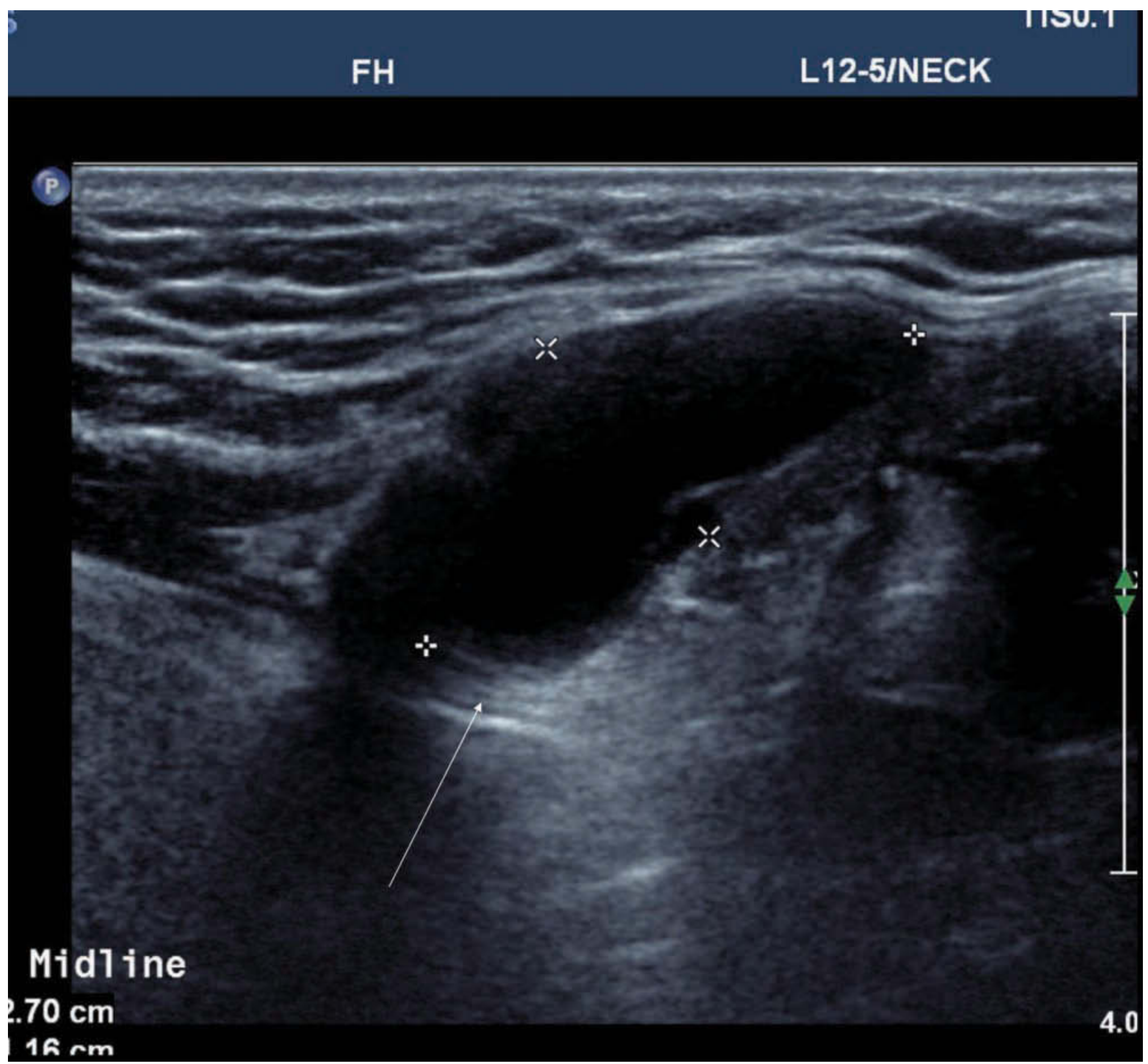
US showing midline infrahyoid homogeneous anechoic cystic mass with posterior wall enhancement (black arrow) - Scott-Brown's Otorhinolaryngology Head & Neck Surgery
- CT/MRI: Useful when US is inconclusive; on MRI - invariably high T2 signal, variable T1 depending on protein content
- Radionuclide scan / thyroid US: Mandatory if a normal thyroid cannot be palpated in its normal position, to rule out lingual thyroid (where the TDC may be the only functioning thyroid tissue)
Absence of orthotopic thyroid on imaging requires careful consideration before surgical excision.
FNA: May be used to rule out malignancy or other masses if clinical doubt exists.
Histopathology
The cyst wall is lined by:
- Stratified squamous epithelium - when near the base of tongue
- Pseudostratified columnar (respiratory) epithelium - in lower cervical locations
- Transitional patterns also occur
The fibrous cyst wall often contains lymphoid aggregates and thyroid follicular remnants.
- Robbins, Cotran & Kumar Pathologic Basis of Disease, p. 694
Complications
- Infection / abscess - most common complication; often triggered by upper respiratory infection
- Fistula formation - if infected cyst ruptures or is inadequately drained; may result in a salivary fistula
- Malignant transformation - rare (~1% incidence); classically papillary thyroid carcinoma (PTC)
- Suspect malignancy when: rapid growth, complex anechoic pattern on US, or calcification
- Finding malignancy in a TDC carries a 6.6% risk of concurrent microcarcinoma in the orthotopic thyroid
- A 2025 systematic review (PMID: 40347268) specifically addressed TDC carcinomas
Treatment
Sistrunk Operation (1920) - Gold Standard
Described by Walter Sistrunk, who recognized that simple cyst excision had unacceptably high recurrence rates because residual duct epithelium along the tract would regenerate.
The Sistrunk procedure includes:
- Complete excision of the cyst
- Removal of the central portion of the hyoid bone (en bloc)
- Excision of the tract from hyoid to the base of the tongue, including a core of genioglossus muscle up to the foramen cecum
Outcomes:
| Procedure | Recurrence Rate |
|---|---|
| Simple cyst excision | ~46% |
| Modified Sistrunk | ~5.8% |
| Full Sistrunk (with pharyngotomy) | <5% |
- Cummings Otolaryngology Head and Neck Surgery, p. 3820
Management of Infected TDC
- Acute phase: Antibiotics +/- incision and drainage (I&D)
- After resolution of infection: Definitive Sistrunk procedure
- Simple I&D without subsequent Sistrunk virtually guarantees recurrence
Factors Predicting Recurrence
- More than 2 infections prior to surgery
- Age under 2 years
- Inadequate initial operation (deviation from Sistrunk technique)
- Schwartz's Principles of Surgery, p. 1738
Malignancy in TDC
- Thyroidectomy is not automatically required for malignant TDC
- The thyroid gland should be closely evaluated and monitored for life
- Management of any thyroid nodules follows standard thyroid cancer guidelines
- A 2026 systematic review on ectopic thyroid cancer (PMID: 42290855) provides updated guidance
Differential Diagnosis of Midline Neck Mass
| Entity | Key Distinguishing Feature |
|---|---|
| Thyroglossal duct cyst | Moves with tongue protrusion, infrahyoid midline |
| Dermoid cyst | Does not move with swallowing/tongue protrusion, doughy consistency |
| Submental lymphadenopathy | Typically multiple nodes, reactive |
| Lingual thyroid | At base of tongue, no orthotopic thyroid |
| Ectopic thyroid | May be the only thyroid tissue |
| Midline ranula | Fluctuant, floor of mouth |
Sources: Sabiston Textbook of Surgery; Schwartz's Principles of Surgery 11e; Scott-Brown's Otorhinolaryngology Head & Neck Surgery; Cummings Otolaryngology Head and Neck Surgery; Robbins, Cotran & Kumar Pathologic Basis of Disease
This is a shared conversation. Sign in to Orris to start your own chat.