
Provide me an image of brain sensitive structures in brain
Reading File
Now let me search the medical library for brain sensitive structures images.
Finding Sources
Finding Sources
Finding Sources
Finding Sources
The best source is Adams and Victor's Principles of Neurology. Let me read the section on "Pain-Sensitive Cranial Structures" and look for images there.
Reading File
Reading File
The textbook text is rich, but I don't see a direct image URL here. Let me search for an image in the Guyton physiology section and also look for image links in these blocks.
Reading File
I found an image. Let me fetch it.
Loading Image
Here is the image from Guyton and Hall's Textbook of Medical Physiology, along with a full explanation of the pain-sensitive structures in the brain.
Pain-Sensitive Structures in the Brain
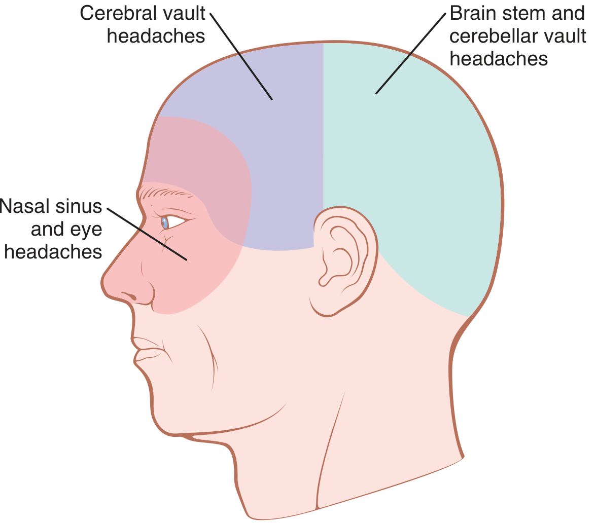
Figure 49.9 - Areas of headache resulting from different causes (Guyton and Hall Textbook of Medical Physiology)
The diagram shows three color-coded zones:
- Pink (front/face) - Nasal sinus and eye headaches
- Purple (upper/front skull) - Cerebral vault headaches
- Teal (posterior skull) - Brainstem and cerebellar vault headaches
Pain-Sensitive vs. Insensitive Structures
Pain-SENSITIVE structures (can cause headache when stimulated):
| Structure | Notes |
|---|---|
| Skin, subcutaneous tissue, scalp muscles | Richly innervated; extracranial |
| Extracranial arteries (e.g., superficial temporal) | Stretching/inflammation causes pain |
| Venous sinuses (dural sinuses) and their large tributaries | Tugging causes intense headache |
| Dura at the base of the brain | Especially sensitive; stimulated by mass lesions |
| Middle meningeal artery | Especially sensitive - surgeons anesthetize specifically |
| Proximal cerebral arteries (anterior, middle cerebral; internal carotid intracranially) | Pain referred to eye/orbitotemporal area |
| Cranial nerves V, IX, X and upper cervical nerves (C1-C3) | Transmit impulses through dura |
| Eye, ear, nasal cavities, paranasal sinuses | Delicate structures with rich pain receptors |
Pain-INSENSITIVE structures (do NOT cause pain when cut/stimulated):
- Brain parenchyma (cortex, white matter) - almost totally insensitive
- Pia-arachnoid (most of it)
- Ependyma and choroid plexuses
- Cerebellum and ventricular walls
Referred Pain Zones (from the diagram)
- Above the tentorium (supratentorial lesions): Pain via CN V (trigeminal) - referred to front half of head (forehead, temporal, orbital regions)
- Below the tentorium (posterior fossa lesions): Pain via CN IX, X, and C1-C3 - referred to occipital/posterior head region
Source: Guyton and Hall Textbook of Medical Physiology, Fig. 49.9; Adams and Victor's Principles of Neurology 12th Ed., "Pain-Sensitive Cranial Structures"
This is a shared conversation. Sign in to Orris to start your own chat.