
Anatomy of vertebrae and spinal cord
Reading File
Now let me search the medical library for vertebrae and spinal cord anatomy, running parallel searches:
Finding Sources
Finding Sources
Finding Sources
Finding Sources
Reading File
Reading File
Finding Sources
Excellent results. Let me read the most anatomy-rich sources in parallel:
Reading File
Reading File
Reading File
Now let me read more on specific vertebrae regions, spinal cord tracts, and fetch key images in parallel:
Reading File
Reading File
Loading Image
Loading Image
Loading Image
Loading Image
Reading File
Loading Image
Loading Image
Finding Sources
I have all the information needed for a comprehensive answer. Let me compile it now.
Anatomy of the Vertebrae and Spinal Cord
Part 1: The Vertebral Column
The vertebral column forms the axial skeleton of the back and has a characteristic S-shaped curve that aligns the trunk over the center of gravity. It consists of 33 vertebrae in 5 regional groups:
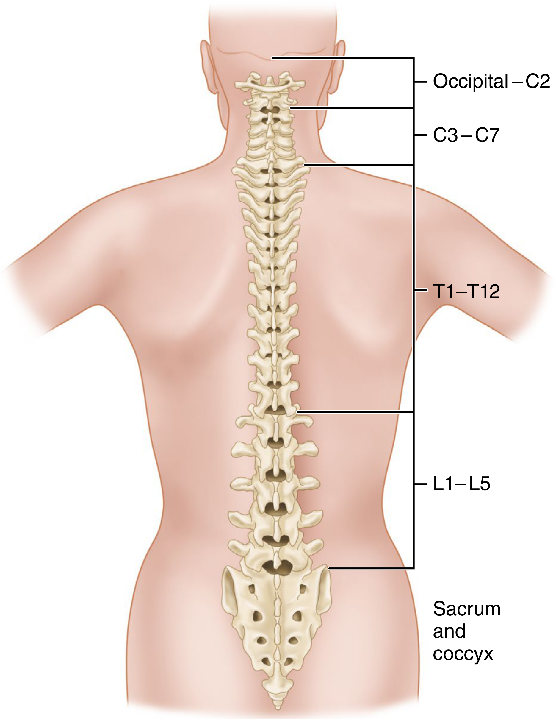
| Region | Number | Notes |
|---|---|---|
| Cervical (C) | 7 | Smallest; foramen transversarium in transverse processes |
| Thoracic (T) | 12 | Articulate with ribs; costal facets |
| Lumbar (L) | 5 | Largest; greatest weight-bearing |
| Sacral (S) | 5 (fused) | Fused into the sacrum in adults |
| Coccygeal | 4 (fused) | Rudimentary coccyx |
Part 2: Structure of a Typical Vertebra
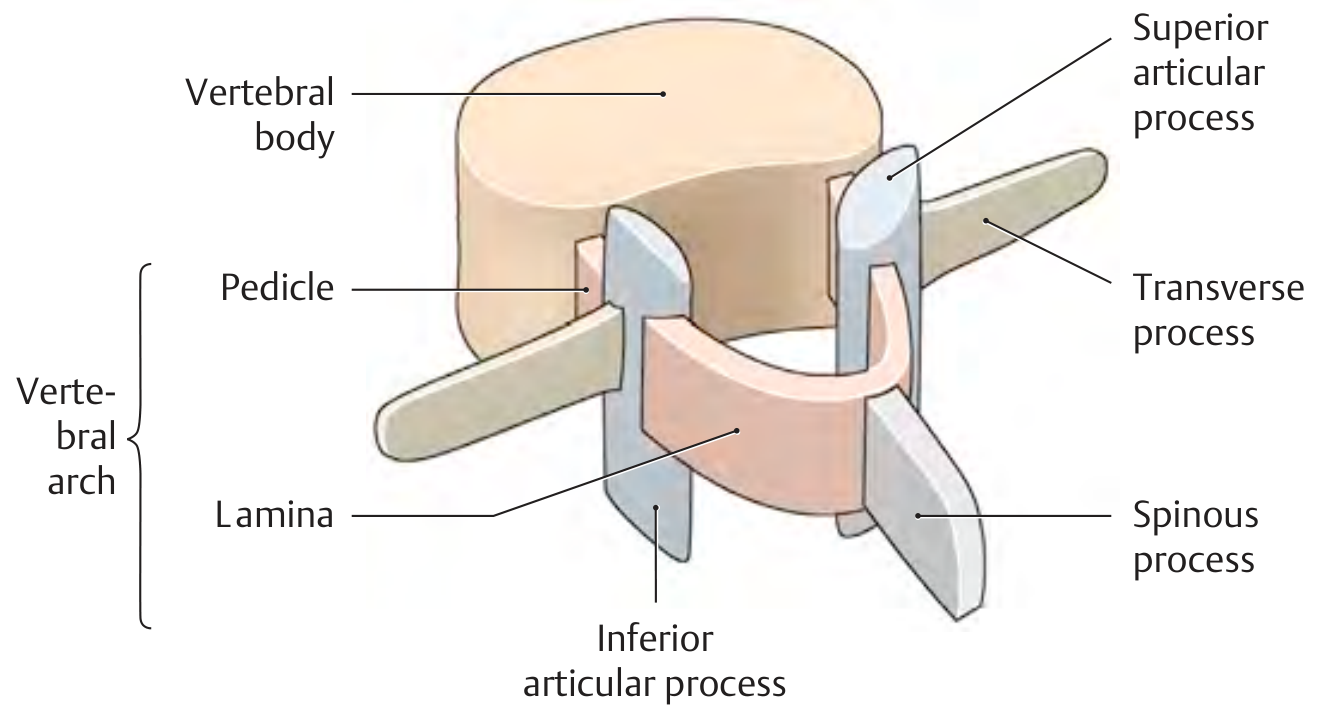
All vertebrae (except atlas and axis) share the same basic elements. - Gray's Anatomy for Students, p. 83-84
Key Structural Components
1. Vertebral Body
- Anterior, ovoid-shaped, weight-bearing part
- Linked to adjacent bodies by intervertebral discs and ligaments
- Size increases from C to L as weight load increases
2. Vertebral Arch (= pedicles + laminae)
- Two pedicles - bony pillars attaching arch to body; their superior/inferior notches form the intervertebral foramina
- Two laminae - flat sheets extending from each pedicle meeting in the midline; form the "roof" of the arch
3. Processes
- Spinous process - projects posteriorly/inferiorly from the junction of laminae; muscle and ligament attachment
- Transverse processes (2) - project posterolaterally from the pedicle-lamina junction; muscle/ligament attachment; articulate with ribs in the thoracic region
- Articular processes (4 total: 2 superior, 2 inferior) - form the facet (zygapophyseal) joints with adjacent vertebrae
4. Vertebral Foramen
- Space enclosed by the vertebral body anteriorly and arch posterolaterally
- All foramina together form the vertebral (spinal) canal, which houses and protects the spinal cord
Part 3: Intervertebral Discs
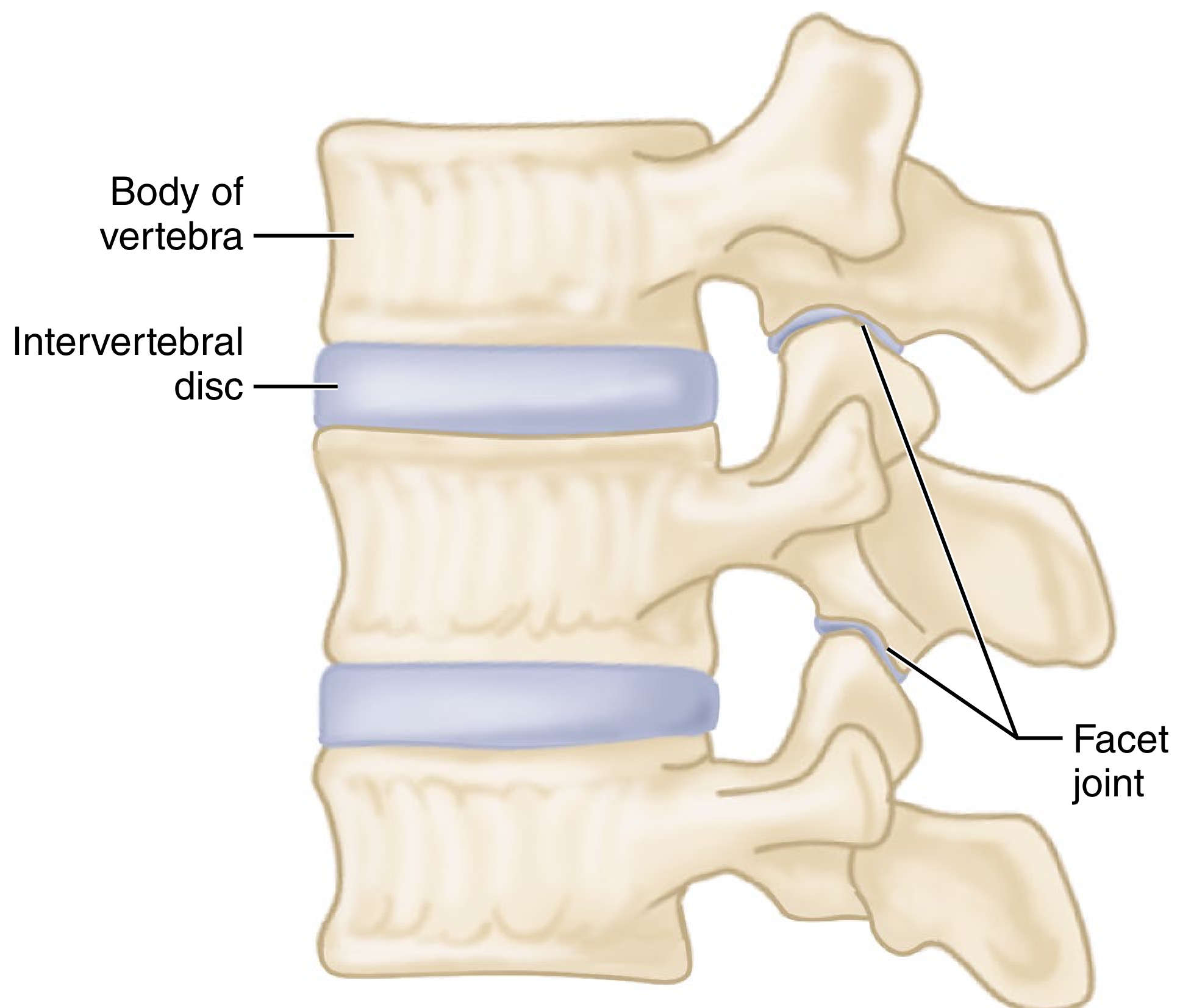
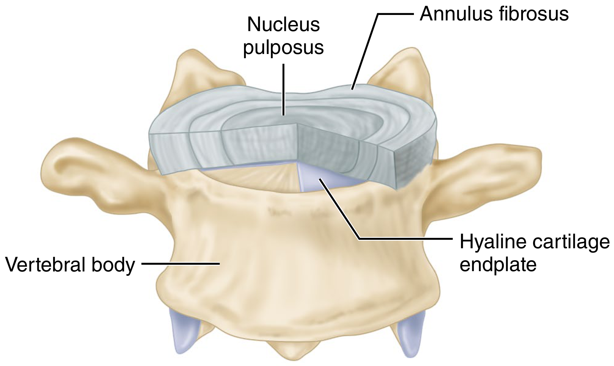
Each disc has:
- Nucleus pulposus - central gelatinous core; shock absorption
- Annulus fibrosus - peripheral ring of fibrocartilage lamellae; resists torsion and compression
- Hyaline cartilage endplates - interface between disc and vertebral body
Part 4: Regional Differences
Cervical Vertebrae (C1-C7)
- Smallest vertebral bodies; square-shaped from above
- Foramen transversarium in each transverse process (transmits vertebral artery)
- Spinous processes are short and bifid (C2-C6)
- Triangular vertebral foramen
- C1 (Atlas) - no vertebral body; ring-shaped; supports the skull; atlas-occipital joints allow nodding ("yes")
- C2 (Axis) - has the dens (odontoid process) projecting superiorly from its body (the fused body of C1); pivot for rotation ("no") at the atlanto-axial joint
Thoracic Vertebrae (T1-T12)
- Heart-shaped bodies; larger than cervical
- Costal facets on vertebral bodies (superior and inferior) and on transverse processes for rib articulation
- Long spinous processes pointing sharply inferiorly
- Circular vertebral foramen
Lumbar Vertebrae (L1-L5)
- Largest, kidney-shaped bodies
- Massive rectangular spinous processes
- Large triangular vertebral foramen
- Costal processes (homologous to rudimentary ribs) instead of true transverse processes
- Greatest range of flexion/extension
Sacrum & Coccyx
- 5 sacral vertebrae fused in adults; articulates with L5 above and coccyx below
- Sacral foramina for anterior and posterior sacral nerve rami
- Coccyx is vestigial and has no clinical relevance
Part 5: Anatomy of the Spinal Cord
General Features
The spinal cord is shorter than the vertebral column:
- In adults, it terminates as the conus medullaris at L1-L2
- In neonates, it ends at approximately L3
- Below the conus, nerve roots form the cauda equina ("horse's tail")
- A fibrous strand called the filum terminale extends from the conus to the coccyx
The spinal cord has two enlargements:
- Cervical enlargement (C5-T1) - gives rise to brachial plexus for the arms
- Lumbosacral enlargement (L1-S2) - gives rise to lumbar/sacral plexus for the legs
Meninges and Spaces
The spinal cord is enclosed in three protective membranes:
- Pia mater - innermost, adherent to cord surface
- Arachnoid mater - middle layer
- Dura mater - outermost, tough fibrous layer
Key spaces:
- Subarachnoid space (between pia and arachnoid) - contains cerebrospinal fluid (CSF)
- Epidural space (between dura and vertebral canal periosteum) - contains fat and venous plexuses; clinically important for epidural anesthesia
Segments
The spinal cord has 31 segments:
- 8 cervical, 12 thoracic, 5 lumbar, 5 sacral, 1 coccygeal
Because the cord is shorter than the column, spinal cord segments do NOT align with their corresponding vertebrae - the discrepancy increases toward the lower segments.
Part 6: Internal Structure of the Spinal Cord
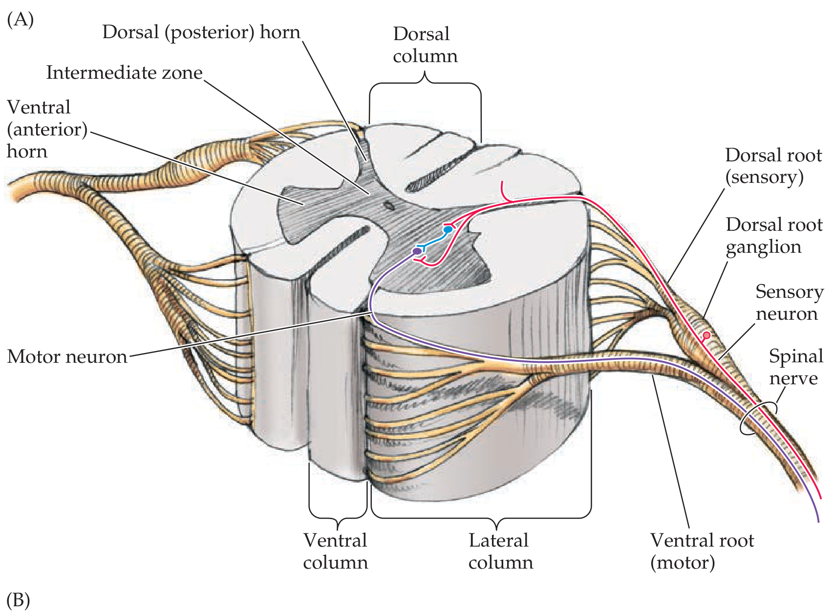
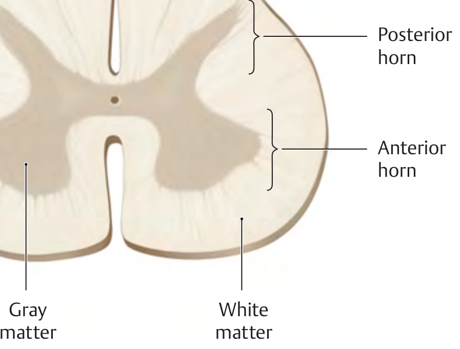
Gray Matter (Central - "Butterfly" or "H" Shape)
| Region | Function |
|---|---|
| Posterior (dorsal) horn | Sensory processing - receives afferent inputs |
| Intermediate zone | Interneurons; autonomic neurons (lateral horn at T1-L2/S2-S4) |
| Anterior (ventral) horn | Motor neurons - sends efferent outputs to muscles |
The gray matter is further divided into Rexed's laminae (I-X), each with specific functional roles.
White Matter (Surrounding Gray Matter - 3 Columns/Funiculi)
| Column | Tracts | Function |
|---|---|---|
| Dorsal (posterior) columns | Fasciculus gracilis & cuneatus | Ipsilateral fine touch, vibration, proprioception |
| Lateral columns | Lateral corticospinal tract (descending); spinothalamic tract (ascending) | Voluntary motor control; contralateral pain and temperature |
| Ventral (anterior) columns | Anterior corticospinal tract; reticulospinal, vestibulospinal tracts | Motor control; posture/balance |
Important somatotopic organization: Within tracts, cervical fibers are located most centrally and sacral fibers most peripherally. This explains why central cord lesions cause disproportionate upper limb deficits (arm > leg), and why syrinx causes a "cape" distribution of sensory loss. - Campbell's Operative Orthopaedics 15th Ed 2026, p. 2225
Part 7: Spinal Nerve Roots
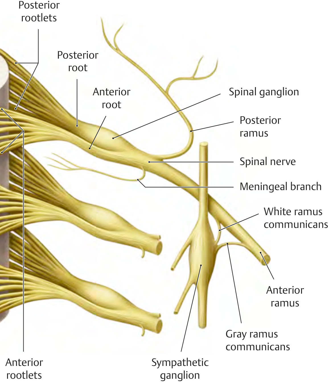
At each segmental level, two roots emerge:
- Dorsal (posterior) root - sensory; contains the dorsal root ganglion (DRG) just outside the cord, which houses sensory neuron cell bodies
- Ventral (anterior) root - primarily motor; lower motor neuron axons
These join in the intervertebral foramen to form the mixed spinal nerve, which then divides into:
- Posterior (dorsal) ramus - back muscles and skin
- Anterior (ventral) ramus - limbs and anterior trunk
Nerve Root Exit Levels (Clinically Important)
- C1-C7: exit above the pedicle of the same number (C6 root exits between C5-C6)
- C8: exits between C7 and T1 (there is no C8 vertebra)
- T1 and below: exit below the pedicle of the same number (L4 root exits between L4-L5)
Summary Table
| Feature | Cervical | Thoracic | Lumbar |
|---|---|---|---|
| Number | 7 | 12 | 5 |
| Body shape | Square | Heart-shaped | Kidney-shaped |
| Foramen shape | Triangular | Circular | Triangular |
| Spinous process | Short, bifid | Long, downward | Short, horizontal |
| Special feature | Foramen transversarium | Costal facets | Largest bodies |
| Cord termination | C1 starts below foramen magnum | - | Conus at L1-L2 |
Sources: - Gray's Anatomy for Students, p. 83-86 | - THIEME Atlas of General Anatomy and Musculoskeletal System, p. 102, 126 | - Neuroanatomy through Clinical Cases 3rd Ed, p. 250 | - Campbell's Operative Orthopaedics 15th Ed 2026, p. 2225 | - Forensic Anthropology: A Comprehensive Introduction, p. 115
Roman Urdu ma aur difficult word ki definition bh batao
Bilkul! Pichli explanation ko Roman Urdu mein dobara likhta hoon, aur mushkil alfaaz ki definitions bhi saath deta hoon.
Vertebrae (ریڑھ کی ہڈی) aur Spinal Cord ki Anatomy
Roman Urdu mein
Hissa 1: Vertebral Column (Ridh ki Haddi ka Silsila)
Vertebral column — yaani ridh ki haddi — poori peeth ka dhanca banati hai. Yeh S-shape mein hoti hai taake jism ka bojh theek se taqseem ho.
Kul 33 vertebrae (haddiyan) hoti hain, 5 hisson mein:
| Hissa | Tadad | Khasoosiyat |
|---|---|---|
| Cervical (gardan) | 7 | Sab se choti haddiyan |
| Thoracic (seena) | 12 | Pasliyon se juri hoti hain |
| Lumbar (kamar) | 5 | Sab se bari, bojh uthane wali |
| Sacral (pelvis) | 5 (juri hui) | Adults mein ek haddi ban jati hain |
| Coccygeal (tailbone) | 4 (juri hui) | Sab se neeche, koi khaas kaam nahi |
Hissa 2: Ek Aam Vertebra ki Banawat
Mushkil Alfaaz ki Definitions:
- Vertebral Body (Vertebral Body) = Haddi ka agla, mota, gol hissa jo bojh uthata hai. Sochein ek drum ki tarah.
- Vertebral Arch (Vertebral Arch) = Pichli taraf ka mehra/darwaza — yeh spinal cord ko dhakta hai.
- Pedicle (Pedicle) = Chhoti sii tang/stalk jo body ko arch se jodti hai. Do hoti hain — daien aur baien.
- Lamina (Lamina) =납ti plate jo dono pedicles ko peeche milati hai aur arch ki "chhat" banati hai.
- Spinous Process (Kanta) = Pichhe ki taraf nikla hua kanta — yahi aap peeth par tangent feel karte hain. Muscles aur ligaments yahan chipakti hain.
- Transverse Process (Side Wala Kanta) = Dono taraf laterally niklta hai — muscles ka attachment point.
- Articular Process (Jor wala hissa) = Upar-neeche ki vertebrae se milne ka hissa — Facet Joint banata hai.
- Vertebral Foramen (Surakh) = Arch aur body ke beech ka khulli jagah jahan spinal cord guzarti hai.
- Vertebral Canal (Nali) = Tamam vertebral foramina mil kar ek nali banati hain — spinal cord is nali mein mehfooz hoti hai.
Hissa 3: Intervertebral Disc (Do Haddiyon ke Darmiyan Takiya)
Har do vertebrae ke darmiyan ek disc hoti hai — yeh jism ka shock absorber hai.
- Nucleus Pulposus = Disc ka beech wala geeli mitti/jelly jaisa part. Pressure absorb karta hai.
- Annulus Fibrosus = Bahari sakht ring jo nucleus ko ghyerti hai. Fiber ki tahen hoti hain. Jab yeh phate to slip disc hoti hai.
- Hyaline Cartilage Endplate = Disc aur haddi ke beech ki chikni teh.
Hissa 4: Alag Alag Ilaqon ki Vertebrae
Cervical (Gardan) — C1 se C7
- Sab se chhoti haddiyan
- Foramen Transversarium = Transverse process mein ek chota surakh jis se Vertebral Artery (gardan ki khoon ki nali) guzarti hai
- Spinous process bifid (kanta do hisson mein banta hai) hota hai C2 se C6 tak
- Atlas (C1) = Sab se upar wali haddi, iska koi body nahi hota — sirf ring hai. Sar ko agay pichay hila ne deta hai ("haan" wali harkat)
- Axis (C2) = Iske upar ek keel jaisi haddi niklti hai jise Dens/Odontoid Process kehte hain. Sar ko ghumane deta hai ("naa" wali harkat)
Thoracic (Seena) — T1 se T12
- Costal Facets = Pasliyon se jorne ke liye chote jod (facets) hote hain
- Spinous processes lambe aur neeche ki taraf jhuke hote hain
Lumbar (Kamar) — L1 se L5
- Sab se bari aur moti vertebrae
- Sab se zyada movement (jhukna, seedha khara hona)
- Koi pasli wala hissa nahi
Hissa 5: Spinal Cord (Ridh ki Nali ke Andar ki Naas)
Aam Jaankari
- Spinal cord dimaag se shuru hoti hai aur L1-L2 tak khatam hoti hai (adults mein)
- Wahan ek makhrooti nok banati hai jise Conus Medullaris kehte hain
- Uske baad nerve roots ka guchha hota hai jise Cauda Equina ("ghore ki dum") kehte hain
- Ek patli dhaga Filum Terminale conus se coccyx tak jata hai
Spinal Cord ke 2 Motape (Enlargements)
- Cervical Enlargement (C5-T1) = Baazuon ke liye nerves yahan se nikalti hain (Brachial Plexus)
- Lumbosacral Enlargement (L1-S2) = Taangon ke liye nerves yahan se nikalti hain (Lumbar/Sacral Plexus)
Meninges (Jhalliyan — Spinal Cord ki Hifazat)
Spinal cord 3 jhilliyon mein lapti hoti hai:
| Jhilli | Matlab | Khasoosiyat |
|---|---|---|
| Pia Mater | "Pio Mater" = Nazuk Maa | Sab se andar, cord se chipki |
| Arachnoid Mater | "Makdi wali" | Beech wali |
| Dura Mater | "Sakhti wali Maa" | Sab se bahar, sakht |
- Subarachnoid Space = Pia aur Arachnoid ke darmiyan jagah — CSF (Cerebrospinal Fluid) yahan hota hai
- CSF = Dimaag aur spinal cord ke aas paas bahnay wala saaf paani jaisa maaya — hifazat karta hai aur ghiza pohanchata hai
- Epidural Space = Dura ke bahar ki jagah — epidural injection yahin di jati hai (dard kam karne ke liye)
Hissa 6: Spinal Cord ka Andar ka Dhanca
Gray Matter (Bhoori Mitti wala Hissa — Butterfly Shakal)
Yeh beech mein hota hai, titli ya H ki shakal ka:
| Hissa | Kaam |
|---|---|
| Posterior Horn (Pichla kanta) | Sensory — Mahsoos karna (dard, temperature, touch) |
| Intermediate Zone (Beech) | Interneurons + Autonomic neurons (khud kar kaam karne wale) |
| Anterior Horn (Agla kanta) | Motor — Muscles ko hukum dena |
- Rexed's Laminae = Gray matter ko aur bhi chote hisson (I se X) mein taqseem kiya gaya hai — har lamina ka alag kaam hai
White Matter (Safed Hissa — 3 Columns)
Yeh gray matter ke chaaro taraf hota hai aur 3 hisson (columns/funiculi) mein banta hai:
| Column | Aham Tracts | Kaam |
|---|---|---|
| Dorsal Column (Pichla) | Fasciculus Gracilis, Fasciculus Cuneatus | Touch, vibration, proprioception (apne jism ki position ka ilm) — usi taraf |
| Lateral Column (Kinara) | Corticospinal tract (motor), Spinothalamic tract (sensory) | Voluntary harkat; dard aur temperature — ulti taraf |
| Ventral Column (Agla) | Anterior corticospinal, Reticulospinal | Motor control, balance |
Mushkil Alfaaz ki Definitions:
- Tract = Naason ka rasta ya raah — jaise motorway par ek lane
- Ascending Tract (Upar wala rasta) = Mehsoosat (sensory signals) jism se dimaag ki taraf jati hain
- Descending Tract (Neeche wala rasta) = Hukum (motor signals) dimaag se jism ki taraf aata hai
- Dorsal Column = Pichhe wala column — fine touch, vibration, proprioception carry karta hai
- Spinothalamic Tract = Dard aur temperature ki information carry karta hai — pehle ulti taraf jaata hai phir upar
- Corticospinal Tract = Dimaag se voluntary muscle movement ka hukum laata hai
- Proprioception = Apne jism ki position aur harkat ka andazi ilm — aankhein band ho tab bhi pata hota hai haath kidhar hai
- Somatotopic Organization = Cord ke andar har body part ki apni jagah hoti hai — cervical (gardan wali) naasein center mein, sacral (neeche wali) bahar ki taraf — isliye central cord injury mein zyada haath mutaasir hote hain taangon se
Hissa 7: Nerve Roots (Naason ki Jarein)
Har level par 2 roots hoti hain:
- Dorsal Root (Sensory Root) = Mahsoosat ki jad — jism se cord mein daakhil hoti hai. Iska ek Dorsal Root Ganglion (DRG) hota hai jahan sensory neurons ke cell bodies hote hain. Ganglion = Naason ke cell bodies ka guchha
- Ventral Root (Motor Root) = Harkat ki jad — cord se nikal kar muscles ko signal deti hai
Yeh dono mil kar Spinal Nerve banate hain:
- Posterior Ramus = Peeth ki muscles aur skin
- Anterior Ramus = Haath, paon aur aage ka hissa
Klinical Niqta — Nerve kahan se nikalti hai?
- C1-C7: Apni vertebra ke UPAR se nikalti hai (C6 root C5-C6 ke darmiyan se)
- C8: C7 aur T1 ke darmiyan (C8 vertebra hoti hi nahi!)
- T1 se neeche sab: Apni vertebra ke NEECHE se nikalti hain (L4 root L4-L5 ke darmiyan)
Mukhtasar Matlab — Mushkil Alfaaz Ek Jagah
| Lafz | Matlab |
|---|---|
| Vertebra | Ridh ki ek haddi |
| Vertebral Canal | Haddiyon ki nali jis mein spinal cord hai |
| Intervertebral Disc | Do haddiyon ke beech ka takiya/cushion |
| Nucleus Pulposus | Disc ka geeli mitti wala beech |
| Annulus Fibrosus | Disc ki sakht bahari ring |
| Foramen | Koi bhi surakh haddi mein |
| Pedicle | Arch ko body se jodne wali tang |
| Lamina | Arch ki납ti chhat |
| Facet Joint | Do vertebrae ke articular process ka jod |
| Conus Medullaris | Spinal cord ki nok — L1/L2 par |
| Cauda Equina | Nok ke baad nerve roots ka guchha |
| Filum Terminale | Conus se coccyx tak dhaga |
| Meninges | Spinal cord ki teen hifazati jhalliyan |
| CSF | Cord ke aas paas bahnay wala saaf maaya |
| Gray Matter | Cord ka andar wala hissa — neuron bodies |
| White Matter | Cord ka bahar wala hissa — axon raste |
| Dorsal Horn | Pichla kanta — sensory |
| Ventral Horn | Agla kanta — motor |
| Tract | Naason ka ek khaas rasta cord mein |
| Dermatome | Jism ki wo jagah jis ki sensation ek cord segment se aati hai |
| Myotome | Muscles ka wo group jo ek cord segment control karta hai |
| Proprioception | Jism ki position ka binaaa dekhay pata hona |
Agar koi aur mushkil lafz samajh nahi aaya ya kisi hisse ki aur detail chahiye to poochhain! 😊
This is a shared conversation. Sign in to Orris to start your own chat.