
Liver physiology
"liver"[MeSH Terms] AND "physiology"[Subheading]
liver lobule hepatocytes sinusoids bile canaliculi diagram
Liver physiology
- Costanzo Physiology, 7th ed: it processes absorbed substances, makes bile acids, handles bilirubin, takes part in carbohydrate, protein and lipid metabolism, and detoxifies and excretes waste.
- Schwartz's Principles of Surgery, 11th ed, Liver Physiology: it has an extraordinary range of functions, including storage, metabolism, production and secretion.
1. Blood supply and structure
- Most of the blood supply is venous blood from the stomach, intestines, pancreas and spleen, delivered by the portal vein. This puts the liver in position to take up absorbed nutrients and detoxify harmful substances (Costanzo Physiology, 7th ed).
- From general physiology knowledge (not retrieved here): the liver receives about 1.5 L/min, roughly 25% of cardiac output.
- The portal vein supplies about 75% of the flow and the hepatic artery about 25%.
- Each supplies about half of the liver's oxygen.
- The hepatic artery buffers a fall in portal flow (the hepatic arterial buffer response).
- Blood flows through sinusoids, which have fenestrated endothelium and Kupffer cells (resident macrophages), and then drains into the central veins.
- Hepatocytes along the sinusoids are organized into acini with three zones.
- Zone 1 (periportal) is the most oxygen-rich. It handles gluconeogenesis, urea synthesis and beta-oxidation.
- Zone 3 (pericentral) is the most vulnerable to ischemia. It handles glycolysis, lipogenesis and cytochrome P450 drug metabolism.
2. Carbohydrate metabolism
- The liver keeps glucose in a normal range over both short and long periods.
- In the fasting state it supplies glucose to the CNS by glycogenolysis and by gluconeogenesis from lactate, amino acids and glycerol.
- After a meal, excess glucose is removed by glycogen synthesis, glycolysis and lipogenesis (Schwartz's Principles of Surgery).
- Hepatic glycogen lasts roughly 12 to 24 hours of fasting, after which gluconeogenesis dominates. This is general knowledge, not from the retrieved text.
3. Lipid metabolism
- The liver forms bile and produces cholesterol and fatty acids (Schwartz's).
- From general knowledge:
- It makes VLDL, which carries triglycerides out.
- It makes lipoproteins and apolipoproteins.
- It performs beta-oxidation and, in prolonged fasting, ketogenesis.
- It clears LDL through LDL receptors.
4. Protein metabolism
- Amino acid deamination produces ammonia, and the liver synthesizes a variety of amino acids (Schwartz's).
- The liver converts ammonia to urea through the urea cycle. This is general knowledge, and it is why liver failure leads to hyperammonemia and encephalopathy.
- It synthesizes most circulating plasma proteins:
- albumin (the main determinant of oncotic pressure) and carrier proteins;
- coagulation and fibrinolytic factors (Schwartz's).
- Vitamin K-dependent factors II, VII, IX and X are made in the liver. This is the reason the INR/PT is used as a marker of synthetic function.
5. Bilirubin metabolism
- The reticuloendothelial system (RES) breaks down senescent red cells. Heme becomes biliverdin (green), which becomes unconjugated bilirubin (yellow).
- Unconjugated bilirubin travels bound to albumin to the liver, where hepatocytes take it up.
- In hepatic microsomes, UDP-glucuronyl transferase conjugates it with glucuronic acid. The product is water soluble (Costanzo Physiology, 7th ed).
- Conjugated bilirubin is secreted into the bile canaliculi and then into the small intestine. A small amount goes to the urine (Schwartz's).
- Intestinal bacteria convert it to urobilinogens. The intestinal mucosa is relatively impermeable to conjugated bilirubin but permeable to unconjugated bilirubin and urobilinogens, so some is reabsorbed into the portal circulation (Schwartz's).
6. Bile formation and the enterohepatic circulation
- Bile is an alkaline fluid containing water, electrolytes, bile pigments, bile salts, phospholipids (such as lecithin) and cholesterol. The human liver produces about 1 L per day (Schwartz's).
- Bile has two roles (Schwartz's):
- aiding digestion and absorption of lipids and fat-soluble vitamins;
- eliminating waste products such as bilirubin and cholesterol.
- Hepatocytes synthesize bile acids from cholesterol. The bile is stored and concentrated in the gallbladder between meals and released into the duodenum when food arrives.
- Bile acids return from the ileum to the liver in the enterohepatic circulation (Costanzo).
- From general knowledge:
- The primary bile acids are cholic acid and chenodeoxycholic acid. The rate-limiting enzyme is cholesterol 7-alpha-hydroxylase.
- They are conjugated with glycine or taurine.
- About 95% is reabsorbed in the terminal ileum.
7. Detoxification, drug metabolism and immune function
- Drug metabolism and immune responses (through the RES and Kupffer cells) both occur in the liver (Schwartz's).
- From general knowledge:
- Phase I reactions (oxidation by cytochrome P450) are followed by phase II conjugation (glucuronidation, sulfation).
- Drugs with high hepatic extraction undergo first-pass metabolism, so their clearance depends on liver blood flow.
8. Other functions (general knowledge)
- Storage of glycogen, vitamins A, D and B12, iron (ferritin) and copper.
- Synthesis of hepcidin, angiotensinogen, thrombopoietin and IGF-1.
- Inactivation of hormones such as insulin, estrogens and aldosterone.
- Hormone activation: 25-hydroxylation of vitamin D.
9. Clinical assessment of liver function
| Function | Test |
|---|---|
| Hepatocellular injury | AST, ALT |
| Cholestasis | ALP, GGT |
| Synthetic function | Albumin, PT/INR |
| Excretory function | Bilirubin (total and direct) |
Recent literature check
Difference between conjugated and unconjugated bile
Conjugated vs unconjugated bilirubin
| Feature | Unconjugated (indirect) | Conjugated (direct) |
|---|---|---|
| Source | Heme breakdown in the reticuloendothelial system | Made inside hepatocytes from unconjugated bilirubin |
| Chemistry | Lipid soluble, insoluble in water | Bilirubin diglucuronide, water soluble |
| Transport in blood | Tightly bound to albumin | Only loosely bound to albumin |
| Enzyme step | None (substrate) | UDP-glucuronyl transferase adds glucuronic acid |
| Renal excretion | Not filtered when albumin-bound, so absent from urine | Filtered, so it appears in urine ("dark urine") |
| Toxicity | Can cross the blood-brain barrier and cause kernicterus | Nontoxic |
| Gut handling | Intestinal mucosa is permeable to it | Intestinal mucosa is relatively impermeable to it |
| Lab fraction | Indirect (total minus direct) | Direct (reacts with the diazo reagent without accelerator) |
- Robbins & Kumar Basic Pathology, Pathophysiology of Jaundice: unconjugated bilirubin can accumulate in the brain and cause severe neurologic damage (kernicterus). Conjugated bilirubin is water soluble, nontoxic and only loosely bound to albumin.
- Harrison's Principles of Internal Medicine, 22nd ed, Evaluation of Liver Function: the unconjugated fraction is insoluble in water and bound to albumin. The conjugated fraction is water soluble and can be excreted by the kidney.
- Schwartz's Principles of Surgery, 11th ed: conjugation happens in the liver. The conjugated bilirubin is excreted into the bile canaliculi.
Direct and conjugated are not identical
Why it matters clinically
- Mainly unconjugated (indirect) hyperbilirubinemia points to a problem before or during conjugation.
- Examples are hemolysis, ineffective erythropoiesis, Gilbert syndrome, Crigler-Najjar syndrome and neonatal jaundice. Neonates have low UDP-glucuronyl transferase activity right after birth.
- Urine bilirubin is absent (acholuric jaundice).
- Mainly conjugated (direct) hyperbilirubinemia points to a problem with excretion.
- Examples are hepatocellular disease, intrahepatic cholestasis, biliary obstruction (gallstone, pancreatic head cancer), Dubin-Johnson syndrome and Rotor syndrome.
- Urine is dark because of bilirubinuria. Stools are pale if bile flow is blocked.
Conjugated vs unconjugated bile acids
- Primary bile acids (cholic acid and chenodeoxycholic acid) are made from cholesterol in hepatocytes. They are then conjugated with glycine or taurine and secreted into the bile canaliculus (Harrison's, The Bile Acids).
- The main function of conjugation is to decrease passive diffusion of bile acids across cell membranes during their enterohepatic circulation (Sleisenger and Fordtran's, Bile Acid Synthesis and Metabolism).
- In adults the glycine to taurine ratio is about 3:1. In fetal life and infancy, taurine conjugates make up about 80% of biliary bile acids (Yamada's Textbook of Gastroenterology).
- Conjugated bile acids are more water soluble and are fully ionized at intestinal pH. This makes them better detergents for micelle formation and fat digestion, and they are retained in the gut lumen until absorbed in the terminal ileum.
- Intestinal bacteria deconjugate bile acids and dehydroxylate them into the secondary bile acids deoxycholic acid and lithocholic acid (Medical Physiology).
- Unconjugated bile acids are more lipophilic. They are more toxic to cells and are partly absorbed passively in the colon.
How do I differentiate the causes of jaundice clinically?
"jaundice"[MeSH Terms] AND "diagnosis"[MeSH Terms]
jaundice scleral icterus
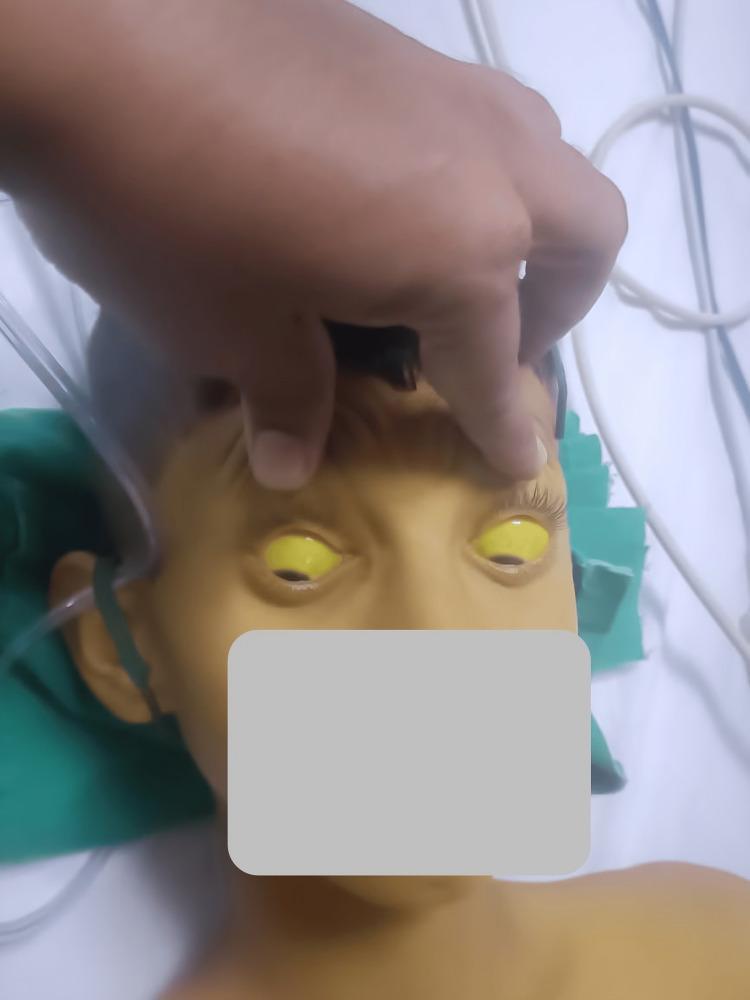
This clinical photograph displays a pediatric patient demonstrating severe systemic jaundice. The primary diagnostic feature is profound scleral icterus, characterized by a bright yellow discoloration of the entire visible sclera in both eyes. The upper eyelids are manually elevated to fully reveal the extent of the icterus. Additionally, the patient's facial skin, forehead, and upper chest exhibit a marked yellowish hue (jaundice), consistent with hyperbilirubinemia. The patient appears to be in a clinical setting, as evidenced by medical tubing and linens visible in the background. This image is an educational example of the physical manifestations of liver dysfunction or hemolytic processes, such as hepatitis, Wilsonian crisis, or acute toxicity, which can lead to significant accumulation of bilirubin in mucosal surfaces and skin.
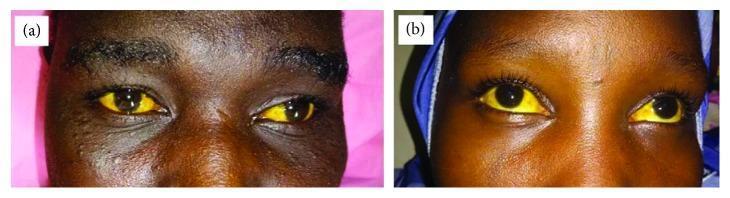
This clinical photograph consists of two panels, (a) and (b), displaying a close-up view of the eyes of two different patients. Both images demonstrate significant conjunctival icterus (scleral jaundice), characterized by a deep, uniform yellow discoloration of the sclera in both eyes. The intensity of the pigmentation is marked, standing in stark contrast to the dark irises. Aside from the jaundice, there are no visible signs of conjunctival injection, chemosis, or discharge. This finding is a clinical hallmark of hyperbilirubinemia, which in these cases is contextually associated with blackwater fever, a severe complication of Plasmodium falciparum malaria characterized by intravascular hemolysis. The visual documentation serves as an educational tool for recognizing advanced jaundice in patients with dark skin tones, where mucosal and scleral examination is critical for detecting hyperbilirubinemia.
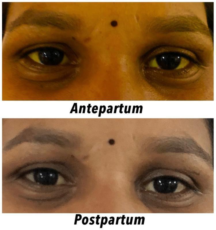
This clinical photograph provides a side-by-side comparison of facial features in a female patient, illustrating the resolution of icterus (jaundice) from the antepartum to the postpartum period. The top panel, labeled 'Antepartum,' displays bilateral scleral icterus, characterized by a distinct yellow discoloration of the ocular sclera. Additionally, the periorbital and facial skin exhibits a generalized yellowish hue. The bottom panel, labeled 'Postpartum,' shows the same patient after delivery, demonstrating a significant clinical improvement. The sclerae have returned to a normal white color, and the skin tone has transitioned back to a typical pigmentation, indicating a reduction in serum bilirubin levels. This comparison is used to visualize the clinical manifestation and subsequent resolution of hepatobiliary or hemolytic conditions during pregnancy, such as intrahepatic cholestasis of pregnancy (IHCP) or other pregnancy-related liver disorders. The image serves as an educational tool for identifying physical signs of hyperbilirubinemia and monitoring treatment response in an obstetric context.
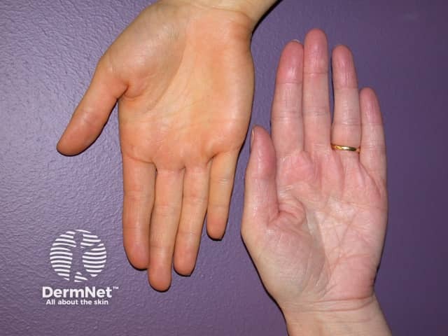
Clinical photograph of the palmar surfaces of both hands showing differential coloration: the left palm exhibits yellow-orange pigmentation consistent with carotenoderma; the right palm demonstrates normal pink skin. The imaging modality is standard clinical photography using a color-calibrated camera; no staining or contrast agents were used. The palmar skin is a target area for beta-carotene deposition due to high keratin content in thick epidermis. Notable features include diffuse, uniform coloration on the left palm with well-defined borders between pigmented and non-pigmented skin; no edema, scaling, or erythema; the palmar creases remain visible. The sclera and mucous membranes are not yellow, helping differentiate from icterus. Potential etiologies include dietary excess of carotene-rich foods (carotenoderma), supplementation, or metabolic conditions affecting carotenoid clearance; less likely is hepatic jaundice, which would typically involve scleral icterus. Clinically this finding is benign and reversible with dietary modification; no invasive workup is required unless accompanied by systemic symptoms. Relevance spans dermatology, clinical photography, and differential diagnosis education; this image supports teaching about color variation in palmar skin, methodical assessment of pigmentary disorders, and patient counseling on diet-related skin changes. Useful in cases of pediatric or adult dermatology, nutrition counseling, and medical training. Clinical note: observe scleral color.
Clinical differentiation of jaundice
- History, examination and screening labs.
- A working differential.
- Specialized tests to narrow it.
- A plan for treatment or further testing.
Step 1: Confirm jaundice and exclude mimics
Step 2: Fractionate the bilirubin
| Pattern | Level of problem | Main causes |
|---|---|---|
| Unconjugated (indirect) predominant | Prehepatic or hepatic uptake and conjugation | Hemolysis, ineffective erythropoiesis, hematoma resorption, Gilbert syndrome, Crigler-Najjar syndrome, neonatal jaundice |
| Conjugated (direct) predominant | Hepatocellular or cholestatic | Viral, alcoholic or drug-induced hepatitis, cirrhosis, biliary obstruction, primary biliary cholangitis (PBC), primary sclerosing cholangitis (PSC), Dubin-Johnson and Rotor syndromes |
Step 3: Clinical pointers
- RUQ pain, fever and prior biliary surgery suggest obstruction with cholangitis.
- A viral prodrome (anorexia, malaise, myalgia) with infectious exposure suggests acute viral hepatitis.
- Ask about alcohol, medications (including over-the-counter drugs and herbal supplements) and environmental hepatotoxins.
- A family history of jaundice or liver disease suggests hereditary hyperbilirubinemia or genetic liver disease.
- These clues must be interpreted with caution, because fever and pain occur in other conditions and the diseases can coexist.
- Fever or RUQ tenderness suggests cholangitis.
- A palpable abdominal mass suggests a neoplastic cause of obstruction.
- Signs of chronic liver disease point to cirrhosis: ascites, splenomegaly, prominent abdominal veins, spider telangiectasias, gynecomastia and asterixis.
- Kayser-Fleischer rings are pathognomonic of Wilson disease.
| Feature | Hemolytic (prehepatic) | Hepatocellular | Cholestatic or obstructive |
|---|---|---|---|
| Urine | Normal, no bilirubin | Dark | Dark |
| Stool | Normal or dark | Normal or pale | Pale (clay-colored) |
| Pruritus | No | Variable | Prominent |
| Palpable gallbladder | No | No | Courvoisier sign suggests a malignant distal obstruction |
| Other | Pallor, splenomegaly | Stigmata of chronic liver disease | Xanthelasma in chronic cholestasis |
Step 4: Liver panel pattern
| Hepatocellular | Cholestatic | Hemolytic | |
|---|---|---|---|
| AST/ALT | Markedly raised (often over 500 U/L in acute hepatitis) | Mild to moderate rise | Normal |
| ALP and GGT | Mild rise | Markedly raised | Normal |
| Bilirubin | Mixed | Mainly conjugated | Mainly unconjugated, usually below 5 mg/dL |
| PT/INR | May be prolonged and does not correct with vitamin K if synthetic function is poor | Prolonged from vitamin K malabsorption, corrects with vitamin K | Normal |
- In alcoholic hepatitis, AST is typically greater than ALT, with a ratio above 2.
- Normal ALP with a raised GGT does not suggest a bone source. A raised ALP with a normal GGT suggests bone or placental origin.
- Hemolysis also shows raised LDH and reticulocytes, low haptoglobin and a positive Coombs test in autoimmune cases.
Step 5: Imaging
- Ultrasound is the first test for biliary dilation. It shows the level of obstruction reliably, but the exact cause in only about two-thirds of patients. Sensitivity for intraductal stones is about 21 to 63% (Current Surgical Therapy, 14th ed).
- CT is highly sensitive for pancreatic tumors, especially those over 2 cm.
- MRCP has about 95% sensitivity for biliary obstruction and is the noninvasive test of choice for bile duct stones. It is not therapeutic.
- ERCP is both diagnostic and therapeutic for stones and strictures.
- Endoscopic ultrasound (EUS) detects tumors at the hilum, bile ducts, ampulla and pancreatic head, and allows fine-needle aspiration. For distal strictures, sensitivity is 84 to 91% and specificity 71 to 100% (Current Surgical Therapy, 14th ed).
- If the ducts are not dilated, look for intrahepatic cholestasis, using serology and autoimmune markers. Examples are AMA for PBC, ANA and anti-smooth muscle antibody for autoimmune hepatitis, and viral hepatitis serology.
- Liver biopsy is used for unresolved cases.
Simple algorithm
- Is the bilirubin mostly unconjugated? If yes, consider hemolysis, Gilbert syndrome or Crigler-Najjar syndrome. Check the blood film, reticulocytes, LDH and haptoglobin. If the rest of the liver panel is normal, Gilbert syndrome is likely.
- If mostly conjugated, is ALT/AST disproportionately raised?
- If yes, this is hepatocellular. Check viral serology, alcohol and drug history, autoimmune markers, and ceruloplasmin and iron studies where relevant.
- If ALP/GGT is disproportionately raised, this is cholestatic. Do an ultrasound.
- If the ultrasound shows dilated ducts, this is extrahepatic obstruction. Proceed to MRCP, CT, EUS or ERCP. If the ducts are not dilated, this is intrahepatic cholestasis.
Red flags needing urgent action
- Cholangitis: fever, RUQ pain and jaundice (Charcot triad) need urgent biliary decompression.
- Acute liver failure: jaundice with encephalopathy or coagulopathy.
- Painless progressive jaundice with weight loss suggests malignancy.