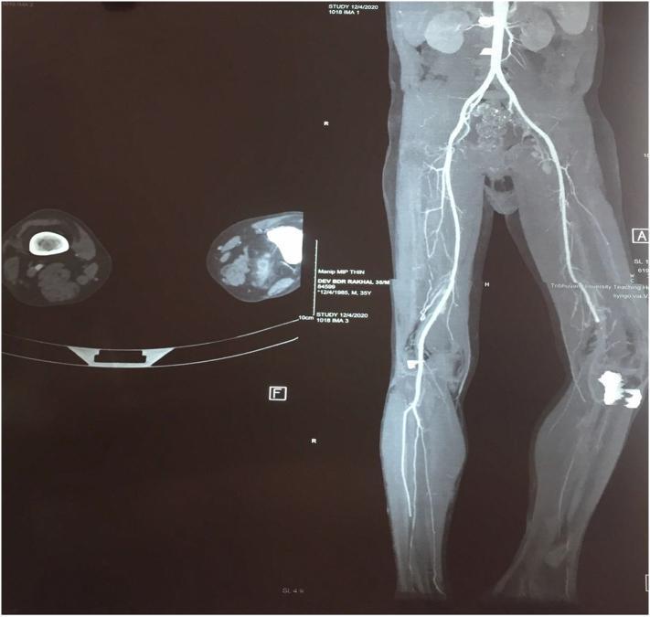
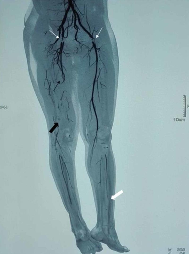
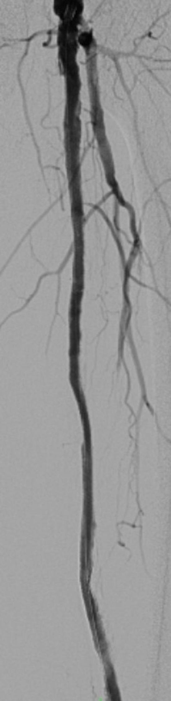
I now have all the content needed to compile comprehensive notes. Note: The "Sapiens" textbook is not available in the library - the closest authoritative anatomy references are the THIEME Atlas of Anatomy (General Anatomy and Musculoskeletal System) and Gray's Anatomy for Students. I'll compile full notes now.
Arteries of the Lower Limb
Reference note: The "Sapiens" anatomy textbook is not available in the library. These notes are compiled from the closest equivalent authoritative sources available: THIEME Atlas of Anatomy - General Anatomy and Musculoskeletal System and Gray's Anatomy for Students, supplemented by Imaging Anatomy: Bones, Joints, Vessels and Nerves (Vol. 3) and Color Atlas of Human Anatomy Vol. 2.
Overview
The arterial supply of the lower limb is derived from the external iliac artery, which is a terminal branch of the common iliac artery. The vessel continues distally under the inguinal ligament and is renamed at each major transition point:
External iliac artery → Femoral artery → Popliteal artery → Anterior tibial artery + Posterior tibial artery
Additional vessels - the superior gluteal, inferior gluteal, and obturator arteries (branches of the internal iliac artery) - contribute to the gluteal region and upper thigh, forming anastomotic networks around the hip joint.
(THIEME Atlas of Anatomy - General Anatomy and Musculoskeletal System, p. 549)
1. External Iliac Artery
- Arises along with the internal iliac artery from the common iliac artery
- Descends along the medial border of the psoas major through the lacuna vasorum
- Becomes the femoral artery at the level of the inguinal ligament
Branches:
| Branch | Distribution |
|---|
| Inferior epigastric artery | Anterior abdominal wall |
| Cremasteric artery | Cremaster muscle (male) |
| Artery of round ligament of uterus | Round ligament (female) |
| Pubic branch | Pubic region |
| Deep circumflex iliac artery | Iliac fossa, anterior abdominal wall |
(THIEME Atlas, p. 549)
2. Femoral Artery
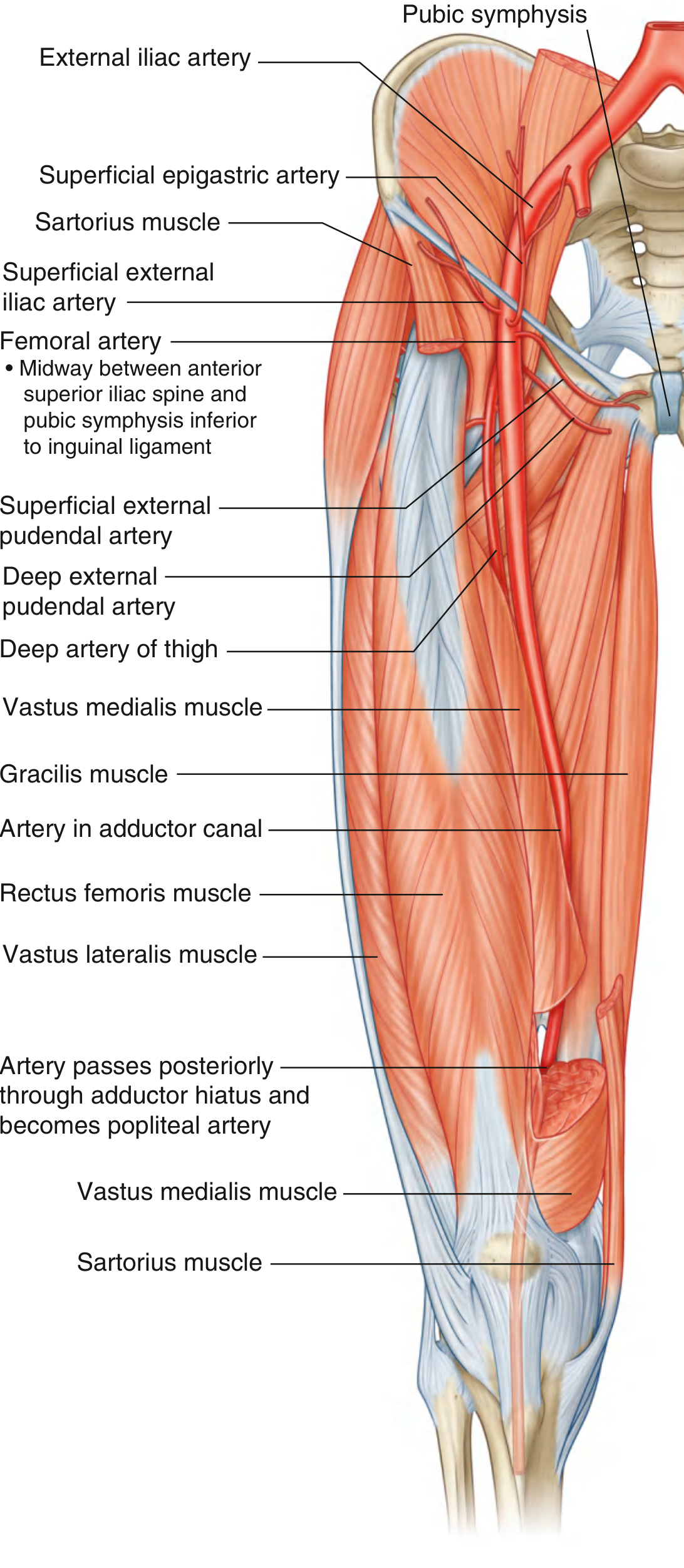
(Fig. 6.65 Femoral Artery - Gray's Anatomy for Students)
Origin and Course
- The femoral artery is the direct continuation of the external iliac artery, beginning as the vessel passes under the inguinal ligament to enter the femoral triangle
- It is palpable in the femoral triangle just inferior to the inguinal ligament, midway between the anterior superior iliac spine (ASIS) and the pubic symphysis - an important clinical landmark
- It runs vertically through the femoral triangle, then continues down the thigh in the adductor canal (Hunter's canal)
- It exits the canal through the adductor hiatus in the adductor magnus muscle and becomes the popliteal artery behind the knee
Femoral Triangle Relations
- Lateral: Femoral nerve
- Middle: Femoral artery
- Medial: Femoral vein
- (The femoral nerve lies outside the femoral sheath; the artery and vein are within it)
(Gray's Anatomy for Students, p. 703)
Branches of the Femoral Artery
| Branch | Notes |
|---|
| Superficial epigastric artery | Supplies lower abdominal wall skin |
| Superficial circumflex iliac artery | Supplies skin over iliac region |
| Superficial external pudendal artery | Supplies skin of perineum and scrotum/labia |
| Deep external pudendal artery | Supplies deeper perineal skin |
| Deep artery of thigh (profunda femoris) | Largest branch; major supply to thigh |
| Descending genicular artery | Contributes to anastomosis around knee |
(THIEME Atlas, p. 549; Gray's Anatomy for Students, p. 703)
2a. Deep Artery of the Thigh (Profunda Femoris)
- The largest branch of the femoral artery; the principal blood supply to the thigh muscles
- Originates from the lateral side of the femoral artery in the femoral triangle
- Passes posteriorly between pectineus and adductor longus, then between adductor longus and adductor brevis, and finally through adductor magnus to connect with the popliteal artery branches
Branches of the profunda femoris:
i. Lateral circumflex femoral artery
- Originates from the lateral side of the profunda (may arise directly from femoral artery)
- Passes deep to sartorius and rectus femoris; divides into three branches:
- Ascending branch - ascends under tensor fasciae latae; circles the femoral neck (with medial circumflex) to supply the femoral head and neck
- Descending branch - descends under rectus femoris to supply the anterior thigh; contributes to the anastomosis around the knee
- Transverse branch - passes laterally through vastus lateralis to the greater trochanter
ii. Medial circumflex femoral artery
- Originates from the medial or posterior side of the profunda
- Passes posteriorly between the pectineus and iliopsoas and around the femoral neck
- Critical blood supply to the femoral head (damage during femoral neck fractures causes avascular necrosis)
- Gives an ascending branch that circles the neck of the femur and supplies the neck and head
iii. Perforating branches (3-4)
- Pierce the adductor magnus to supply the posterior compartment muscles of the thigh
(Gray's Anatomy for Students, p. 703)
3. Popliteal Artery
- The popliteal artery is the continuation of the femoral artery after it passes through the adductor hiatus
- It runs through the popliteal fossa behind the knee
- Passes between the gastrocnemius and popliteus muscles
- Passes under the tendinous arch of the soleus muscle and enters the deep posterior compartment
- Divides at the inferior border of the popliteus into its two terminal branches: anterior tibial artery and posterior tibial artery
Branches of the Popliteal Artery
| Branch | Notes |
|---|
| Posterior tibial recurrent artery | Contributes to knee anastomosis |
| Medial superior genicular artery | Part of arterial plexus around the knee |
| Lateral superior genicular artery | Part of arterial plexus around the knee |
| Sural arteries (×2) | Supply gastrocnemius, soleus, plantaris |
| Middle genicular artery | Supplies ligaments and capsule of knee joint |
| Medial inferior genicular artery | Part of arterial plexus around the knee |
| Lateral inferior genicular artery | Part of arterial plexus around the knee |
Note: The paired superior and inferior genicular arteries (medial + lateral) form the arterial anastomotic network around the knee joint, which is important for collateral circulation when the popliteal artery is occluded.
(THIEME Atlas, p. 549; Gray's Anatomy for Students, p. 731)
4. Anterior Tibial Artery
- Passes forward through the aperture (opening) in the upper part of the interosseous membrane to enter the anterior compartment of the leg
- Descends between the tibialis anterior and extensor hallucis longus muscles
- Continues as the dorsal pedal (dorsalis pedis) artery distal to the extensor retinaculum on the dorsum of the foot
Branches
| Branch | Notes |
|---|
| Anterior tibial recurrent artery | Contributes to knee anastomosis |
| Anterior lateral malleolar artery | Anastomoses around lateral malleolus |
| Anterior medial malleolar artery | Anastomoses around medial malleolus |
| Dorsal pedal (dorsalis pedis) artery | Continuation onto dorsum of foot |
(THIEME Atlas, p. 549)
5. Dorsal Pedal (Dorsalis Pedis) Artery
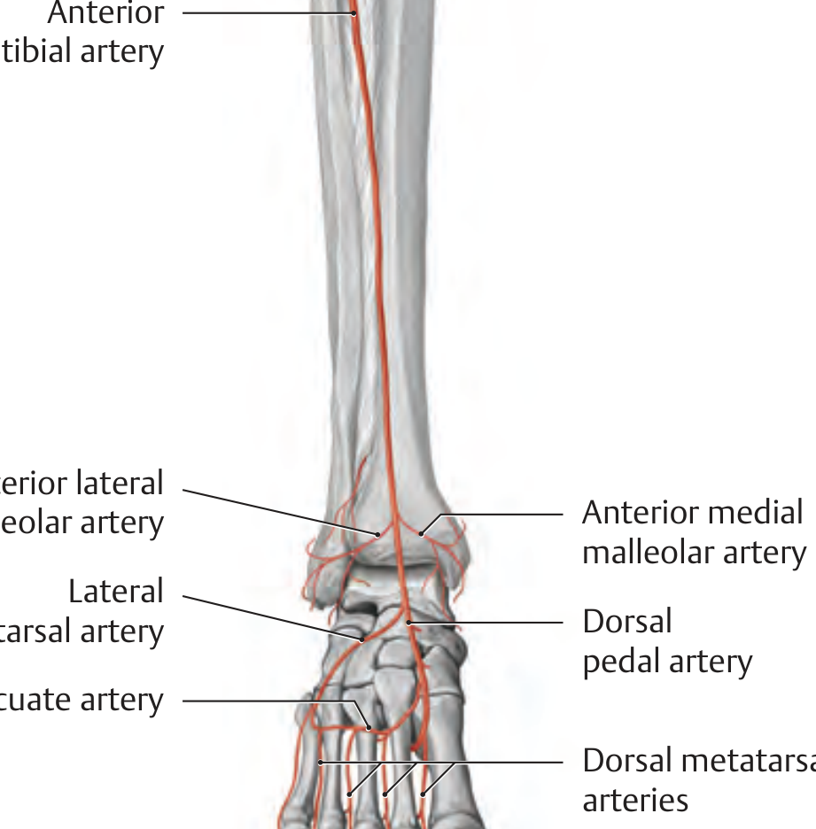
(THIEME Atlas - Arteries at the ankle and dorsum of foot, anterior view)
- Direct continuation of the anterior tibial artery distal to the extensor retinaculum
- Runs on the dorsum of the foot
Branches:
| Branch | Notes |
|---|
| Anterior lateral malleolar artery | Ankle anastomosis |
| Anterior medial malleolar artery | Ankle anastomosis |
| Lateral tarsal artery | Lateral tarsal bones |
| Arcuate artery | Arches across dorsal metatarsals |
| Dorsal metatarsal arteries | From arcuate artery; supply dorsum of toes |
| Deep plantar artery | Passes between 1st and 2nd metatarsals into sole; joins deep plantar arch |
(THIEME Atlas, p. 549)
6. Posterior Tibial Artery
- The direct continuation of the popliteal artery (the larger terminal branch)
- Enters the flexor (posterior) compartment of the leg
- Descends through the deep region of the posterior compartment on the surfaces of tibialis posterior and flexor digitorum longus
- Passes through the tarsal tunnel behind the medial malleolus
- Divides at the medial malleolus into its two terminal branches: medial plantar artery and lateral plantar artery
Branches
| Branch | Notes |
|---|
| Circumflex fibular artery | Passes laterally through soleus around the neck of fibula; connects with knee anastomosis |
| Fibular (peroneal) artery | Major branch; descends along posterior fibula |
| Medial plantar artery | Terminal branch; enters sole of foot (medial side) |
| Lateral plantar artery | Terminal branch; enters sole of foot (lateral side); forms deep plantar arch |
(Gray's Anatomy for Students, p. 731; THIEME Atlas, p. 549)
6a. Fibular (Peroneal) Artery
- Largest branch of the posterior tibial artery
- Descends along the lateral side of the posterior compartment, adjacent to the medial crest of the posterior fibula (between tibialis posterior and flexor hallucis longus)
- Supplies posterior compartment muscles and bone
- Also gives branches through the intermuscular septum to supply the fibularis (peroneal) muscles in the lateral compartment
- A perforating branch passes anteriorly through the inferior aperture in the interosseous membrane to anastomose with a branch of the anterior tibial artery
(Gray's Anatomy for Students, p. 731)
7. Arteries of the Sole of the Foot
The posterior tibial artery terminates behind the medial malleolus into:
Medial Plantar Artery
- Smaller terminal branch
- Runs along the medial side of the sole
- Supplies the medial aspect of the sole and great toe
Lateral Plantar Artery
- Larger terminal branch
- Crosses the sole obliquely to reach the base of the 5th metatarsal
- Then turns medially, forming the deep plantar arch (with the deep plantar artery from dorsalis pedis)
- The deep plantar arch gives off plantar metatarsal arteries, which divide into common and proper plantar digital arteries to supply the toes
(THIEME Atlas, p. 549)
8. Anastomoses - Summary
| Region | Vessels Involved |
|---|
| Hip and upper thigh | Femoral, inferior gluteal, superior gluteal, obturator, internal pudendal branches |
| Around the knee | Superior and inferior genicular arteries (medial and lateral), middle genicular, popliteal, descending branch of lateral circumflex femoral, descending genicular |
| Ankle region | Anterior and posterior malleolar branches (anterior tibial, posterior tibial, fibular) |
| Sole of the foot | Deep plantar arch (lateral plantar + deep plantar branch of dorsalis pedis) |
(Gray's Anatomy for Students, p. 636, 669)
9. Summary Table - Arterial Segments
| Artery | Origin | Termination | Key Feature |
|---|
| External iliac artery | Common iliac a. | Femoral a. (at inguinal ligament) | Gives deep circumflex iliac a. and inferior epigastric a. |
| Femoral artery | External iliac a. | Popliteal a. (at adductor hiatus) | Palpable midway ASIS-pubic symphysis |
| Profunda femoris | Femoral a. | Perforating branches to posterior thigh | Main supply to thigh muscles |
| Popliteal artery | Femoral a. (adductor hiatus) | Anterior + posterior tibial a. | Divides at lower border of popliteus |
| Anterior tibial artery | Popliteal a. | Dorsalis pedis a. | Crosses interosseous membrane anteriorly |
| Posterior tibial artery | Popliteal a. | Medial + lateral plantar a. | Passes behind medial malleolus |
| Fibular artery | Posterior tibial a. | Perforating branches at ankle | Main supply to lateral leg compartment |
| Dorsalis pedis | Anterior tibial a. | Deep plantar arch | Palpable on dorsum of foot |
| Lateral plantar artery | Posterior tibial a. | Deep plantar arch | Larger of the two plantar arteries |
| Medial plantar artery | Posterior tibial a. | Digital branches (medial sole) | Smaller of the two plantar arteries |
Clinical Notes
- Femoral pulse is palpable at the mid-inguinal point (midway between ASIS and pubic symphysis), just below the inguinal ligament. This is used for IV access and cardiac catheterization.
- Avascular necrosis (AVN) of the femoral head follows disruption of the medial circumflex femoral artery, which is the dominant supply to the femoral head; it is commonly seen after displaced femoral neck fractures.
- Popliteal artery aneurysm is the most common peripheral artery aneurysm; it is associated with atheromatous disease and may cause acute limb ischemia.
- Peripheral arterial disease (PAD) most commonly affects the superficial femoral artery at the adductor canal, and the popliteal artery; it presents with intermittent claudication.
- Dorsalis pedis pulse is palpable on the dorsum of the foot lateral to the extensor hallucis longus tendon; the posterior tibial pulse is felt behind the medial malleolus. Both are routinely assessed in peripheral vascular disease.
- Collateral circulation around the knee and hip protects against ischemia when individual vessels are occluded.
Sources:
- General Anatomy and Musculoskeletal System - THIEME Atlas of Anatomy (ISBN 9781626237186), pp. 549-550
- Gray's Anatomy for Students (ISBN 9780323934237), pp. 669-670, 703-706, 731
- Imaging Anatomy: Bones, Joints, Vessels and Nerves, Vol. 3 (ISBN 9781626239845), p. 359-360
- Color Atlas of Human Anatomy, Vol. 2 (ISBN 9783132424487)
Note on "Sapiens" textbook: The Sapiens anatomy textbook is not present in the available library. These notes draw from the four THIEME/Elsevier anatomy textbooks listed above, which cover the same content at an equivalent or greater depth. If you have a specific edition of Sapiens in mind, please share it and the notes can be updated accordingly.