
Sympathomimetic drugs
sympathomimetic drugs classification adrenergic receptors
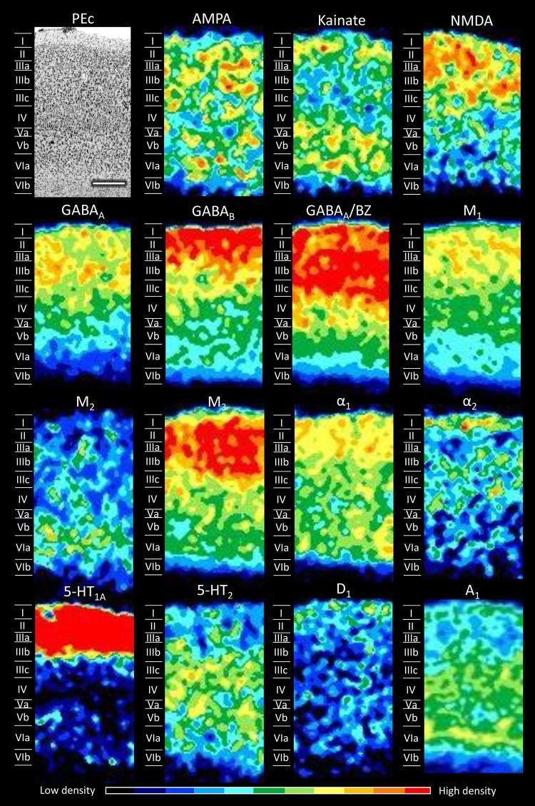
This composite figure illustrates the cyto- and receptor-architecture of cortical area PEc in the macaque monkey superior parietal lobule. The top-left panel shows a Nissl-stained section detailing the laminar organization from the pial surface (Layer I) to the white matter border (Layer VIb). Accompanying this are 15 color-coded receptor autoradiographs demonstrating the laminar distribution densities for various neurotransmitter receptors, including glutamatergic (AMPA, Kainate, NMDA), GABAergic (GABAA, GABAB, GABAA/BZ), muscarinic (M1, M2, M3), adrenergic (alpha1, alpha2), serotonergic (5-HT1A, 5-HT2), dopaminergic (D1), and purinergic (A1) types. A pseudocolor scale at the bottom indicates receptor density, ranging from blue (low) to red (high). Notable patterns include the high density of 5-HT1A receptors in supragranular layers I-II, the relatively homogeneous distribution of AMPA receptors across all layers, and the infragranular concentration of Kainate and A1 receptors. This comparative visualization serves as a 'receptor fingerprint' for neuroanatomical classification and functional mapping of the parietal cortex.
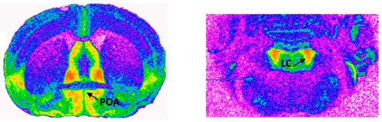
This figure presents two pseudocolor autoradiograms illustrating the distribution and density of alpha2-adrenergic receptors in rodent brain sections. The left image shows a coronal section at the level of the preoptic area (POA), where high receptor density is indicated by warm colors (red and orange) in a centrally located, elongated region along the ventral aspect. The surrounding neural tissue exhibits lower binding density, represented by cooler colors (green, blue, and purple). The right image displays a more posterior section at the level of the locus coeruleus (LC). In this section, high alpha2-adrenergic receptor density is concentrated in a compact, localized area within the LC itself, demarcated in bright orange and red. These images demonstrate neuroanatomical variations in receptor expression, with the POA showing a more diffuse ventral distribution compared to the highly focused concentration in the LC. Such visual data is essential for understanding catecholaminergic signaling pathways and the pharmacological mapping of adrenergic receptors in the central nervous system.
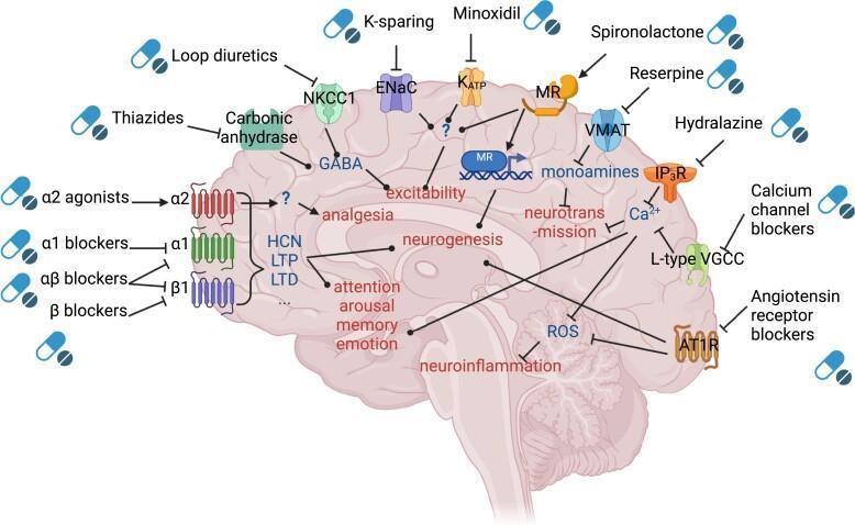
This pathophysiology diagram illustrates the main mechanisms of action for anti-hypertensive drugs within the brain. Set against a sagittal cross-section of the human brain, the illustration maps various pharmacological classes to their molecular targets and downstream physiological effects. Major drug classes identified include alpha-2 agonists, alpha-1 blockers, alpha-beta blockers, and beta blockers acting on adrenergic receptors; diuretics such as thiazides and loop diuretics targeting carbonic anhydrase and NKCC1; and calcium channel blockers (CCBs) acting on L-type voltage-gated calcium channels (VGCC). The diagram also features K-sparing diuretics, spironolactone, minoxidil, and angiotensin receptor blockers (ARBs) targeting AT1R. Key downstream mechanisms include modulation of neurotransmission, neurogenesis, neuroinflammation, and excitability. Specific pathways shown include the inhibition of Reactive Oxygen Species (ROS) by AT1R blockers and CCBs, and the influence of various agents on cognitive domains like memory, emotion, arousal, and attention. The schematic uses standard BioRender iconography to distinguish between stimulatory (sharp arrows) and inhibitory (flat-headed arrows) interactions, highlighting the complex neurobiological impact of systemic anti-hypertensives on central nervous system function.
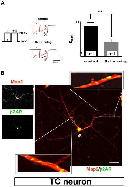
This composite educational image illustrates the modulation and localization of ̢2-adrenergic receptors (̢2AR) in thalamocortical (TC) neurons of the dorsal lateral geniculate nucleus (dLGN). Section A presents electrophysiological data, including a double-pulse voltage protocol (holding at -40 mV, pulses to +10 mV) and representative current traces. A corresponding bar graph quantifies the degree of calcium-dependent inactivation (Dinact), showing a statistically significant reduction (p<0.01) from control conditions when treated with the ̢2AR agonist salmeterol plus ̢1 and ̢3 antagonists. Section B displays immunocytochemical analysis of a cultured TC neuron. Individual grayscale panels show Map2 (a dendritic marker) and ̢2AR staining. The large merged fluorescence image shows Map2 in red and ̢2AR in green. Co-expression is evidenced by yellow signals, primarily localized in the soma (indicated by an arrow) and proximal dendrites. High-magnification insets further detail the punctate co-localization of ̢2AR along the dendritic shafts, highlighting its role in modulating local calcium channel activity.
Sympathomimetic Drugs
1. Definition and Overview
- Katzung's Basic and Clinical Pharmacology, 16th Edition
- Goodman & Gilman's Pharmacological Basis of Therapeutics
2. Classification
By Mechanism of Action
| Category | Mechanism | Examples |
|---|---|---|
| Direct-acting | Bind and activate adrenergic receptors directly | Epinephrine, norepinephrine, phenylephrine, isoproterenol, dobutamine, albuterol |
| Indirect-acting | Cause release of stored NE from nerve terminals | Amphetamine, tyramine |
| Mixed-acting | Both direct receptor activation AND NE release | Ephedrine, pseudoephedrine |

3. Adrenergic Receptor Subtypes and Their Effects
| Receptor | Location | Effect when activated |
|---|---|---|
| α1 | Vascular smooth muscle, pupil dilator, urethra | Vasoconstriction, mydriasis, urinary retention |
| α2 | Presynaptic nerve terminals, CNS, platelets | Inhibits NE release (negative feedback), CNS sedation, platelet aggregation |
| β1 | Heart, kidney (JGA) | Increased HR, contractility, AV conduction; renin release |
| β2 | Bronchial/uterine smooth muscle, skeletal muscle vessels | Bronchodilation, vasodilation, tocolysis, glycogenolysis |
| β3 | Adipose tissue, bladder | Lipolysis, bladder relaxation |
| D1 | Renal/mesenteric vasculature | Vasodilation, natriuresis |
| D2 | Presynaptic; pituitary, CNS | Modulates NE release; inhibits prolactin |
4. Structure-Activity Relationships (SAR)
- Greatest sympathomimetic activity occurs when two carbon atoms separate the ring from the amino group
- Catecholamines = compounds with -OH groups at positions 3 and 4 of the benzene ring (NE, EPI, DA, isoproterenol)
- Amino group substitution: Larger alkyl substituents increase β-receptor activity (isoproterenol is a pure β agonist). Smaller amino substitution favors α activity
- Aromatic ring substitution: Maximal α and β activity requires -OH at positions 3 and 4. -OH at positions 3 and 5 with large amino group confers β2 selectivity (e.g., terbutaline)
- Lipophilicity: Absence of polar hydroxyl groups increases CNS penetration (amphetamine, ephedrine cross the blood-brain barrier readily)
- Oral bioavailability: Catecholamines are metabolized by COMT in gut/liver - poor oral bioavailability. Non-catecholamines are not COMT substrates, so they have better oral bioavailability and longer duration
5. Specific Drugs
Endogenous Catecholamines
- Agonist at α1, α2, β1, and β2 receptors
- Low dose: β2 effects predominate (vasodilation in skeletal muscle, bronchodilation, fall in diastolic BP)
- High dose: α effects predominate (vasoconstriction, rise in both systolic and diastolic BP)
- Uses: Anaphylaxis (first-line), cardiac arrest (CPR), local vasoconstriction with local anesthetics, acute asthma
- Route: IV, IM, SC, inhalation, topical
- Agonist at α1, α2, and β1 receptors; minimal β2 activity
- Increases both systolic and diastolic BP, increases peripheral resistance
- Compensatory baroreflex counters direct chronotropic effect (net: reflex bradycardia)
- Uses: Vasopressor of first choice in septic shock (provides balance of α and modest β1 activity)
- Dose-dependent effects:
- Low dose (2-5 mcg/kg/min): D1 receptor activation → renal and mesenteric vasodilation, natriuresis
- Medium dose (5-15 mcg/kg/min): β1 stimulation → increased cardiac output
- High dose (>15 mcg/kg/min): α1 stimulation → vasoconstriction
- Note: Dopamine is now NOT preferred over norepinephrine in septic shock due to higher incidence of arrhythmias and mortality
Synthetic Catecholamines
- Predominantly β1 agonist with some β2 and weak α1 activity
- Increases myocardial contractility (positive inotropy) with less tachycardia than epinephrine
- Uses: Acute heart failure, pharmacologic stress testing (dobutamine stress echo)
- Agent of choice when increased cardiac output is needed
- Pure β agonist (β1 + β2); no α activity
- Increases HR and contractility; causes vasodilation → fall in diastolic BP
- Now rarely used; replaced by selective β2 agonists
Non-Catecholamine Sympathomimetics
- Pure α1 agonist; not a catecholamine (not inactivated by COMT - longer duration)
- Uses: Nasal decongestant, mydriasis (eye drops), blood pressure support in anesthesia
- Raises BP, causes reflex bradycardia
- Prodrug → desglymidodrine (selective α1 agonist) after enzymatic hydrolysis
- Orally active; peak effect ~1 hour
- Use: Orthostatic hypotension (autonomic failure)
- Caution: Causes supine hypertension
- Selective β2 agonist (short-acting)
- Use: Acute bronchospasm in asthma and COPD; hyperkalemia (IV)
- Best delivered by inhalation (greatest local bronchial effect, minimal systemic toxicity)
- 80-90% of inhaled dose deposits in mouth/pharynx - optimal particle size 2-5 μm
- Long-acting β2 agonists (LABAs)
- Use: COPD maintenance; asthma (always combined with inhaled corticosteroid - LABA monotherapy in asthma associated with increased mortality)
- Indacaterol, olodaterol, vilanterol: ultra-long-acting, once-daily dosing in COPD
- Mixed-acting (both direct β2 activity + indirect NE release)
- Oral activity; CNS stimulant (no ring hydroxyl → crosses BBB)
- Uses: Nasal decongestion, orthostatic hypotension, bronchospasm
- Rebound congestion ("rhinitis medicamentosa") can follow nasal decongestants
- Direct α agonist; long-acting nasal decongestant (OTC)
- Risk of rebound hyperemia with prolonged use
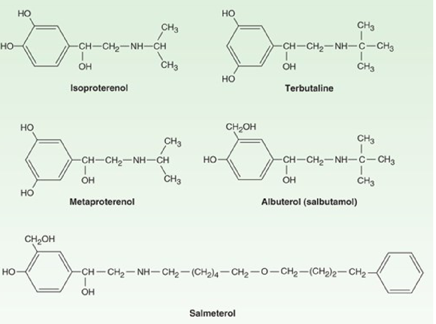
6. Clinical Uses
| Indication | Drug(s) of Choice | Receptor |
|---|---|---|
| Anaphylaxis | Epinephrine (IM/IV) | α1, β1, β2 |
| Septic shock (vasopressor) | Norepinephrine (first-line) | α1 > β1 |
| Cardiogenic shock / acute HF | Dobutamine | β1 |
| Cardiac arrest | Epinephrine | β1, α1 |
| Acute asthma | Albuterol (inhaled) | β2 |
| COPD maintenance | LABA (salmeterol, indacaterol) | β2 |
| Orthostatic hypotension | Midodrine, droxidopa | α1 |
| Nasal decongestion | Oxymetazoline, phenylephrine | α1 |
| ADHD / narcolepsy | Amphetamine, methylphenidate | Indirect (NE + DA release) |
| Mydriasis / ophthalmology | Phenylephrine | α1 |
| Adjunct to local anesthetics | Epinephrine 1:100,000 - 1:200,000 | α1 |
7. Mechanism: β2 Agonists and Bronchodilation
8. Adverse Effects
| Effect | Mechanism |
|---|---|
| Tachycardia, palpitations | β1 stimulation |
| Hypertension | α1 stimulation |
| Arrhythmias | β1 excess (especially with non-selective agents) |
| Tremor | β2 stimulation in skeletal muscle |
| Hypokalemia | β2 → K+ shift into cells |
| CNS stimulation, insomnia, anxiety | CNS-penetrant agents (amphetamine, ephedrine) |
| Rebound nasal congestion | Prolonged nasal α1 agonist use |
| Supine hypertension | Midodrine |
9. Pharmacokinetics Summary
| Property | Catecholamines | Non-catecholamines |
|---|---|---|
| Oral bioavailability | Poor (COMT + MAO in gut/liver) | Good |
| Duration of action | Short | Longer |
| COMT substrate | Yes | No |
| BBB penetration | Poor (polar) | Better (non-polar) |
| CNS effects | Minimal | More (ephedrine, amphetamine) |
- Katzung's Basic and Clinical Pharmacology, 16th Edition, pp. 233-238, 550-551
- Goodman & Gilman's Pharmacological Basis of Therapeutics, pp. 272-275
- Guyton and Hall Textbook of Medical Physiology
- Barash, Cullen, and Stoelting's Clinical Anesthesia, 9e
Introduction to ans
Introduction to the Autonomic Nervous System (ANS)
1. Overview and Definition
- Sympathetic (thoracolumbar)
- Parasympathetic (craniosacral)
- Enteric (intrinsic nervous system of the gut)
- Costanzo Physiology, 7th Edition
2. General Organization - Two-Neuron Chain
- Preganglionic neuron - cell body in CNS; axon travels to an autonomic ganglion
- Postganglionic neuron - cell body in the ganglion; axon innervates the effector organ
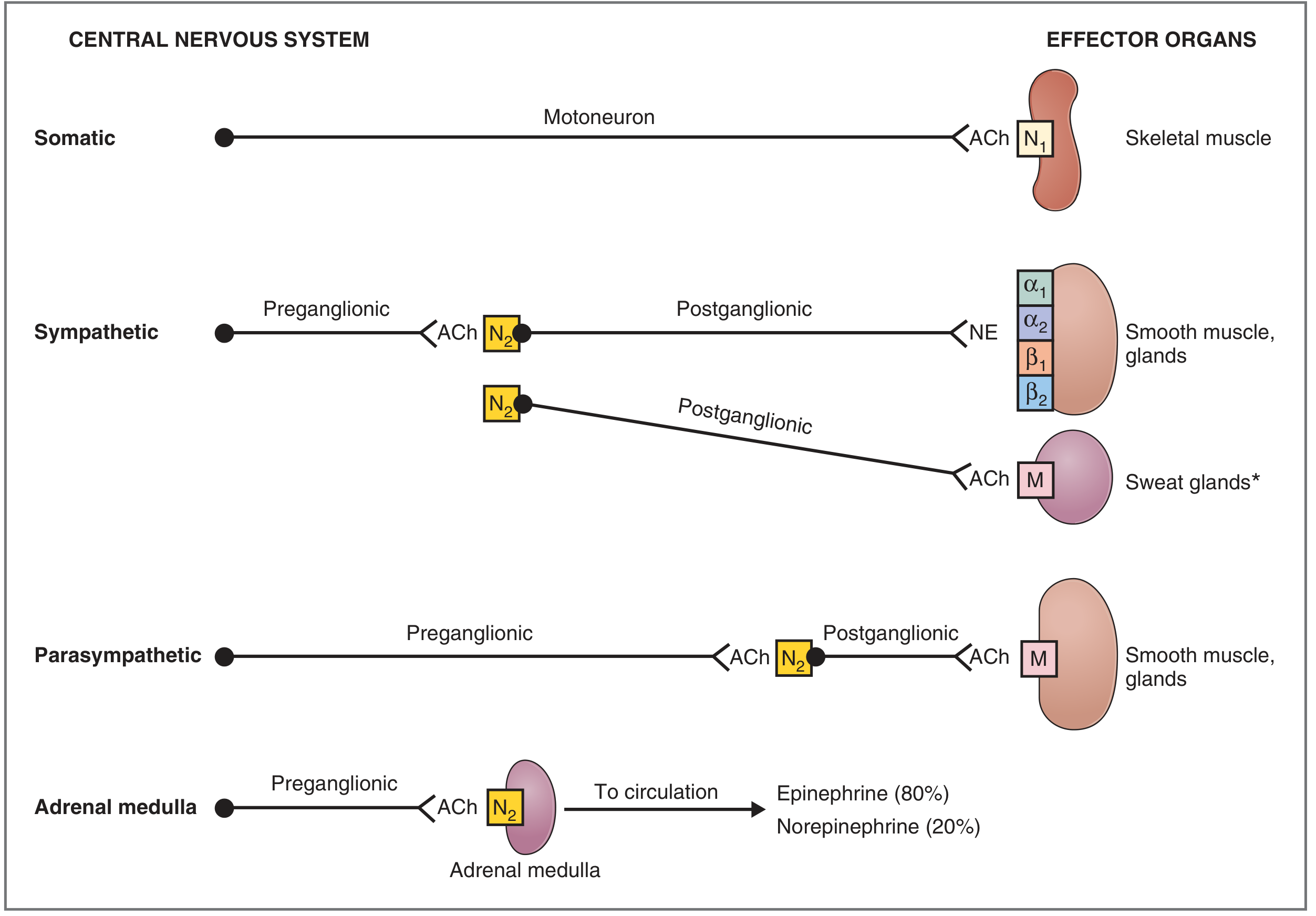
3. Sympathetic vs. Parasympathetic: Anatomical Comparison
| Characteristic | Sympathetic | Parasympathetic | Somatic |
|---|---|---|---|
| Origin of preganglionic neurons | T1-L3 (thoracolumbar spinal cord) | CN III, VII, IX, X; S2-S4 (craniosacral) | Ventral horn motor neurons |
| Location of ganglia | Paravertebral (sympathetic chain) or prevertebral | In or near effector organs | - |
| Preganglionic axon length | Short | Long | - |
| Postganglionic axon length | Long | Short | - |
| Effector organs | Smooth muscle, cardiac muscle, glands | Smooth muscle, cardiac muscle, glands | Skeletal muscle |
| Ganglionic neurotransmitter | ACh (nicotinic N2 receptor) | ACh (nicotinic N2 receptor) | - |
| Postganglionic neurotransmitter | Norepinephrine (except sweat glands - ACh) | ACh | ACh |
| Effector organ receptors | α1, α2, β1, β2 (adrenoreceptors) | Muscarinic (M1-M5) | Nicotinic (N1) |
- Costanzo Physiology, Table 2.1
4. Neurotransmitters and Receptor Terminology
- Cholinergic neurons release acetylcholine (ACh)
- Adrenergic neurons release norepinephrine (NE)
Key Rule (memorize this):
ALL preganglionic neurons (sympathetic AND parasympathetic) release ACh → act on nicotinic (N2) receptors in ganglia
Postganglionic sympathetic neurons release NE → act on α or β adrenoreceptors (EXCEPTION: sweat glands and some blood vessels get cholinergic sympathetic innervation → muscarinic receptors)
Postganglionic parasympathetic neurons release ACh → act on muscarinic receptors
Adrenal medulla = modified sympathetic ganglion. Preganglionic ACh → chromaffin cells release epinephrine (80%) + NE (20%) directly into circulation
- Costanzo Physiology, p. 55-57
5. Sympathetic Nervous System
Anatomy
- Preganglionic neurons originate in the intermediolateral cell column (lateral horn) of spinal cord segments T1-L3
- Axons exit through ventral roots → form the paravertebral sympathetic chain (22 pairs of ganglia running alongside the vertebral column)
- Some fibers pass through the chain without synapsing and synapse in prevertebral ganglia (celiac, superior mesenteric, inferior mesenteric) to reach abdominal viscera
Function - "Fight or Flight"
-
↑ Arterial blood pressure
-
↑ Heart rate and contractility
-
↑ Blood flow to skeletal muscles
-
↑ Blood glucose (glycogenolysis)
-
↑ Mental alertness
-
Bronchodilation
-
Pupil dilation (mydriasis)
-
Inhibition of GI motility
-
Costanzo Physiology, p. 55
6. Parasympathetic Nervous System
Anatomy (Craniosacral)
-
Cranial component - preganglionic fibers travel in 4 cranial nerves:
- CN III (oculomotor) → Edinger-Westphal nucleus → ciliary ganglion → pupil constriction, accommodation
- CN VII (facial) → lacrimal & salivatory nuclei → pterygopalatine / submandibular ganglia → lacrimal, nasal, submandibular/sublingual glands
- CN IX (glossopharyngeal) → inferior salivatory nucleus → otic ganglion → parotid gland
- CN X (vagus) → dorsal motor nucleus → ganglia in/near target organs → heart, lungs, GI tract (down to splenic flexure)
-
Sacral component (S2-S4) → pelvic splanchnic nerves → pelvic ganglia → descending colon, rectum, urinary bladder, genitalia
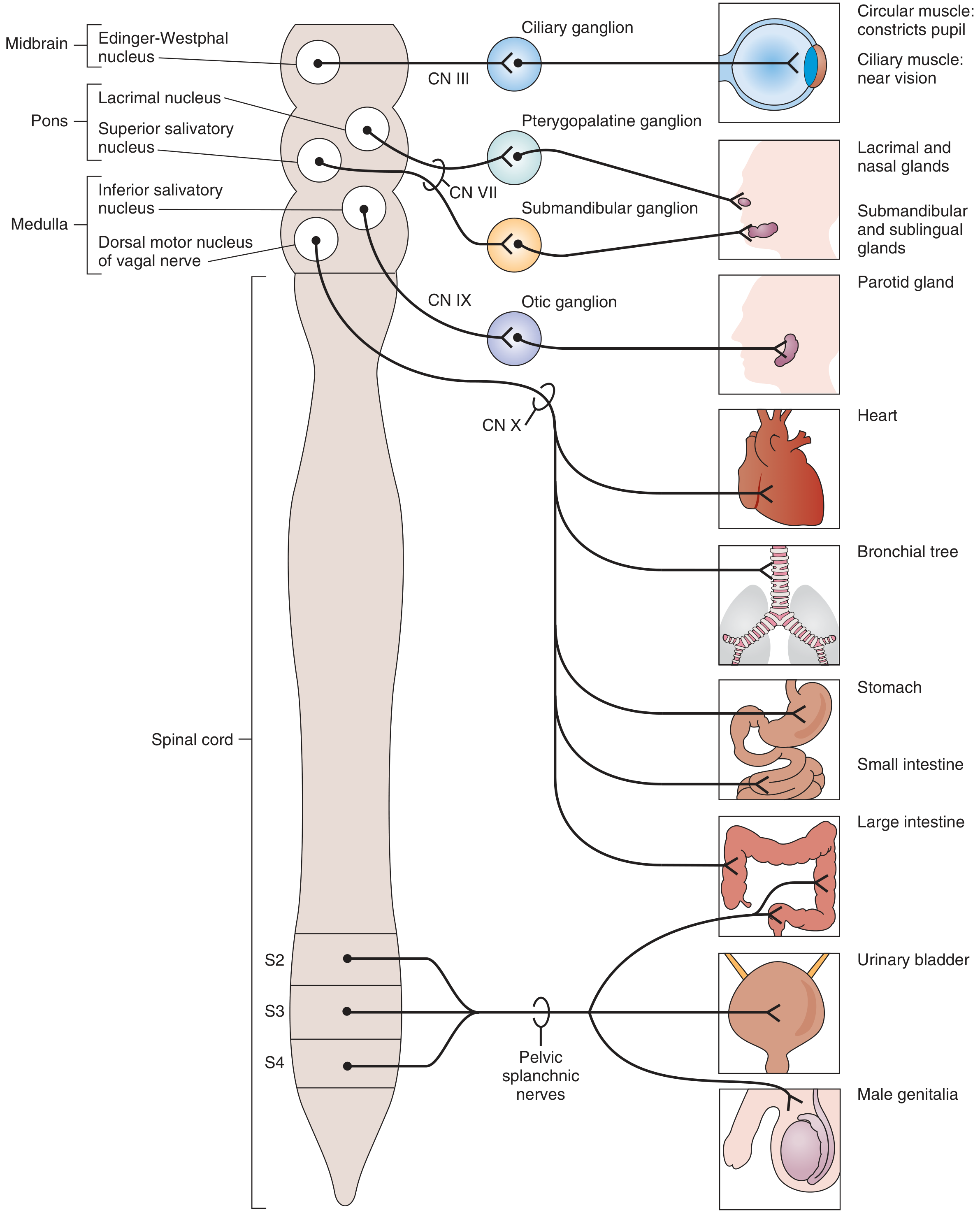
Function - "Rest and Digest"
- ↓ Heart rate and contractility (vagal tone)
- ↑ GI motility and secretion
- Pupil constriction (miosis)
- Increased salivation, lacrimation, bronchial secretion
- Bronchoconstriction
- Bladder detrusor contraction (micturition)
- Penile erection
7. Effects on Major Organ Systems
| Organ | Sympathetic Effect | Parasympathetic Effect |
|---|---|---|
| Heart rate | ↑ (β1) | ↓ (M2) |
| Myocardial contractility | ↑ (β1) | ↓ slight (M2) |
| Blood vessels | Vasoconstriction (α1) | Vasodilation (some, via NO) |
| Bronchi | Bronchodilation (β2) | Bronchoconstriction (M3) |
| GI motility | ↓ (α2, β2) | ↑ (M) |
| GI sphincters | Contracts (α1) | Relaxes (M) |
| Urinary bladder (detrusor) | Relaxes (β2) | Contracts (M) |
| Bladder internal sphincter | Contracts (α1) | Relaxes (M) |
| Pupil | Dilates - mydriasis (α1, radial muscle) | Constricts - miosis (M3, circular muscle) |
| Ciliary muscle | Relaxes - far vision (β2) | Contracts - near vision (M3) |
| Salivary glands | Thick, viscous secretion (α1) | Profuse, watery secretion (M) |
| Sweat glands | ↑ sweating (Ach → M - sympathetic cholinergic!) | - |
| Liver | Glycogenolysis ↑ (β2, α1) | - |
| Adipose tissue | Lipolysis ↑ (β3) | - |
| Male genitalia | Ejaculation (α1) | Erection (M) |
| Kidney (JGA) | ↑ Renin (β1) | - |
| Uterus | Relaxation (β2) / Contraction (α1) | Variable |
8. Neuroeffector Junctions
- Varicosities - beaded swellings along postganglionic axon branches that contain neurotransmitter in vesicles
- Diffuse, branching networks - overlap from multiple postganglionic neurons can innervate the same target tissue (en passant transmission)
- No motor end plate - receptors are distributed widely over the target tissue surface, not concentrated in a specialized region
- Costanzo Physiology, p. 55
9. Receptors: Summary Table
| Receptor | Type | Location | Agonist | Antagonist |
|---|---|---|---|---|
| α1 | Gq-coupled GPCR | Vascular smooth muscle, iris (radial), bladder sphincter | NE, phenylephrine | Prazosin, phenoxybenzamine |
| α2 | Gi-coupled GPCR | Presynaptic nerve terminals, CNS, platelets | Clonidine | Yohimbine |
| β1 | Gs-coupled GPCR | Heart (SA node, ventricles), kidney JGA | NE, EPI, dobutamine | Metoprolol, propranolol |
| β2 | Gs-coupled GPCR | Bronchial smooth muscle, skeletal muscle vessels, uterus | EPI, albuterol | Propranolol |
| β3 | Gs-coupled GPCR | Adipose tissue, bladder | NE, EPI | - |
| Nicotinic (N1) | Ligand-gated ion channel | Neuromuscular junction | ACh, nicotine | Tubocurarine |
| Nicotinic (N2) | Ligand-gated ion channel | Autonomic ganglia | ACh, nicotine | Hexamethonium |
| Muscarinic (M1-M5) | GPCR | Effector organs of parasympathetic system, sweat glands | ACh, muscarine | Atropine |
- Costanzo Physiology, Table 2.2
10. Enteric Nervous System
11. Central Control of the ANS
- Hypothalamus - the principal integrating center; regulates temperature, thirst, hunger, and cardiovascular/endocrine homeostasis
- Medulla oblongata - cardiovascular and respiratory centers; dorsal motor nucleus of vagus
- Spinal cord - contains preganglionic neurons in the intermediolateral column (T1-L3 for sympathetic; S2-S4 for parasympathetic)
- Limbic system and prefrontal cortex - emotional and behavioral influence on ANS
12. Key Mnemonic: Sympathetic vs. Parasympathetic
| Sympathetic | Parasympathetic | |
|---|---|---|
| Functional state | "Fight or Flight" | "Rest and Digest" |
| Origin | Thoracolumbar (T1-L3) | Craniosacral (CN III, VII, IX, X; S2-S4) |
| Ganglion location | Far from organ | Near/in organ |
| Pre-ganglionic fiber | Short | Long |
| Post-ganglionic fiber | Long | Short |
| Post-ganglionic NT | Norepinephrine | Acetylcholine |
| Effector receptor | Adrenoreceptors (α, β) | Muscarinic |
- Costanzo Physiology, 7th Edition, pp. 54-65
- Guyton and Hall Textbook of Medical Physiology
- Katzung's Basic and Clinical Pharmacology, 16th Edition
- Goodman & Gilman's Pharmacological Basis of Therapeutics