
explain physiology and pathophysiology of fever in detail mechanism each and everything im a medical student
fever AND pathophysiology
fever pathophysiology hypothalamic set point prostaglandin E2 Merck Manual
diagram of fever pathogenesis endogenous pyrogens hypothalamus prostaglandin E2 set point
https://www.merckmanuals.com/professional/infectious-disease…
Fever: physiology and pathophysiology
- Fever: hypothalamic set point is raised, usually through cytokine-driven prostaglandin E2 (PGE2). The patient may shiver and have cold skin during the rising phase.
- Hyperthermia: set point remains normal, but heat production or heat gain overwhelms heat dissipation. Examples include heat stroke, malignant hyperthermia, serotonin syndrome, and anticholinergic toxicity. Antipyretics do not reliably work in hyperthermia.
1. Normal thermoregulation: the physiology you need first
Normal core temperature
- Circadian rhythm: lowest in early morning, highest in late afternoon or evening, often by about 0.5°C.
- Ovulatory cycle: progesterone raises basal temperature after ovulation.
- Exercise, age, environment, food intake, and measurement site.
Heat production
- Basal metabolic activity, especially in liver, brain, heart, and skeletal muscle
- Skeletal-muscle activity and exercise
- Shivering thermogenesis
- Sympathetic stimulation of metabolism
- Non-shivering thermogenesis, particularly brown adipose tissue in neonates
Heat loss
- Radiation: infrared heat loss to cooler surroundings
- Convection: transfer to moving air or water
- Conduction: direct transfer to objects in contact with skin
- Evaporation: sweat evaporation and respiratory water loss
2. Thermoregulatory control system
| Component | Function |
|---|---|
| Sensors | Detect skin and core temperature |
| Integrator/comparator | Preoptic-anterior hypothalamus compares actual temperature with set point |
| Effectors | Alter heat production, conservation, loss, and behavior |
A. Thermoreceptors
Peripheral thermoreceptors
Central thermoreceptors
- Preoptic area and anterior hypothalamus
- Spinal cord
- Deep abdominal and thoracic tissues
- Great veins and some viscera
B. Hypothalamic response to temperature error
If actual temperature is below the set point
- Cutaneous vasoconstriction
- Reduced sweating
- Shivering
- Increased sympathetic activity and metabolism
- Behavioral responses: seeking warmth, adding clothes, curling up
If actual temperature is above the set point
- Cutaneous vasodilation
- Sweating through sympathetic cholinergic fibers
- Reduced muscle tone and thermogenesis
- Behavioral cooling: removing clothing, seeking shade, drinking fluids
- Costanzo Physiology, 7th ed., regulation of body temperature and fever section.
- Guyton and Hall Textbook of Medical Physiology, 14th ed., pp. 900-901.
3. What raises the set point? Pyrogens
A. Exogenous pyrogens
- Gram-negative bacterial lipopolysaccharide, LPS or endotoxin
- Peptidoglycan and lipoteichoic acid from Gram-positive organisms
- Bacterial exotoxins and superantigens
- Viral nucleic acids and proteins
- Fungal cell-wall products
- Parasitic products
B. Endogenous pyrogens, now called pyrogenic cytokines
- Interleukin-1 beta, IL-1β
- Interleukin-6, IL-6
- Tumor necrosis factor-alpha, TNF-α
- Interferons, especially during viral infection
- Ciliary neurotrophic factor, less clinically emphasized
- Monocytes and macrophages
- Dendritic cells
- Neutrophils
- Endothelial cells
- Kupffer cells
- Microglia, especially in CNS inflammation/injury
- Trauma and surgery
- Myocardial infarction
- Stroke or intracranial hemorrhage
- Autoimmune and autoinflammatory disease
- Malignancy
- Transfusion reactions
- Vaccination
- Drug reactions
4. The molecular pathway: infection to fever
High-yield sequence
Step 1: pathogen recognition and innate immune signaling
- IL-1β
- TNF-α
- IL-6
- COX-2
- Other inflammatory mediators
Step 2: communication between blood and brain
- Organum vasculosum of the lamina terminalis, OVLT
- Median preoptic area
- Other vascular structures near the anterior hypothalamus
- Peripheral inflammation and LPS can stimulate visceral afferents, particularly the vagus nerve.
- Hepatic Kupffer-cell activation may contribute to early fever signaling.
- This pathway may help explain rapid fever onset before large systemic cytokine elevations.
5. PGE2: the final mediator of fever
A. Synthesis
- Phospholipase A2, which releases arachidonic acid from membrane phospholipids
- Cyclooxygenase-2, COX-2, which converts arachidonic acid into prostaglandin intermediates
- Prostaglandin E synthase, especially microsomal PGE synthase-1
- Formation of PGE2
B. Action in the preoptic area
6. Phases of fever and their mechanisms
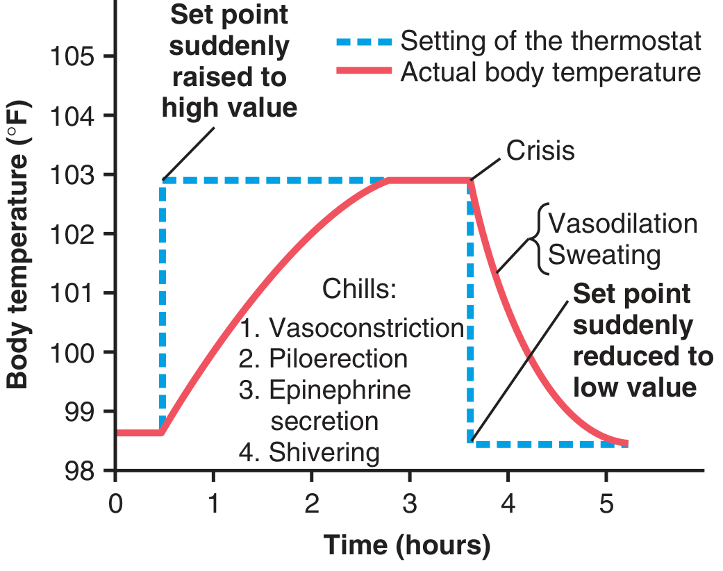
Phase 1: onset or chill phase
What happens?
Physiological responses
- Sympathetic cutaneous vasoconstriction
- Cool, pale skin
- Reduced heat loss
- Shivering
- Piloerection, minor in humans
- Increased skeletal muscle tone
- Behavioral heat seeking: blanket, warm room, curled posture
Symptoms
- Feeling cold despite an elevated measured temperature
- Chills and rigors
- Cold extremities
- Teeth chattering
- “Goosebumps”
The patient feels cold because their current temperature is below the new set point, not because their core temperature is actually low.
Phase 2: plateau or fastigium
- Chills stop
- Patient may feel neither hot nor cold, or feel warm
- Skin often becomes warmer
- Fever remains relatively stable while cytokine/PGE2 drive persists
Phase 3: defervescence or fever break
Physiological responses
- Cutaneous vasodilation
- Sweating
- Increased evaporative heat loss
- Warm, flushed skin
- Increased respiratory heat loss
- Behavioral cooling
Symptoms
- Sweating
- Flushing
- Feeling hot
- Fall in temperature
- Lysis: gradual temperature reduction over days
- Crisis: rapid fall in temperature over hours, often with marked sweating and vasodilation
7. Diagram: set point change explains chills and sweating
8. Why fever causes systemic symptoms
| Manifestation | Main mechanism |
|---|---|
| Fever | Hypothalamic PGE2 and raised set point |
| Malaise, fatigue, somnolence | Cytokine effects on CNS circuits |
| Myalgia and arthralgia | Peripheral PGE2, cytokines, nociceptor sensitization |
| Anorexia | Hypothalamic and cytokine-mediated sickness behavior |
| Tachycardia | Increased metabolic demand and sympathetic activation |
| Tachypnea | Increased metabolism, CO2 production, and heat dissipation |
| Leukocytosis | IL-1, IL-6, colony-stimulating effects |
| Increased CRP, fibrinogen, hepcidin | Predominantly IL-6-induced hepatic acute-phase response |
Cardiovascular and metabolic effects
- Basal metabolic rate
- Oxygen consumption
- Carbon dioxide production
- Insensible water loss
- Catabolism
- Cardiac workload
- Heart failure or ischemic heart disease
- Severe lung disease
- Sepsis
- Limited physiological reserve
- Neurologic injury
- Infants and frail older adults
9. Is fever beneficial or harmful?
Potential host benefits
- Reduce replication or growth of some pathogens
- Enhance neutrophil migration and phagocytic activity
- Improve some lymphocyte and T-cell responses
- Increase acute-phase and innate immune activity
- Induce heat-shock proteins that can protect cells from stress
Potential harms
- Increased metabolic and oxygen demand
- Dehydration and electrolyte loss
- Delirium or worsened encephalopathy
- Increased myocardial workload
- Increased cerebral metabolic demand, which is particularly problematic after stroke, traumatic brain injury, or cardiac arrest
- Febrile seizures in susceptible children, though these are not caused by direct neuronal “damage” from routine fever
10. Fever versus hyperthermia: essential exam distinction
| Feature | Fever | Hyperthermia |
|---|---|---|
| Hypothalamic set point | Raised | Normal |
| Main mediator | Cytokines and PGE2 | Excess heat load or uncontrolled heat production |
| Chills in rising phase | Common | Usually absent |
| Sweating | Often appears when set point falls | Can be absent, especially in heat stroke or anticholinergic toxicity |
| Antipyretics | Usually reduce temperature | Usually ineffective |
| Examples | Infection, autoimmune disease, malignancy, drug fever | Heat stroke, malignant hyperthermia, NMS, serotonin syndrome, thyrotoxic crisis |
Why antipyretics fail in hyperthermia
11. Mechanism of antipyretics
NSAIDs and aspirin
- Vasodilation
- Sweating
- Heat loss
- Defervescence
Paracetamol/acetaminophen
Glucocorticoids
12. Important special forms of temperature elevation
Central or hypothalamic fever
- Intracerebral hemorrhage
- Subarachnoid hemorrhage
- Traumatic brain injury
- Hypothalamic trauma, tumor, or inflammation
Drug fever
- Hypersensitivity reaction
- Cytokine release
- Direct central thermoregulatory effects
- Drug-induced hyperthermic syndromes, which must be distinguished from true fever
Neuroleptic malignant syndrome and serotonin syndrome
Malignant hyperthermia
13. One-line mechanism for exams
Exogenous pyrogens activate innate immune cells, causing release of IL-1β, IL-6, and TNF-α. These act at hypothalamic circumventricular endothelial sites, particularly the OVLT, inducing COX-2-dependent PGE2 synthesis. PGE2 acts mainly via EP3 receptors in the preoptic-anterior hypothalamus, raises the thermoregulatory set point, and activates vasoconstriction, shivering, increased thermogenesis, and heat-seeking behavior until core temperature reaches the new set point.
14. Fast recall summary
Fever pathway
Phases
- Set point rises: patient feels cold, vasoconstricts, shivers.
- Plateau: core temperature equals raised set point.
- Set point falls: patient feels hot, vasodilates, sweats.
Key distinction
Compare fever, hyperthermia, and heat stroke
heat stroke AND pathophysiology
heat-related illness heat stroke clinical features cooling CDC 2025
Fever vs hyperthermia vs heat stroke
| Feature | Fever (pyrexia) | Hyperthermia | Heat stroke |
|---|---|---|---|
| Definition | Regulated rise in core temperature due to increased hypothalamic set point | Unregulated rise in core temperature because heat gain/production exceeds heat loss | Severe hyperthermia with CNS dysfunction after heat exposure or exertion |
| Hypothalamic set point | Raised | Normal | Normal, but thermoregulatory mechanisms have failed |
| Main mechanism | Cytokines, mainly IL-1β, IL-6, TNF-α, induce COX-2 and PGE2 | Excess external heat, excess endogenous heat production, impaired heat loss, or drug toxicity | Extreme environmental/exertional heat load plus failed heat dissipation and systemic inflammatory injury |
| Typical causes | Infection, autoimmune disease, malignancy, tissue injury, vaccination, drug fever | Heat exposure, exercise, endocrine/drug syndromes, malignant hyperthermia, NMS, serotonin syndrome | Classic or exertional heat exposure |
| Chills/rigors | Common during rising phase | Usually absent | Usually absent |
| Skin during onset | Often cool or cold due to vasoconstriction | Often hot | Hot. May be dry or sweaty |
| Sweating | Reduced during chill phase; marked during defervescence | Variable | May be absent in classic heat stroke, but often present in exertional heat stroke |
| Antipyretic response | Usually responds to paracetamol/acetaminophen or NSAIDs | Usually does not respond | Does not respond adequately |
| Primary treatment | Treat cause; antipyretic for comfort/select indications | Remove cause, active cooling, syndrome-specific therapy | Immediate aggressive cooling and emergency supportive care |
| Organ injury | Usually limited in ordinary fever | Depends on severity/cause | Common: CNS, renal, hepatic, skeletal muscle, coagulation, cardiovascular system |
1. Fever
Mechanism
- Cutaneous vasoconstriction
- Shivering
- Increased muscle tone and thermogenesis
- Heat-seeking behavior
- Vasodilation
- Sweating
- Flushing
- Heat loss and defervescence
Guyton and Hall Textbook of Medical Physiology, 14th ed., pp. 900-901.
2. Hyperthermia
Definition
Major mechanisms
-
Excess environmental heat
- High ambient temperature
- High humidity, which prevents sweat evaporation
- Poor ventilation
- Excess clothing or protective equipment
-
Increased endogenous heat production
- Strenuous exercise
- Seizures
- Severe agitation
- Thyroid storm
- Sympathomimetic toxicity
-
Impaired heat loss
- Dehydration or hypovolemia
- Anticholinergic drugs impairing sweating
- Skin vasoconstriction or poor cardiovascular reserve
- High humidity preventing evaporation
-
Drug or toxin syndromes
- Neuroleptic malignant syndrome
- Serotonin syndrome
- Malignant hyperthermia
- Sympathomimetic toxicity
- Anticholinergic toxicity
Why antipyretics do not work
3. Heat stroke
Definition
- Confusion
- Delirium
- Ataxia
- Agitation
- Seizures
- Coma
Types
A. Classic, nonexertional heat stroke
- Older adults
- Infants
- People with chronic heart, lung, renal, or neurologic disease
- People without air conditioning
- Those taking diuretics, anticholinergics, antipsychotics, antihistamines, or some cardiovascular drugs
B. Exertional heat stroke
- Athletes
- Military personnel
- Manual workers
- People exercising in heat and humidity
4. Heat stroke pathophysiology
Stepwise process
Important injury mechanisms
- Direct protein denaturation and enzyme dysfunction
- Mitochondrial failure and ATP depletion
- Endothelial injury and capillary leak
- Splanchnic hypoperfusion and increased gut permeability
- Translocation of endotoxin or bacterial products from gut
- Systemic inflammatory response resembling sepsis
- Coagulation activation and possible DIC
- Rhabdomyolysis, especially in exertional heat stroke
- Acute kidney injury from dehydration, shock, myoglobin, and direct thermal injury
- Hepatic injury or acute liver failure
- Cerebral dysfunction due to direct heat injury, hypoperfusion, edema, and inflammation
5. Key clinical differences
| Finding | Fever | Hyperthermia | Heat stroke |
|---|---|---|---|
| Patient feels cold despite raised temperature | Common in onset phase | Uncommon | Uncommon |
| Rigors | Possible | Unusual | Unusual |
| Recent infection/inflammatory illness | Often present | Not necessary | Not necessary |
| Hot environment/exertion history | Not required | May be present | Usually present |
| Mental status change | Depends on underlying disease | Variable | Hallmark of severe disease |
| Core temperature | Often 38-40°C | Variable, may be very high | Often ≥40°C, but diagnosis is clinical |
| CK elevation/rhabdomyolysis | Not typical from uncomplicated fever | Depends on cause | Common in exertional heat stroke |
| Hepatic injury/DIC/AKI | Unusual in routine fever | Possible if severe | Suggests severe heat stroke |
| Antipyretic effect | Temperature falls | Little/no effect | Little/no effect |
| Definitive immediate action | Identify/treat cause | Active cooling/cause-specific treatment | Rapid cooling first, emergency care |
6. Management contrast
Fever
- Assess and treat the underlying cause.
- Use fluids as appropriate.
- Antipyretics can improve comfort and reduce metabolic demand in selected patients.
- Do not assume every fever requires suppression.
Hyperthermia
- Stop the heat source or causative drug.
- Begin external cooling.
- Give supportive care and address the specific syndrome.
- Examples: dantrolene for malignant hyperthermia, sedation and supportive treatment in severe sympathomimetic toxicity, syndrome-specific treatment for serotonin syndrome or NMS.
Suspected heat stroke
- Remove from heat and remove excess clothing.
- Begin rapid cooling immediately, ideally cold-water immersion where feasible.
- Call emergency services and arrange hospital care.
- Monitor airway, circulation, glucose, electrolytes, renal function, CK, liver function, and coagulation.
- Do not delay cooling to obtain labs or transfer.
Exam pearl
Fever is a thermostat problem: the hypothalamic set point is deliberately raised by PGE2.
Hyperthermia is a heat-balance problem: the set point is normal, but the body cannot get rid of heat.
Heat stroke is hyperthermia with CNS dysfunction and potential multiorgan failure.
clinical review
heat stroke AND management
CDC Yellow Book heat stroke diagnosis management rapid cooling core temperature altered mental status
https://www.cdc.gov/yellow-book/hcp/environmental-hazards-ri…
Clinical review: fever, hyperthermia, and heat stroke
1. First clinical question
Is this fever with a raised hypothalamic set point, or hyperthermia with failed heat dissipation?
- Fever: treat the underlying inflammatory cause. Antipyretics can help.
- Hyperthermia/heat stroke: start active cooling immediately. Antipyretics are ineffective.
2. Bedside definitions
| Condition | Clinical definition |
|---|---|
| Fever | Regulated elevation of temperature caused by a raised hypothalamic set point, usually cytokine and PGE2 mediated |
| Hyperthermia | Uncontrolled temperature elevation with an unchanged set point, caused by excess heat generation/gain or inadequate heat loss |
| Heat stroke | Severe hyperthermia, usually core temperature ≥40°C, plus CNS dysfunction in the setting of heat exposure or exertion |
3. Clinical assessment of a hot patient
A. Immediate priorities: ABCDE
A and B
- Airway protection if reduced consciousness, recurrent seizures, or vomiting.
- Oxygen only if hypoxemic or critically ill.
- Assess respiratory rate, work of breathing, and aspiration risk.
C
- Heart rate, BP, peripheral perfusion, capillary refill.
- Establish IV access in suspected heat stroke or severe systemic illness.
- ECG for significant hyperthermia, electrolyte abnormalities, chest symptoms, or instability.
D
- Measure GCS/mental status.
- Check bedside glucose early.
- Look for agitation, delirium, ataxia, confusion, seizures, rigidity, clonus, or focal deficits.
E
- Remove excess clothing.
- Inspect skin for sweating, rash, trauma, injection marks, cellulitis, meningism, or signs of infection.
- Assess hydration and environmental exposure.
B. Measure the right temperature
4. Focused history
History suggesting fever
- Cough, dysuria, abdominal pain, diarrhea, headache, neck stiffness
- Rash, joint pain, sore throat, focal pain
- Recent infection, travel, animal or insect exposure
- Immunosuppression, malignancy, indwelling devices
- Recent surgery, trauma, infarction, thrombosis
- New medications, antibiotics, anticonvulsants, biologics, illicit drugs
- Infective or inflammatory symptoms
- Chills/rigors
- Cold extremities in the rising phase
- No clear heat exposure or exertional trigger
- Temperature falls after antipyretic treatment
History suggesting hyperthermia or heat stroke
- Hot or humid environment
- Exercise, military training, manual labor, sport
- Duration of heat exposure
- Fluid intake, vomiting, diarrhea, alcohol use
- Clothing or personal protective equipment
- Lack of air conditioning or inability to leave a hot environment
- Drug use: amphetamines, cocaine, MDMA, anticholinergics, antipsychotics, serotonergic drugs
- Anaesthetic exposure, particularly volatile agents or succinylcholine
- Thyroid disease and symptoms of thyrotoxicosis
- Collapse during exertion or after sustained environmental heat exposure
- Altered behavior, confusion, seizures, or coma
- Marked tachycardia, hypotension, and hyperventilation
- Very high core temperature
- No chill phase
- Poor response to antipyretics
5. Examination patterns
| Feature | Fever | Hyperthermia | Heat stroke |
|---|---|---|---|
| Set point | Raised | Normal | Normal |
| Chills/rigors | Common, especially during onset | Unusual | Unusual |
| Skin | May be cool early due to vasoconstriction | Warm/hot | Hot; dry in some classic cases, but sweaty in exertional cases |
| Sweating | Common when fever breaks | Variable | May be absent or present |
| CNS dysfunction | Not typical from ordinary fever itself | Depends on cause | Defining feature |
| Trigger | Infection/inflammation commonly | Heat, drugs, endocrine or metabolic causes | Heat exposure or exertion |
| Antipyretic response | Often present | No meaningful effect | No meaningful effect |
| Main treatment | Treat cause ± antipyretic | Cooling + treat cause | Immediate rapid cooling + resuscitation |
6. Recognizing heat stroke
- Irritability or behavioral change
- Confusion and delirium
- Ataxia
- Collapse or syncope
- Seizures
- Coma
- Tachycardia and hypotension
- Tachypnea
- Nausea/vomiting
- Muscle cramps or weakness
- Rhabdomyolysis, especially exertional cases
- Acute kidney injury
- Transaminitis or acute hepatic failure
- Coagulopathy or DIC
7. Differentials for hyperthermia plus altered mental status
| Diagnosis | Helpful clues |
|---|---|
| Sepsis/meningitis/encephalitis | Infective focus, hypotension, rash, meningism, immunosuppression |
| Heat stroke | Heat/exertion exposure, high core temperature, CNS dysfunction |
| Serotonin syndrome | Serotonergic medication, clonus, hyperreflexia, agitation, diarrhea |
| Neuroleptic malignant syndrome | Dopamine antagonist or dopamine withdrawal, lead-pipe rigidity, hyporeflexia, elevated CK |
| Malignant hyperthermia | During/soon after anaesthesia, hypercapnia, rigidity, acidosis, rapidly rising temperature |
| Sympathomimetic toxicity | Cocaine/amphetamine/MDMA, agitation, mydriasis, diaphoresis, hypertension |
| Anticholinergic toxicity | Dry hot skin, mydriasis, urinary retention, ileus, delirium |
| Thyroid storm | Thyrotoxic symptoms, goiter, tremor, atrial fibrillation, GI symptoms |
| Exercise-associated hyponatremia | Prolonged exercise, excessive hypotonic fluid intake, confusion/seizures, often lower temperature than heat stroke |
| Status epilepticus | Witnessed seizures or prolonged postictal state, elevated CK/lactate |
8. Investigations
In uncomplicated fever
- CBC with differential
- CRP/ESR
- Urea, creatinine, electrolytes
- LFTs
- Urinalysis and urine culture
- Blood cultures before antibiotics when bacteremia/sepsis is suspected
- Chest radiograph, viral testing, malaria testing, lumbar puncture, imaging, etc., based on the syndrome
In suspected heat stroke or severe hyperthermia
- Rectal core temperature
- Bedside glucose
- CBC
- Electrolytes, urea, creatinine
- Calcium, phosphate, magnesium
- LFTs and bilirubin
- CK and urinalysis for myoglobinuria
- Venous or arterial blood gas and lactate
- Coagulation profile, fibrinogen, D-dimer if severe illness
- ECG and cardiac biomarkers if indicated
- Blood cultures and infection evaluation if sepsis remains possible
- Toxicology testing when history or signs suggest poisoning
- AKI
- Hyperkalemia or other electrolyte abnormalities
- Metabolic acidosis
- High CK and myoglobinuria
- Elevated aminotransferases, sometimes delayed
- Thrombocytopenia, prolonged PT/INR, DIC
9. Management
A. Fever
- Identify and treat the cause.
- Give oral or IV fluids when clinically needed.
- Use paracetamol/acetaminophen or an NSAID mainly for discomfort or high metabolic burden, if not contraindicated.
- Treat sepsis, meningitis, malaria, inflammatory disease, drug reaction, etc., according to the working diagnosis.
B. Hyperthermia
- Stop exposure and remove the triggering drug if possible.
- Undress patient and begin active external cooling.
- Give supportive care, including fluids when hypovolemia is present.
- Treat the specific cause.
C. Suspected heat stroke: emergency sequence
1. Cool first
- Remove from heat and remove clothing.
- Start rapid cooling at once.
- Cold-water immersion is preferred for exertional heat stroke when feasible.
- If immersion is unavailable: spray/mist with tepid water and use high-flow fans, applying ice packs to neck, axillae, and groin as adjuncts.
- Continue cooling until core temperature is around 39°C.
2. Support physiology
- Airway protection if coma/severe agitation.
- IV isotonic crystalloid guided by perfusion, volume status, urine output, and cardiac/renal risk.
- Correct hypoglycemia and clinically important electrolytes.
- Treat seizures with benzodiazepines.
- Consider benzodiazepines for severe agitation or shivering that impairs cooling.
- Monitor cardiac rhythm, urine output, CK, renal function, LFTs, and coagulation.
3. Avoid
- Delaying cooling to complete diagnostic workup
- Routine antipyretics
- Using shivering-inducing cooling methods without managing shivering
- Assuming dry skin is required for the diagnosis
- Giving large volumes of hypotonic fluids in a patient who may have exercise-associated hyponatremia
10. Heat exhaustion versus heat stroke
| Feature | Heat exhaustion | Heat stroke |
|---|---|---|
| Core temperature | Normal to moderately raised | Usually markedly raised |
| CNS function | May have headache, fatigue, dizziness; severe persistent CNS signs absent | Delirium, ataxia, seizures, coma, or significant altered behavior |
| Organ injury | Usually absent | May include AKI, rhabdomyolysis, liver injury, DIC |
| Management | Rest, cooling, oral/IV rehydration, reassess | Emergency rapid cooling, hospital-level resuscitation and monitoring |
11. High-yield viva answers
Because PGE2 raises the hypothalamic set point. The current core temperature becomes lower than the new target, so the body activates heat production and conservation.
The set point falls to normal, often after inflammation resolves or PGE2 is inhibited. The current body temperature is then above the set point, causing vasodilation and sweating.
Heat stroke is hyperthermia with an unchanged set point, not PGE2-mediated set-point elevation.
Hyperthermia with central nervous system dysfunction in an appropriate heat or exertion context.
Immediate rapid active cooling.
clinical review on fever and pyrexia and hyperthermia
fever AND clinical management
Merck Manual fever evaluation adults clinical approach hyperthermia antipyretics
Clinical review: fever (pyrexia) and hyperthermia
1. Definitions
Fever / pyrexia
- Usually caused by infection or sterile inflammation.
- Mediated by cytokines, especially IL-1β, IL-6, and TNF-α, which induce hypothalamic PGE2.
- The patient may experience chills and shivering because, after the set point rises, the existing core temperature is perceived as too low.
Hyperthermia
- Hypothalamic set point is not raised.
- The body may already be attempting to lose heat, but cannot keep up.
- It can progress rapidly to cellular injury and multiorgan failure.
- Antipyretics do not treat the primary mechanism.
2. Normal temperature and definitions of abnormal temperature
- Fever: oral temperature approximately ≥38.0°C, or rectal/core temperature ≥38.0°C.
- Hyperpyrexia: very high fever, usually >41°C or >41.5°C depending on the definition used.
- Hyperthermia: elevated temperature without hypothalamic set-point elevation.
- Heat stroke: severe hyperthermia with CNS dysfunction, typically with core temperature around or above 40°C.
3. Clinical physiology of fever
Pyrogenic pathway
- Harrison’s Principles of Internal Medicine, 22nd ed., p. 178.
- Costanzo Physiology, 7th ed., pp. 1090-1098.
Clinical phases
| Phase | Set point compared with actual temperature | Clinical features |
|---|---|---|
| Onset/chill phase | Set point suddenly rises above actual core temperature | Feeling cold, chills, rigors, vasoconstriction, pale/cool skin, shivering |
| Plateau/fastigium | Actual temperature reaches raised set point | Chills stop; patient may feel hot or relatively comfortable; temperature remains high |
| Defervescence | Set point falls toward normal but core temperature remains high | Vasodilation, warm flushed skin, sweating, temperature falls |
Important bedside implication
4. Causes of fever
A. Infection
- Viral syndromes, influenza, COVID-19
- Pneumonia
- Urinary tract infection and pyelonephritis
- Meningitis, encephalitis
- Intra-abdominal infection
- Tuberculosis
- Malaria and other travel-associated infections
- Infective endocarditis
- Skin, soft tissue, bone, joint, and device-related infections
- Sepsis
B. Noninfectious inflammatory disease
- SLE and vasculitis
- Adult-onset Still disease
- Rheumatoid disease flare
- Sarcoidosis
- Inflammatory bowel disease
- Gout or pseudogout
- Pericarditis
- Pancreatitis
C. Malignancy
- Lymphoma
- Leukemia
- Renal-cell carcinoma
- Hepatocellular carcinoma
- Advanced solid malignancy
D. Other causes
- Drug fever
- Transfusion reaction
- Pulmonary embolism or DVT
- Myocardial infarction
- Tissue injury, trauma, surgery, burns
- Thyroiditis
- CNS injury, for example hemorrhage or stroke
- Vaccination
5. Clinical approach to a patient with fever
Step 1: Assess severity first
- Hypotension, shock, poor perfusion
- Hypoxia or respiratory distress
- Altered mental state
- Meningism, seizures, focal neurologic deficits
- Non-blanching rash or purpura
- Severe headache or photophobia
- Severe abdominal pain
- Neutropenia or immunocompromise
- Pregnancy/postpartum status
- Indwelling vascular catheter, prosthetic valve, joint, or device
- Extremes of age
- Recent surgery or invasive procedure
Step 2: Focused history
Time course
- Onset, duration, maximum recorded temperature
- Continuous, intermittent, remittent, relapsing, or periodic pattern
- Chills or true rigors
- Response to antipyretics
Localizing symptoms
- Respiratory: cough, sputum, dyspnea, pleuritic pain
- Urinary: dysuria, frequency, flank pain
- CNS: headache, photophobia, neck stiffness, confusion
- GI: vomiting, diarrhea, abdominal pain
- Skin: rash, wound discharge, cellulitis
- Musculoskeletal: monoarthritis, back pain, myalgia
- Constitutional: weight loss, night sweats, fatigue
Exposure history
- Travel, freshwater exposure, insect or tick bite
- Animal contact
- Sick contacts
- Food and sexual history
- Injection drug use
- Occupational exposure
- Recent hospital admission or antimicrobial use
Host factors
- Diabetes, CKD, cirrhosis, HIV
- Chemotherapy, steroids, transplant drugs, biologics
- Cancer and neutropenia
- Prosthetic material or implanted devices
- Pregnancy
Drug history
Step 3: Examination
- Vital signs, including repeat temperature and oxygen saturation
- Hydration, perfusion, mental state
- Skin and mucosa: rash, petechiae, eschar, cellulitis, IV sites
- Lymph nodes
- Oral cavity and dentition
- Heart: murmurs, peripheral endocarditis stigmata
- Lungs
- Abdomen, hepatosplenomegaly, costovertebral angle tenderness
- Joints, spine, muscles
- Neurologic examination and meningism
- Rectal, pelvic, or prostate examination only when clinically indicated
6. Investigation strategy in fever
Common initial tests in an unwell adult
- CBC with differential
- Urea, creatinine, electrolytes, glucose
- Liver function tests
- CRP, with or without procalcitonin depending on local use
- Urinalysis and urine culture if urinary symptoms/risk
- Blood cultures if sepsis, bacteremia, endocarditis, neutropenia, or unexplained significant fever is suspected
- Chest radiograph if respiratory features or unclear systemic illness
- Lactate and blood gas if sepsis/shock is possible
- Respiratory viral testing when epidemiologically or clinically relevant
Targeted tests
| Clinical clue | Potential next test |
|---|---|
| Meningism or altered sensorium | CT first if indicated, then lumbar puncture and CSF studies |
| Travel/tropical exposure | Malaria smear or rapid antigen test, dengue/enteric fever tests as appropriate |
| New murmur, embolic phenomena, IV drug use | Multiple blood cultures and echocardiography for endocarditis |
| Persistent back pain | MRI if vertebral osteomyelitis/epidural abscess suspected |
| Monoarthritis | Urgent arthrocentesis for Gram stain, culture, crystals |
| Immunocompromise | Broader microbiology and early imaging based on risk |
| Persistent/recurrent fever | CT, autoimmune evaluation, PET-CT or biopsy directed by clues |
Fever of unknown origin, FUO
- Temperature >38.3°C on several occasions
- Illness lasting >3 weeks
- No diagnosis after appropriate evaluation
- Infections
- Malignancy
- Noninfectious inflammatory disease
- Miscellaneous causes, including drug fever and thromboembolism
7. Fever treatment
Treat the cause
- Antibiotics for a proven or strongly suspected bacterial infection
- Antimalarial therapy when indicated
- Source control for abscess or infected device
- Immunomodulatory therapy only after appropriate exclusion of infection in inflammatory disease
- Stop a likely culprit medication in drug fever
Antipyretics
Paracetamol/acetaminophen
NSAIDs
- AKI or CKD
- Peptic ulcer disease or GI bleeding
- Thrombocytopenia
- Anticoagulation
- Heart failure
- Some viral illnesses and dehydration states
8. Hyperthermia: clinical recognition
Causes
Environmental and exertional
- Heat exposure
- High humidity
- Exercise in heat
- Dehydration
- Heavy protective clothing
- Poor ventilation or no access to air conditioning
Drug and toxicologic
- Anticholinergic toxicity
- Sympathomimetics: cocaine, amphetamines, MDMA
- Serotonin syndrome
- Neuroleptic malignant syndrome, NMS
- Malignant hyperthermia after anaesthesia
Endocrine/metabolic and neurologic
- Thyroid storm
- Pheochromocytoma crisis
- Status epilepticus
- Severe agitation
- CNS injury affecting thermoregulation
Red flags for hyperthermia
- Clear heat/exertion or toxicologic context
- Temperature rising despite antipyretics
- Lack of chills
- Marked tachycardia, hypotension, dehydration
- Altered mental status
- Severe rigidity, clonus, hyperreflexia, or seizures
- Very elevated CK, metabolic acidosis, hyperkalemia, AKI, or liver injury
9. Fever vs hyperthermia: practical comparison
| Feature | Fever / pyrexia | Hyperthermia |
|---|---|---|
| Hypothalamic set point | Raised | Unchanged |
| Core mechanism | Cytokines → COX-2 → PGE2 | Excess heat gain/production or impaired heat loss |
| Chills and rigors | Common, especially early | Usually absent |
| Skin | Cool/pale in chill phase; sweaty when defervescing | Often hot; sweat may be present or absent |
| Typical cause | Infection or inflammation | Heat, exertion, drugs, toxins, endocrine/metabolic disease |
| Antipyretics | Often effective | Usually ineffective |
| Cooling | Adjunct in selected severe fever | Essential, immediate in severe cases |
| Major danger | Usually underlying disease | Direct thermal injury and multiorgan failure |
10. Hyperthermic syndromes: distinguishing features
| Syndrome | Typical trigger | Neuromuscular findings | Skin/GI findings | Initial priorities |
|---|---|---|---|---|
| Heat stroke | Exertion/environmental heat | CNS dysfunction; seizures possible | Hot skin, sweating may be absent or present | Rapid active cooling |
| Serotonin syndrome | Serotonergic drugs/interactions | Hyperreflexia and inducible/spontaneous clonus, often lower limbs | Diarrhea, hyperactive bowel sounds, diaphoresis | Stop drug, benzodiazepines, cooling, supportive care |
| NMS | Dopamine antagonist or dopamine withdrawal | Lead-pipe rigidity, hyporeflexia/bradyreflexia | Autonomic instability | Stop drug, cooling, supportive care, consider specific therapy |
| Malignant hyperthermia | Volatile anaesthetic/succinylcholine | Rigidity, rising CO2, acidosis | Usually intraoperative/perioperative | Stop trigger, dantrolene, active cooling |
| Anticholinergic toxicity | Atropine-like drugs | Delirium, no clonus | Hot, dry skin; mydriasis, urinary retention, ileus | Cooling, benzodiazepines, toxicology management |
| Sympathomimetic toxicity | Cocaine/amphetamine/MDMA | Agitation, tremor, seizures | Diaphoresis, mydriasis, hypertension | Benzodiazepines, cooling, supportive care |
| Thyroid storm | Severe thyrotoxicosis | Tremor/agitation | Goiter, AF, GI symptoms | Antithyroid therapy, beta-blockade when appropriate, steroids, cooling |
11. Heat stroke: emergency clinical review
- Exertional heat stroke: young, healthy, active individual; collapse during exertion; sweating is often profuse.
- Classic/nonexertional heat stroke: older patient with comorbidity, chronic illness, medications, or prolonged heat-wave exposure; sweating may be absent.
Immediate management
- Start cooling immediately. Do not wait for laboratory results, transport, or diagnostic certainty.
- Remove clothing and relocate to a cool environment.
- Use cold-water immersion if feasible, especially in exertional heat stroke.
- If immersion is impractical, use evaporative cooling: mist with water plus fans, with ice packs as adjuncts.
- Monitor rectal core temperature and stop aggressive cooling near 39°C.
- Support airway, circulation, glucose, seizures, and agitation.
- Monitor for AKI, rhabdomyolysis, hepatic injury, DIC, electrolyte disorders, and shock.
12. When fever needs urgent escalation
- Sepsis physiology: hypotension, tachypnea, hypoxia, high lactate, altered mentation
- Suspected meningitis or encephalitis
- Purpura, non-blanching rash, or rapidly progressive skin lesions
- Severe headache, neck stiffness, seizures, or focal neurologic signs
- Neutropenia or significant immunosuppression
- Pregnancy/postpartum fever
- Fever after surgery, invasive procedure, transplant, or central line
- Persistent fever with weight loss, night sweats, lymphadenopathy, or hepatosplenomegaly
- Fever plus acute monoarthritis
- Fever with severe abdominal, back, or flank pain
- Temperature >41°C or suspected hyperthermia
- Any hyperthermia with altered mental status
13. Exam and ward-round summary
Fever or pyrexia is a regulated rise in body temperature due to cytokine-induced PGE2 elevation of the hypothalamic set point. It may present with chills during onset, plateau at the new set point, and sweating during defervescence.Hyperthermia is an unregulated rise in temperature with an unchanged set point, due to excessive heat load or failed heat loss. It requires physical cooling and treatment of the cause, not antipyretics.Heat stroke is hyperthermia with CNS dysfunction and possible multiorgan injury. Treat it as a time-critical emergency: cool first, investigate in parallel.
approach to fever in opd
acute febrile illness AND outpatient
adult acute fever ambulatory outpatient evaluation red flags clinical guideline
Approach to fever in OPD
- Patients needing urgent emergency referral/admission
- A likely anatomical or syndromic source
- Patients in whom outpatient observation and review are safe
1. Confirm that it is fever
- Ask for the measured highest temperature, site of measurement, timing, and antipyretic use.
- Measure and document temperature in clinic, along with:
- Pulse
- Blood pressure
- Respiratory rate
- SpO₂
- Mental status
- Capillary refill/perfusion
- Weight when relevant for follow-up or dosing
2. Triage first: is the patient safe for OPD?
Refer urgently to ED / admit if any danger sign is present
Sepsis or organ dysfunction
- SBP <90 mmHg, poor perfusion, syncope, cold extremities
- New confusion, drowsiness, delirium, or reduced GCS
- Respiratory distress, cyanosis, SpO₂ low, or RR ≥22/min
- Persistent tachycardia, oliguria, high lactate if available
- Inability to take oral fluids or persistent vomiting
CNS or severe infection features
- Severe headache, neck stiffness, photophobia
- Seizure, focal neurologic deficit, altered sensorium
- Purpura/petechiae or a rapidly spreading rash
- Suspected meningitis, encephalitis, malaria with complications, or severe dengue depending on local epidemiology
High-risk host
- Neutropenia or recent chemotherapy
- Significant immunosuppression: high-dose steroids, transplant drugs, biologics, advanced HIV
- Pregnancy or early postpartum period
- Frail older adult, especially with functional decline
- Cirrhosis, advanced CKD, uncontrolled diabetes, significant cardiac or lung disease
- Prosthetic valve, vascular catheter, implanted device, recent surgery
Other urgent syndromes
- Fever with acute monoarthritis: exclude septic arthritis
- Fever with severe abdominal pain, back pain, or peritonism
- Fever with rapidly progressive skin/soft-tissue infection
- Core temperature >41°C, suspected hyperthermia, or heat stroke
- Any febrile patient who appears toxic despite a “normal” initial BP
If unstable, obtain cultures and basic investigations where this does not delay care, begin appropriate resuscitation/empiric treatment as indicated, and arrange referral.
3. Structured OPD history
A. Characterize the fever
- Duration: acute <7 days, subacute 1-3 weeks, prolonged >3 weeks
- Continuous, intermittent, remittent, relapsing, or periodic
- Highest documented temperature
- Chills versus true rigors
- Night sweats, weight loss, anorexia, fatigue
- Response to paracetamol/NSAIDs
- Previous similar episodes
B. Find the focus: symptom-directed review
| System | Key questions and clinical possibilities |
|---|---|
| Respiratory | Cough, sputum, dyspnea, pleuritic pain, sore throat, rhinorrhea, sinus pain |
| Urinary | Dysuria, frequency, urgency, flank pain, suprapubic pain |
| GI/hepatobiliary | Diarrhea, vomiting, abdominal pain, jaundice, GI bleeding |
| CNS | Headache, meningism, photophobia, confusion, seizures |
| Skin/soft tissue | Rash, abscess, cellulitis, wounds, eschar, insect bite |
| MSK | Acute monoarthritis, back pain, focal bone pain, myalgia |
| Cardiac | New murmur, embolic symptoms, prosthetic valve, IV drug use |
| Genital/pelvic | Vaginal discharge, pelvic pain, postpartum symptoms, testicular pain |
C. Exposure and epidemiology
- Sick contacts and outbreak exposure
- Recent hospital stay or antibiotics
- Travel and residence, including rural area or endemic region
- Mosquito/tick exposure, freshwater exposure, animal contact
- Food and water exposure
- Tuberculosis contact
- Occupation, especially healthcare, farming, abattoir work, laboratory exposure
- Sexual history where relevant
- Injection drug use
D. Drug and host history
- New medications, antibiotics, anticonvulsants, allopurinol, biologics
- Recent vaccination or transfusion
- Diabetes, CKD, liver disease, HIV, malignancy
- Steroids, chemotherapy, immunosuppressants
- Devices: central line, urinary catheter, prosthetic heart valve/joint
- Pregnancy and postpartum status
4. Examination: do not skip the full examination
General examination
- Toxic appearance, dehydration, pallor, icterus, cyanosis
- Lymphadenopathy
- Rash: petechiae, purpura, maculopapular rash, vesicles, eschar
- Peripheral edema, signs of DVT
- Mental state and hydration status
Focused examination
- ENT/oral cavity: tonsillitis, dental infection, sinus tenderness
- Chest: crackles, bronchial breathing, wheeze, pleural rub
- Cardiovascular: new murmur, peripheral stigmata of endocarditis
- Abdomen: focal tenderness, hepatosplenomegaly, CVA tenderness
- Skin and lines: cellulitis, abscess, IV-site infection, pressure sores
- Joints/spine: hot swollen joint, vertebral tenderness
- Neurologic exam: meningism, focal deficit, cerebellar signs
- Pelvic/rectal/prostate exam: only if clinically indicated and with consent/chaperone as appropriate
5. Decide the clinical syndrome
A. Fever with a clear focus
- URI/pharyngitis
- Community-acquired pneumonia
- UTI/pyelonephritis
- Cellulitis
- Acute gastroenteritis
- Dental infection
- Suspected dengue, malaria, enteric fever, or leptospirosis based on setting
B. Acute undifferentiated fever
- Local epidemiology and season
- Day of illness
- CBC and focused tests only when results will affect management
- Review within 24-48 hours if diagnosis remains uncertain
- Viral infection
- Dengue or malaria in endemic settings
- Early enteric fever
- COVID/influenza when circulating
- Leptospirosis/rickettsial illness when exposure pattern fits
- Early bacterial infection before focal signs develop
C. Prolonged fever
- Tuberculosis
- Endocarditis
- Abscess
- Malignancy, especially lymphoma
- Connective-tissue disease/vasculitis
- Drug fever
- HIV-related opportunistic infection
- Travel-related infection
6. Rational investigations in OPD
Do not use “fever panel” testing indiscriminately
Basic tests if the patient is significantly symptomatic, has no clear focus, or has risk factors
- CBC with differential and platelet count
- Urinalysis, with urine culture when UTI/pyelonephritis is suspected
- Renal function, electrolytes, glucose
- Liver enzymes and bilirubin if systemic illness, jaundice, suspected dengue/leptospirosis/hepatitis, or drug toxicity
- CRP can support assessment but should not replace clinical reasoning
- Blood culture before antibiotics if bacteremia, endocarditis, enteric fever, sepsis, or significant unexplained fever is suspected
Syndrome-directed testing
| Presentation | Appropriate tests |
|---|---|
| Cough/dyspnea or abnormal chest exam | Chest radiograph, viral testing where relevant, sputum testing selectively |
| Dysuria/flank pain | Urinalysis and urine culture |
| Acute undifferentiated fever in endemic area | Malaria rapid test/smear; dengue testing based on day of illness and local protocol |
| Meningism/altered sensorium | Emergency referral for imaging/LP pathway, blood cultures, empiric therapy when indicated |
| Suspected enteric fever | Blood culture before antibiotics; targeted tests according to local guidelines |
| Jaundice/AKI/myalgia/conjunctival suffusion | Consider leptospirosis testing and urgent evaluation |
| Eschar/rash with compatible exposure | Consider rickettsial testing/treatment according to local protocol |
| New murmur or embolic phenomena | Multiple blood cultures and echocardiography referral |
| Acute hot swollen joint | Arthrocentesis urgently, not just CBC/CRP |
| Immunosuppression | Lower threshold for cultures, imaging, and hospital referral |
7. Treatment in OPD
General measures
- Oral fluids and nutrition as tolerated
- Rest
- Advice on monitoring temperature and urine output
- Avoid alcohol and unnecessary exertion
- Explain that the underlying cause, not the temperature alone, determines severity
Antipyretics
- Paracetamol/acetaminophen is often first choice if no contraindication.
- NSAIDs can be considered selectively but avoid or use caution with:
- Dehydration, AKI, CKD
- Gastritis/peptic ulcer/GI bleed
- Heart failure
- Anticoagulation or thrombocytopenia
- Possible dengue, where bleeding risk and local guidance matter
Antibiotics
- Simple viral URI
- Uncomplicated acute bronchitis
- Nonspecific fever without a bacterial focus in a stable patient
8. Disposition and safety-netting
- Drink and take oral medications
- Understand advice
- Access follow-up promptly
- Return immediately if worsening occurs
Give explicit return precautions
- Breathlessness, chest pain, persistent tachypnea
- Confusion, fainting, seizure, severe headache, neck stiffness
- Persistent vomiting or inability to drink
- Reduced urine output, dizziness, severe weakness
- New rash, bleeding, petechiae, jaundice
- Severe abdominal pain, worsening back pain, or acute joint swelling
- Fever persisting beyond the planned review, recurrent fever, or clinical deterioration despite treatment
Review timing
- 24-48 hours: undifferentiated fever, borderline findings, suspected early illness, or pending tests
- Earlier/same day: high-risk patients or uncertain disposition
- Immediate referral: any danger sign, unstable vital sign, or suspected serious infection/hyperthermia
9. One-page OPD algorithm
Patient with fever/history of fever
        |
        v
1. Record vitals, SpO2, mental state, hydration
        |
        +--> Unstable / red flags / high-risk host?
        |        |
        |        +--> Yes: resuscitate, targeted samples, urgent referral/admission
        |
        v
2. Focused history + full examination
        |
        +--> Clear focus?
        |        |
        |        +--> Yes: syndrome-specific tests and treatment
        |
        v
3. No focus: acute undifferentiated fever
        |
        +--> Use local epidemiology, day of illness, exposure history
        +--> Targeted CBC/urine/malaria-dengue or other relevant tests
        +--> Avoid blind antibiotics
        +--> Review in 24-48 h
        |
        v
4. Fever persistent/recurrent/prolonged
        |
        +--> Reassess for TB, endocarditis, abscess, malignancy,
             inflammatory disease, drug fever, travel-related disease
        +--> Directed imaging, cultures, serology, specialist referral
Final practical pearl
In OPD, treat the patient and syndrome, not the thermometer reading.
A well-appearing, stable patient with a likely viral illness often needs supportive care and review. A patient with fever plus abnormal vital signs, altered mental status, immunosuppression, focal red flags, or inability to maintain oral intake needs escalation.