
Megaloblastic anemia and it's lab diagnosis
Reading File
Megaloblastic anemia is caused by impaired DNA synthesis (usually B12 or folate deficiency), leading to large, immature RBCs. Lab diagnosis includes peripheral smear (macro-ovalocytes, hypersegmented neutrophils), elevated MCV, low B12/folate levels, elevated homocysteine, and elevated MMA (in B12 deficiency specifically).
Searching sources...
Finding Sources
Searching PubMed
megaloblastic anemia laboratory diagnosis
Good. Now let me read the most relevant sections - Henry's Clinical Diagnosis (lab methods book) and Robbins Pathology:
Reading File
Reading File
Excellent content. Let me read more from both sources - especially laboratory diagnosis sections:
Reading File
Reading File
Now let me get the detailed lab diagnosis section from Henry's and image content from Robbins:
Reading File
Reading File
Now let me read the cobalamin lab diagnosis section and look for images in Robbins related to megaloblastic anemia:
Finding Sources
Reading File
Now let me get the cobalamin lab diagnosis section and search for images in the textbooks:
Reading File
Finding Sources
I have comprehensive data. Let me also fetch the key images and get the cobalamin lab diagnosis values:
Loading Image
Reading File
Now let me get the key lab values table comparing B12 vs folate deficiency, and search for images of the bone marrow findings:
Reading File
Finding Sources
Let me read the Diagnosis of Cobalamin Deficiency section and Robbins morphology section:
Reading File
Reading File
Excellent! I now have very comprehensive data. Let me fetch the bone marrow image from Robbins and the hypersegmented neutrophil image:
Loading Image
Loading Image
I now have everything I need for a comprehensive answer. Let me compile the full response.
Megaloblastic Anemia and Its Laboratory Diagnosis
Definition and Pathogenesis
Megaloblastic anemia is caused by impaired DNA synthesis, leading to defective nuclear maturation while cytoplasmic development continues normally. This nuclear-to-cytoplasmic asynchrony results in abnormally large cells across all rapidly proliferating lines (RBCs, WBCs, platelets). The most common causes are vitamin B12 (cobalamin) or folate deficiency.
The core mechanism: deficiency of either vitamin reduces intracellular 5,10-methylene tetrahydrofolate, which is required by thymidylate synthase to convert deoxyuridine monophosphate (dUMP) → deoxythymidine monophosphate (dTMP) - a reaction essential for normal DNA synthesis. Without dTMP, cells accumulate in S-phase with a prolonged intermitotic resting phase and block in mitosis.
Causes
| Vitamin B12 Deficiency | Folate Deficiency |
|---|---|
| Pernicious anemia (intrinsic factor deficiency) | Poor dietary intake (alcoholism, old age) |
| Gastrectomy (total or subtotal) | Malabsorption (celiac, Crohn's) |
| Ileal resection / ileitis | Increased requirements (pregnancy, hemolysis) |
| Fish tapeworm (D. latum) | Anticonvulsants, oral contraceptives |
| Bacterial overgrowth (blind loop) | Methotrexate, other antifolates |
| Strict veganism | Hemodialysis (increased loss) |
| Nitrous oxide exposure | — |
Causes unresponsive to B12/folate: cytotoxic drugs (6-MP, cytarabine, hydroxyurea, 5-FU), orotic aciduria, myelodysplastic syndromes.
Laboratory Diagnosis
1. Complete Blood Count (CBC)
| Parameter | Finding |
|---|---|
| Hemoglobin | Decreased (often moderate to severe) |
| MCV | Elevated (macrocytic) - often >100 fL, can exceed 120-130 fL |
| MCHC | Normal (not truly hyperchromic despite appearance) |
| WBC | Decreased (leukopenia) |
| Platelets | Decreased (thrombocytopenia) |
| Reticulocyte count | Low (ineffective hematopoiesis) |
Pancytopenia is the rule in advanced disease.
2. Peripheral Blood Smear - Most Important Morphologic Finding
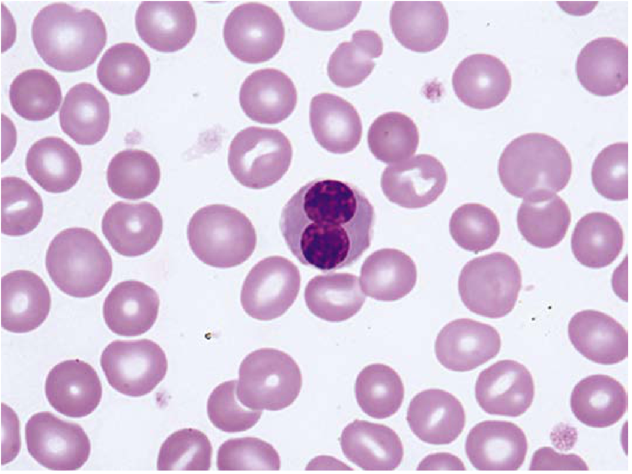
Peripheral blood smear in megaloblastic anemia: macro-ovalocytes, marked anisocytosis/poikilocytosis, and a circulating megaloblast (Henry's Clinical Diagnosis, p. 679)
Key findings:
- Macro-ovalocytes - large, oval RBCs lacking central pallor (highly characteristic)
- Hypersegmented neutrophils - the single most specific finding. Normal neutrophils have 3-4 lobes; hypersegmentation = >5% of neutrophils with 5+ lobes, OR any neutrophil with ≥6 lobes
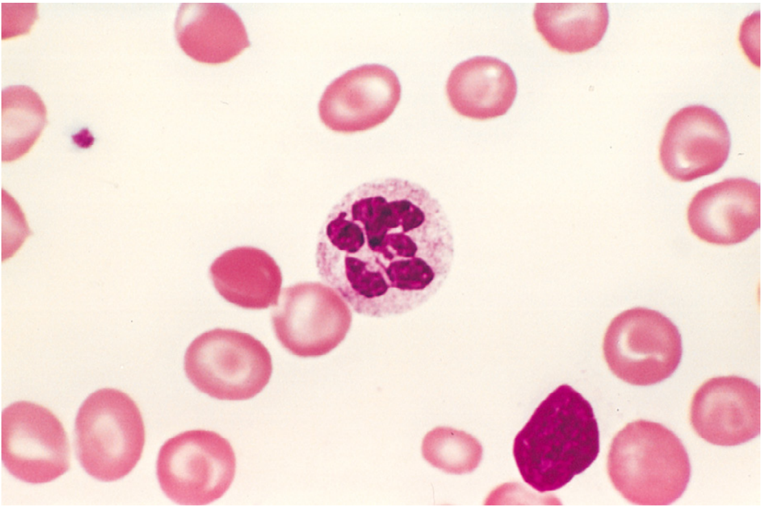
Hypersegmented neutrophil with 6-lobed nucleus (Robbins Pathology, Fig. 14.15)
- Marked anisocytosis and poikilocytosis including red cell fragments
- Microcytes and dacrocytes (teardrop cells) are common
- Basophilic stippling, multiple Howell-Jolly bodies
- Nucleated RBCs with karyorrhexis in severe cases
- Occasional circulating megaloblasts in extreme cases
3. Bone Marrow Examination
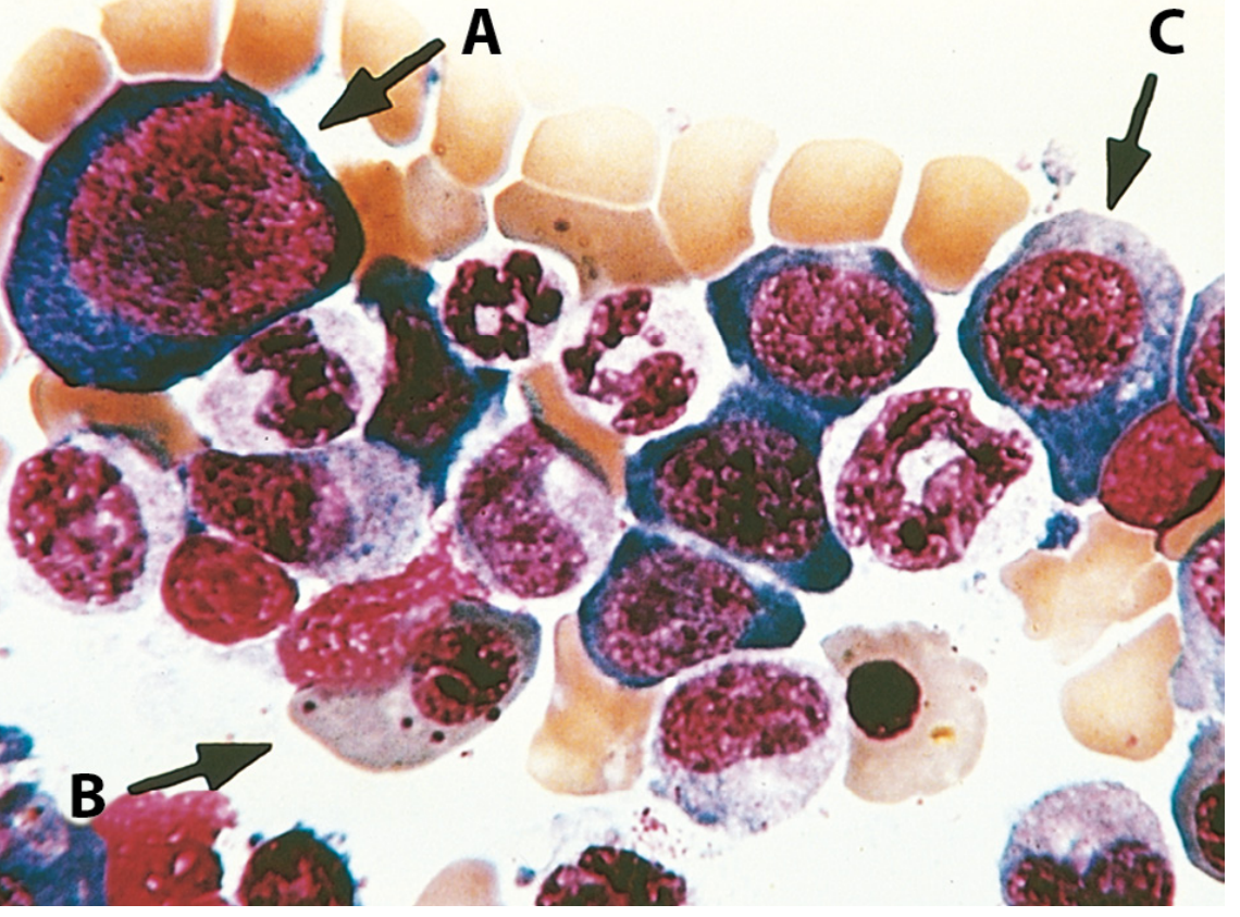
Bone marrow aspirate: (A) promegaloblast with fine chromatin and prominent nucleoli; (B) orthochromatic megaloblast - hemoglobinized cytoplasm but non-pyknotic nucleus; (C) intermediate megaloblast. Also note giant metamyelocytes. (Robbins Pathology, Fig. 14.16)
Findings:
- Hypercellular marrow (responding to high EPO levels)
- Megaloblasts: large erythroid precursors with finely distributed "open" chromatin (like lace) despite advanced cytoplasmic hemoglobinization - nuclear/cytoplasmic asynchrony
- Giant metamyelocytes and band forms (granulocytic dysmaturation)
- Abnormally large, multilobate megakaryocytes
- Increased mitotic figures
- Ineffective hematopoiesis: most precursors undergo apoptosis before release
4. Biochemical Markers (Differentiating B12 vs. Folate)
| Test | B12 Deficiency | Folate Deficiency |
|---|---|---|
| Serum Vitamin B12 | Low (<100 ng/L; normal 200-900 ng/L) | Normal or elevated |
| Serum Folate | Normal or slightly low (~10%) | Low (<3 μg/L) |
| Red Cell Folate | Low in ~2/3 of cases | Low (better reflects stores) |
| Plasma Homocysteine | Elevated (>90% of cases) | Elevated (~75%) |
| Plasma Methylmalonic Acid (MMA) | Elevated (>90% of cases) | Normal |
| Serum LDH | Greatly elevated (ineffective erythropoiesis) | Greatly elevated |
| Serum bilirubin | Elevated (indirect) | Elevated |
| Serum iron | Elevated | Elevated |
Key discriminator: Elevated MMA is specific for B12 deficiency. Both B12 and folate deficiency raise homocysteine, but only B12 deficiency raises MMA. MMA and homocysteine elevations precede the fall in serum B12.
5. Additional Tests for Specific Cause
For Pernicious Anemia (most common cause of B12 deficiency):
- Anti-intrinsic factor (IF) antibodies: Highly specific for pernicious anemia (~sensitivity 50-70%, specificity ~100%). The combination of megaloblastic anemia + low B12 + anti-IF antibodies is essentially diagnostic.
- Anti-parietal cell antibodies: More sensitive but less specific (~73% sensitivity when combined with anti-IF)
- These antibody tests largely replace the Schilling test in modern practice
Holotranscobalamin (HoloTC / "Active B12"):
- Measures TC-bound (biologically active) B12 fraction
- Falls before total serum B12 decreases - early marker of negative B12 balance
- More accurate than total B12 for diagnosing functional deficiency
Schilling Test (historical):
- Two-part test to localize absorption defect (IF deficiency vs. intestinal disease vs. bacterial overgrowth)
- Largely replaced by antibody assays in modern labs
Urinary Formiminoglutamic Acid (FIGLU):
- Increases in urine after oral histidine loading if folate deficiency present
- Useful when serum folate is normal but tissue coenzyme levels are low (antifolate drugs)
Deoxyuridine (dU) Suppression Test:
- Measures marrow cell capacity to utilize deoxyuridine for DNA synthesis in vitro
- Abnormal in both B12 and folate deficiency; corrected by adding the specific missing factor - helps distinguish the two
6. Other Laboratory Features
- Serum LDH (especially LD-1 isoenzyme): markedly elevated due to intramedullary hemolysis (ineffective erythropoiesis)
- Serum indirect bilirubin: elevated
- Serum iron and transferrin saturation: elevated (not being utilized for hematopoiesis)
- Urinary methylmalonate: elevated in B12 deficiency (provided methylmalonic aciduria excluded)
- Serum muramidase (lysozyme): may be elevated, suggesting ineffective granulopoiesis
Summary Diagnostic Algorithm
Macrocytic anemia (elevated MCV) on CBC
↓
Peripheral smear: macro-ovalocytes + hypersegmented neutrophils
↓
Measure: Serum B12 + Serum Folate + Red Cell Folate
↓
If equivocal (B12 100-400 pg/mL): add MMA + Homocysteine
↓
MMA ↑ + Homocysteine ↑ → B12 deficiency
MMA normal + Homocysteine ↑ → Folate deficiency
↓
If B12 deficient: Anti-IF antibody + Anti-parietal cell antibody
↓
Bone marrow biopsy: if diagnosis uncertain or myelodysplasia suspected
Sources:
- Henry's Clinical Diagnosis and Management by Laboratory Methods, pp. 679-683
- Robbins, Cotran & Kumar Pathologic Basis of Disease, pp. 608-612
- Recent review: Harrington DJ et al., Ann Clin Biochem 2025 - laboratory biomarkers for vitamin B12 status evaluation (PMID: 39367523)
For exam
Asking for Preferences
Which exam are you preparing for? · What format do you need?
Here is a high-yield exam-focused summary on Megaloblastic Anemia and its lab diagnosis:
Megaloblastic Anemia - High-Yield Exam Notes
MUST-KNOW ONE-LINER
Megaloblastic anemia = impaired DNA synthesis → nuclear/cytoplasmic asynchrony → large, immature cells; most commonly due to B12 or folate deficiency.
PATHOGENESIS (1 sentence)
Deficiency of B12/folate → reduced 5,10-methylene THF → thymidylate synthase cannot make dTMP from dUMP → defective DNA synthesis → cells grow but can't divide → megaloblasts.
CAUSES - Quick List
B12 deficiency:
- Pernicious anemia (MC cause in adults) - anti-IF antibodies
- Strict veganism
- Gastrectomy (no IF → deficiency in 3-6 yrs)
- Terminal ileum resection/Crohn's
- Fish tapeworm (D. latum)
- Nitrous oxide abuse
Folate deficiency:
- Poor diet / alcoholism (MC cause overall)
- Pregnancy (increased requirement)
- Methotrexate, phenytoin, OCP
- Celiac disease, malabsorption
- Hemodialysis
Drugs causing megaloblastic anemia regardless of B12/folate:
- Hydroxyurea, cytarabine, 6-MP, 5-FU, methotrexate
HIGH-YIELD LAB DIAGNOSIS
Step 1 - CBC
| Finding | Value |
|---|---|
| MCV | >100 fL (often 110-130+) |
| Hb | Low |
| WBC | Low |
| Platelets | Low |
| Reticulocytes | Low (ineffective erythropoiesis) |
Pancytopenia = classic exam finding
Step 2 - Peripheral Blood Smear (Most important)
- Macro-ovalocytes - large, oval RBCs, NO central pallor
- Hypersegmented neutrophils - THE hallmark
-
5% PMNs with ≥5 lobes, OR any PMN with ≥6 lobes
-
- Anisocytosis, poikilocytosis
- Howell-Jolly bodies, basophilic stippling
- Occasional circulating megaloblasts in severe cases
Step 3 - Bone Marrow (if needed)
- Hypercellular marrow
- Megaloblasts - large erythroid precursors with open (lacy) chromatin + mature (pink) cytoplasm = nuclear-cytoplasmic asynchrony
- Giant metamyelocytes (pathognomonic of megaloblastic change)
- Abnormally large megakaryocytes
- Ineffective hematopoiesis → apoptosis in marrow
Step 4 - Serum Biochemistry (KEY TABLE)
| Test | B12 Deficiency | Folate Deficiency |
|---|---|---|
| Serum B12 | LOW (<100 ng/L) | Normal |
| Serum folate | Normal (or mildly low) | LOW (<3 µg/L) |
| RBC folate | Low in 2/3 cases | LOW |
| Homocysteine | HIGH | HIGH |
| MMA (methylmalonic acid) | HIGH ⭐ | NORMAL ⭐ |
| LDH | Very HIGH | Very HIGH |
| Serum bilirubin (indirect) | High | High |
| Serum iron | High | High |
Exam trick: MMA is the KEY differentiator. MMA high = B12 deficiency. MMA normal = folate deficiency.
Step 5 - Specific Tests for Pernicious Anemia
- Anti-intrinsic factor (IF) antibodies - specific (~100%), less sensitive; diagnostic when positive
- Anti-parietal cell antibodies - sensitive but non-specific
- Anti-IF + low B12 + megaloblastic anemia = essentially diagnostic of PA (Schilling test no longer needed)
MNEMONICS
Causes of hypersegmented neutrophils:
"HAMM" - Hypervitaminosis B12 therapy, Anemia (megaloblastic), Myelodysplasia, Medications (hydroxyurea)
B12 vs Folate - only B12 causes:
"B12 = MMA + Neuro"
- MMA elevated only in B12
- Neurological damage (subacute combined degeneration of cord) only in B12
Subacute combined degeneration (B12 only):
- Posterior columns (vibration, proprioception lost first)
- Lateral corticospinal tracts (upper motor neuron signs)
- Spinocerebellar tracts
HIGH-YIELD ONE-LINERS FOR MCQs
- Most common cause of megaloblastic anemia worldwide = Folate deficiency
- Most common cause in adults (developed countries) = Pernicious anemia (B12)
- Most specific peripheral smear finding = Hypersegmented neutrophil
- MMA elevated in = B12 deficiency ONLY (not folate)
- Homocysteine elevated in = BOTH B12 AND folate deficiency
- Neurological complications = B12 deficiency ONLY (not folate)
- Giving folate to B12-deficient patient = Corrects anemia but neurological damage continues/worsens - DANGEROUS
- Serum LDH = Markedly elevated (due to intramedullary hemolysis)
- Reticulocyte response after B12 injection = Peaks day 5-7 (confirms deficiency)
- Schilling test = Historical test to distinguish IF deficiency vs. malabsorption (largely replaced by antibody assays)
- Red cell folate vs serum folate = RBC folate is a better indicator of tissue stores; serum folate fluctuates with recent diet
- Normal serum B12 but megaloblastic anemia = Think TC (transcobalamin) deficiency - serum B12 normal but cells don't get it
QUICK COMPARISON: B12 vs FOLATE DEFICIENCY
| Feature | B12 | Folate |
|---|---|---|
| Neurological damage | YES (SCD of cord) | No |
| MMA | HIGH | Normal |
| Homocysteine | High | High |
| Body stores last | 3-5 years | 3-4 months |
| Absorption site | Terminal ileum | Proximal jejunum |
| Antibodies | Anti-IF, anti-parietal cell | None |
| Treatment | IM cyanocobalamin | Oral folic acid 1-5 mg/day |
EXAM TRAP
- Patient with low MCV + low B12 = Think concurrent iron deficiency (iron deficiency lowers MCV, B12 deficiency raises it - net result can be normal MCV masking megaloblastic anemia). Always check smear for dimorphic picture.
- Alcohol causes macrocytosis WITHOUT megaloblastic marrow (non-megaloblastic macrocytosis) - smear will NOT show hypersegmented neutrophils.
This is a shared conversation. Sign in to Orris to start your own chat.