
Acut cholangitis evidence on management
Reading File
Acute cholangitis is a bacterial infection of the biliary tract, most often from biliary obstruction (gallstones, strictures, malignancy). Management centers on antibiotics, biliary drainage, and supportive care, stratified by severity using the Tokyo Guidelines.
Searching sources...
Finding Sources
Searching PubMed
acute cholangitis management Tokyo guidelines
Reading File
Reading File
Searching PubMed
acute cholangitis management biliary drainage antibiotics
Reading File
Reading File
Reading Article
PMID: 37732816
Reading File
Finding Sources
Loading Image
Finding Sources
Reading File
I now have comprehensive information to provide a thorough evidence-based answer. Let me compile it.
Acute Cholangitis: Evidence-Based Management
Pathophysiology & Background
Acute cholangitis is ascending infection of the biliary tract, arising from two prerequisites: biliary obstruction (causing stasis) and bacterial contamination. Normal protective mechanisms include the sphincter of Oddi (mechanical barrier), laminar bile flow (flushes bacteria), and bacteriostatic bile salts. When obstruction occurs, bacteria - most commonly from duodenal reflux or portal vein spread - proliferate in stagnant bile. Elevated intrabiliary pressure drives cholangiovenous reflux, causing bacteremia and systemic sepsis.
Most common pathogens: E. coli, Klebsiella spp., Enterobacter, Pseudomonas, Citrobacter spp. (gram-negative rods predominate). Enterococcus (gram-positive) is less common but relevant after biliary instrumentation or surgery. Bactibilia is found in up to 90% of patients with choledocholithiasis.
Most common cause: Choledocholithiasis. Other causes include biliary strictures, prior biliary instrumentation, hepatojejunostomy, and indwelling biliary stents. Malignant strictures cause cholangitis less often than benign ones, because complete obstruction limits ascending infection.
Clinical Diagnosis
| Finding | Description |
|---|---|
| Charcot's triad | RUQ pain + fever + jaundice - present in 50-70% |
| Reynolds' pentad | Charcot's triad + hypotension + altered mental status - signifies severe sepsis, mortality approaches 100% without treatment |
Lab findings: leukocytosis, elevated ALP, GGT, bilirubin; transaminases mildly elevated; hyperamylasemia in ~30%.
Tokyo Guidelines Severity Grading (TG18)
The Tokyo Guidelines (2007, updated 2013 and 2018) are the primary international framework for stratifying acute cholangitis:
Moderate (Grade II) - at least 2 of:
| Criterion | Threshold |
|---|---|
| WBC | >12,000/µL or <4,000/µL |
| Fever | ≥39°C |
| Age | ≥75 years |
| Total bilirubin | ≥5 mg/dL |
| Albumin | <0.73 × lower limit of normal |
Severe (Grade III) - any organ dysfunction:
- Cardiovascular: hypotension requiring vasopressors
- Neurological: altered consciousness
- Respiratory: PaO2/FiO2 <300
- Renal: oliguria, creatinine >2.0 mg/dL
- Hepatic: PT-INR >1.5
- Hematologic: platelets <100,000/µL
(Sabiston Textbook of Surgery; Yamada's Textbook of Gastroenterology)
Imaging
- Ultrasound (first-line): identifies biliary dilatation and gallstones, but only ~50% sensitive for CBD stones due to duodenal gas interference.
- CT: better for level of obstruction; 94% accuracy for choledocholithiasis when suspected.
- MRCP/EUS: near-ERCP sensitivity/specificity for CBD stones; used when clinical suspicion is not strong enough to justify immediate ERCP, and provides an anatomical roadmap.
- ERCP: diagnostic and therapeutic in one session - preferred when strong clinical suspicion of severe cholangitis. Do NOT use as a purely diagnostic tool given its complication profile.
Management: Severity-Stratified Approach
Step 1: All Patients - Immediate Resuscitation
- IV fluid resuscitation
- Correction of electrolytes and coagulopathy
- Analgesia
- Blood cultures before antibiotics
- Bile cultures at time of drainage
Step 2: Antibiotic Therapy (All Grades)
Start empirical broad-spectrum IV antibiotics immediately after blood cultures, targeting gram-negative enteric organisms. Tailor to local antimicrobial sensitivity patterns.
Preferred regimens:
- Piperacillin-tazobactam or a fluoroquinolone (e.g., ciprofloxacin) - preferred over older ampicillin + aminoglycoside combinations (latter associated with gram-negative resistance and nephrotoxicity)
- Add anaerobic coverage (e.g., metronidazole) if prior bilioenteric surgery (hepatojejunostomy, Billroth II, choledochoduodenostomy)
- Be alert for ESBL-producing Enterobacteriaceae - carbapenem may be needed based on local resistance data
Duration of antibiotics:
- Standard: 4-7 days after source control achieved
- If bacteremia present: 14 days (risk of endocarditis)
- A 2024 RCT (Srinu et al., PMID 37732816) in 120 patients with moderate-severe cholangitis found 4-day antibiotic courses were non-inferior to 8-day courses in clinical cure rates (78% vs 80%, p=0.82) after successful biliary drainage, with no difference in mortality or recurrence - suggesting shorter courses are appropriate once source control is achieved. Malignant etiology and hypotension at presentation were associated with lower cure rates.
- An older RCT (Maingot's Abdominal Operations) also found that 3-day courses may be sufficient in uncomplicated cases after drainage.
(Clinical Gastrointestinal Endoscopy, 3e; Yamada's Textbook of Gastroenterology)
Step 3: Biliary Drainage - The Definitive Treatment
Drainage of the infected biliary system is the mainstay of treatment. Timing and modality depend on severity:
| Grade | Management |
|---|---|
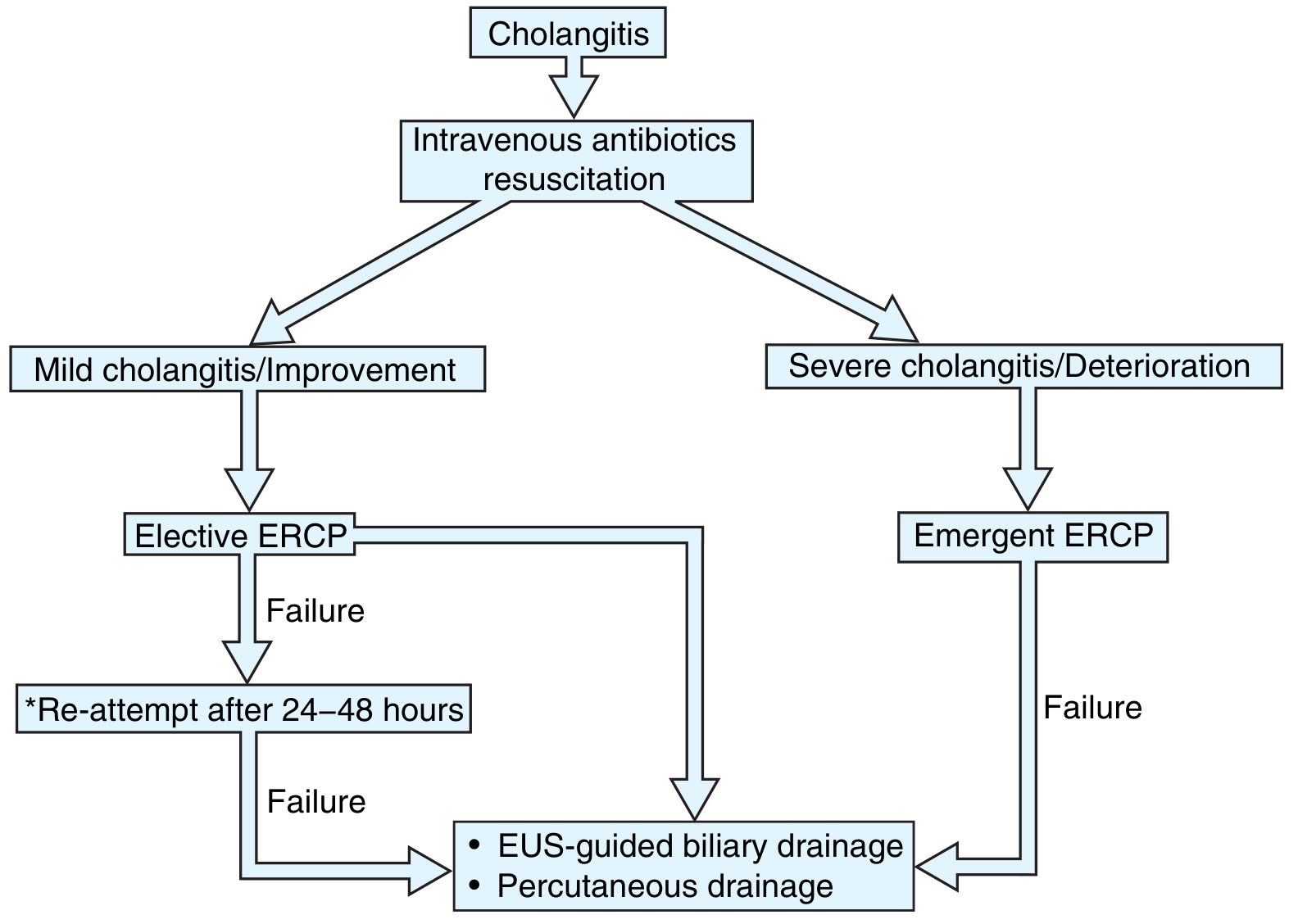
| Grade I (Mild) | Antibiotics alone; biliary drainage if no response within 24-48h |
| Grade II (Moderate) | Early biliary drainage (endoscopic or percutaneous) in addition to antibiotics |
| Grade III (Severe) | Immediate hemodynamic/respiratory stabilization + ICU + urgent biliary drainage as soon as possible |
The management algorithm is illustrated below (from Clinical Gastrointestinal Endoscopy, 3e):
Drainage Modalities
1. ERCP (preferred first-line)
- Transpapillary route; allows stone extraction, sphincterotomy, stent placement, or nasobiliary tube (NBT) placement simultaneously
- In severe/unstable patients: initial placement of biliary stent or NBT to decompress (palliative intent); definitive stone clearance deferred
- Key technique: aspirate infected bile under pressure before injecting contrast - repeated contrast injection into an obstructed system raises intrabiliary pressure and worsens cholangiovenous reflux/sepsis
- Delay >48h to ERCP is associated with higher costs and longer hospital stays; drainage within 24h predicts improved survival
- Plastic stent vs. NBT: both equally effective for initial drainage; plastic stent avoids patient discomfort from NBT
2. EUS-guided biliary drainage (choledochoduodenostomy)
- Used when ERCP fails or anatomy is altered (post-surgical)
- Avoids percutaneous access morbidity; limited by availability of local expertise
- Effective rescue option after failed ERCP
3. Percutaneous transhepatic cholangiography (PTC)/drainage
- Used when ERCP and EUS-guided drainage fail or are unavailable
- Higher morbidity than endoscopic routes
- Particularly useful for hilar or intrahepatic obstruction
4. Surgical drainage (last resort)
- Common bile duct exploration + T-tube placement
- Reserved when endoscopic and percutaneous approaches are unavailable or unsuccessful
- Definitive surgical treatment of the underlying cause (e.g., cholecystectomy) is deferred until:
- Hemodynamic stability restored
- Cholangitis resolved
- Definitive diagnosis confirmed
(Sabiston Textbook of Surgery; Clinical Gastrointestinal Endoscopy, 3e; Maingot's Abdominal Operations)
Subsequent Definitive Management
- After cholangitis resolves, cholecystectomy is indicated in patients with gallstone disease to prevent recurrence - for mild disease, this can be performed before discharge; for severe disease, delayed until full recovery.
- Intraoperative cholangiography should be performed at cholecystectomy when choledocholithiasis is suspected.
- Definitive ERCP for complete stone clearance or stricture management follows once the patient is stable.
Summary Table
| Element | Evidence/Recommendation |
|---|---|
| Diagnosis | Charcot's triad + TG18 criteria; US first, MRCP/EUS if diagnosis uncertain |
| Antibiotics | Immediate IV empirical (gram-negative coverage); tailor to local sensitivity |
| Duration | 4-7 days post source control; 14 days if bacteremia; short courses (3-4 days) likely sufficient (RCT evidence) |
| Mild | Antibiotics ± drainage if no response |
| Moderate | Early ERCP/drainage |
| Severe | ICU stabilization + urgent ERCP/drainage ASAP |
| Drainage preference | ERCP > EUS-guided > PTC > surgery |
| Timing of ERCP | Within 24h for severe; delay >48h worsens outcomes |
| Anaerobic cover | Add if prior bilioenteric surgery |
| ESBL awareness | Consider carbapenem if resistant organisms likely |
Key references:
- Sabiston Textbook of Surgery (22nd ed.)
- Yamada's Textbook of Gastroenterology (7th ed.), pp. 2178-2180
- Clinical Gastrointestinal Endoscopy (3rd ed.), pp. 841-842
- Maingot's Abdominal Operations, pp. 1041-1042
- Srinu et al. (2024). Short vs. conventional antibiotic duration in cholangitis. Am J Gastroenterol. PMID 37732816 - RCT supporting 4-day courses post-drainage
Differential diagnososs
The differential diagnosis of acute cholangitis covers conditions that share RUQ pain, fever, and/or jaundice. The main differentials include acute cholecystitis, gallstone pancreatitis, liver abscess, hepatitis, and peptic ulcer disease.
Searching sources...
Finding Sources
Finding Sources
Reading File
Reading File
Finding Sources
Finding Sources
Reading File
Reading File
Finding Sources
Finding Sources
Reading File
I now have sufficient material for a comprehensive differential diagnosis table. Here is the full answer:
Differential Diagnosis of Acute Cholangitis
Acute cholangitis shares its cardinal features - RUQ pain, fever, and jaundice (Charcot's triad) - with several other conditions. The key diagnostic task is to distinguish cholangitis from conditions that require different (sometimes urgent) management.
Primary Biliary/Hepatic Differentials
1. Acute Cholecystitis
The most common condition confused with cholangitis.
| Feature | Acute Cholangitis | Acute Cholecystitis |
|---|---|---|
| Fever | Yes | Yes |
| RUQ pain | Yes | Yes |
| Jaundice | Yes (common) | Absent or mild |
| Murphy's sign | Absent | Positive |
| CBD dilatation on US | Yes | No (unless Mirizzi's) |
| WBC | Elevated | Elevated |
| Bilirubin | Markedly elevated | Normal or mildly elevated |
| ALP/GGT | Elevated (cholestatic pattern) | Mild rise |
| Septic shock risk | High | Lower |
Diagnostic criteria for cholecystitis require local signs (Murphy's sign, RUQ mass/tenderness) + systemic signs + imaging - with sensitivity 91.2% and specificity 96.9% against surgical pathology. Note: jaundice is the discriminating feature - its presence strongly favors cholangitis over uncomplicated cholecystitis. (Tintinalli's Emergency Medicine)
2. Choledocholithiasis (without cholangitis)
CBD stones can cause biliary colic and mild jaundice without infection. The distinction is that fever and signs of sepsis are absent in simple choledocholithiasis. The moment fever appears alongside CBD obstruction, cholangitis must be assumed.
3. Mirizzi's Syndrome
A large gallstone impacted in the cystic duct or Hartmann's pouch externally compresses the CHD/CBD, causing obstructive jaundice. Can mimic cholangitis closely, including jaundice and RUQ pain. May be complicated by secondary cholangitis. Diagnosis is made on MRCP/ERCP showing external compression of the bile duct, not intrinsic CBD obstruction. (Current Surgical Therapy, 14e)
4. Gallstone Pancreatitis
- Caused by a stone transiently obstructing the ampulla; pancreatic inflammation persists even after stone passage
- Shares RUQ/epigastric pain and fever with cholangitis
- Key distinguishing features:
- Markedly elevated serum amylase/lipase (>3x ULN)
- Pain typically radiates to the back
- Jaundice is less prominent and transient
- Imaging: peripancreatic inflammation on CT, not bile duct dilatation
- Note: when cholangitis and pancreatitis coexist, urgent ERCP is indicated (Yamada's Textbook of Gastroenterology)
5. Pyogenic Liver Abscess
- Presents with fever, RUQ pain, and sometimes jaundice (if biliary involvement)
- Usually more insidious onset with higher, spiking/hectic fevers and diaphoresis
- Jaundice may be absent or mild unless associated with biliary obstruction
- US/CT: hypoechoic or low-density lesion with peripheral ring enhancement - distinguishes it from cholangitis (which shows bile duct changes, not a focal lesion)
- Blood cultures and aspirate culture are the key workup
- Can be a complication of untreated cholangitis (Yamada's Textbook of Gastroenterology)
6. Amebic Liver Abscess
- Fever, RUQ pain; history of travel to or residence in endemic area
- More common in young men
- Serological test for Entamoeba histolytica antibodies is positive in >90%
- Aspirate has characteristic "anchovy paste" appearance (necrotic hepatocytes)
- Single abscess more common than pyogenic (multiple abscesses)
- Jaundice is typically absent (Yamada's Textbook of Gastroenterology)
7. Acute Viral Hepatitis (A, B, E)
- Fever, RUQ pain, jaundice can all be present - can closely mimic cholangitis
- Key distinguishing features:
- Markedly elevated transaminases (ALT/AST in hundreds to thousands) - disproportionate to ALP/GGT
- Bile duct dilatation is absent on imaging
- History of exposure, prodrome of malaise/anorexia/nausea
- Viral serology (anti-HAV IgM, HBsAg, anti-HEV IgM) confirms diagnosis
- In acute hepatic failure: encephalopathy + coagulopathy + jaundice should prompt evaluation for fulminant hepatitis (Rosen's Emergency Medicine)
Extra-Biliary Differentials
8. Peptic Ulcer Disease / Perforated Ulcer
- Epigastric pain radiating to right; can present with fever if perforation occurs
- Perforated PUD: peritonism (board-like rigidity, absent bowel sounds), free air under diaphragm on erect CXR
- Jaundice is absent
- Amylase may be mildly elevated (from gastric leak)
9. Right Lower Lobe Pneumonia / Pleurisy
- Fever + right-sided upper abdominal pain referred from the diaphragm
- No jaundice
- Cough, pleuritic chest pain, reduced air entry on auscultation
- CXR/CT chest confirms consolidation (Current Surgical Therapy, 14e)
10. Empyema (Right-sided)
- Fever + pleuritic right-sided pain; can be confused if referred abdominally
- No jaundice; no biliary changes on imaging (Current Surgical Therapy, 14e)
11. Appendicitis (Atypical/Retrocecal)
- Occasionally presents with RUQ pain (retrocecal or subhepatic appendix)
- Fever and leukocytosis present
- No jaundice; no bile duct changes
- CT abdomen is diagnostic
12. Life-threatening Diagnoses Not to Miss
These conditions can mimic biliary disease but have dramatically different management:
| Condition | Clue |
|---|---|
| Ruptured AAA | Pulsatile mass, hemodynamic collapse, no jaundice |
| Aortic dissection | Tearing chest/back pain, BP differential between arms |
| Inferior MI | ECG changes, troponin rise, no biliary imaging changes |
| Perforated gallbladder | Peritonism, free fluid on US, rapid deterioration |
| Ischemic bowel | Disproportionate pain to exam, metabolic acidosis, CT findings |
(Yamada's Textbook of Gastroenterology, 7e)
Diagnostic Approach Summary
| Investigation | Purpose |
|---|---|
| US abdomen | First-line: CBD dilatation, gallstones, gallbladder wall thickening |
| LFTs (ALP, GGT, bilirubin) | Cholestatic pattern favors biliary cause |
| ALT/AST | If markedly elevated - hepatitis rather than biliary obstruction |
| Amylase/lipase | Elevated >3x - pancreatitis |
| Serology (viral hepatitis) | If transaminases dominant |
| CT abdomen | Better for level of obstruction, abscess, perforation |
| MRCP/EUS | Non-invasive CBD stone/stricture evaluation before ERCP |
| Blood cultures | Positive in 20-70% of cholangitis; not seen in cholecystitis/hepatitis |
| CA19-9 | Can be elevated in biliary obstruction - not specific for malignancy when bilirubin is high |
The key discriminating triad: fever + jaundice + CBD dilatation on imaging = acute cholangitis until proven otherwise. (Current Surgical Therapy, 14e; Tintinalli's Emergency Medicine)
Tokya classification
The Tokyo Guidelines (TG07 → TG13 → TG18) classify acute cholangitis into three severity grades based on organ dysfunction, lab parameters, and clinical features, guiding timing and urgency of biliary drainage.
Searching sources...
Finding Sources
Finding Sources
Reading File
Reading File
Reading File
Finding Sources
Reading File
Finding Sources
I have all the key data from the textbooks. Here is the complete answer:
Tokyo Guidelines (TG18): Classification of Acute Cholangitis
The Tokyo Guidelines were first published in 2007 (TG07), then updated in 2013 (TG13) and 2018 (TG18). They provide a standardised framework for diagnosis, severity grading, and treatment timing of acute cholangitis.
Part 1 - Diagnostic Criteria (TG18)
Diagnosis requires at least one criterion from each applicable domain:
| Domain | Criteria |
|---|---|
| A. Systemic Inflammation | Fever and/or rigors; AND/OR lab evidence of inflammatory response (elevated WBC, CRP) |
| B. Cholestasis | Jaundice (total bilirubin ≥2 mg/dL); AND/OR abnormal LFTs (elevated ALP, GGT, AST, ALT) |
| C. Imaging | Biliary dilatation; AND/OR evidence of aetiology on imaging (stone, stricture, stent, etc.) |
Interpretation:
- Suspected cholangitis: 1 criterion from A + 1 criterion from B or C
- Definite cholangitis: 1 criterion from A + 1 from B + 1 from C
(Note: Charcot's triad alone has high specificity but only ~26% sensitivity, hence the structured TG18 criteria)
Part 2 - Severity Grading (TG18)
Grade I - Mild Cholangitis
- Does not meet criteria for Grade II or Grade III at initial presentation
- No organ dysfunction
- Responds to initial medical treatment (IV antibiotics + fluids)
Grade II - Moderate Cholangitis
Any two or more of the following:
| Criterion | Threshold |
|---|---|
| WBC (leukocytosis or leukopenia) | >12,000/mm³ or <4,000/mm³ |
| Fever | ≥39°C |
| Age | ≥75 years |
| Total bilirubin | ≥5 mg/dL |
| Albumin | <lower limit of normal × 0.7 |
Grade III - Severe Cholangitis
Dysfunction of at least one organ system:
| Organ System | Criterion |
|---|---|
| Cardiovascular | Hypotension requiring vasopressors |
| Neurological | Disturbance of consciousness |
| Respiratory | PaO₂/FiO₂ ratio <300 |
| Renal | Oliguria; serum creatinine >2 mg/dL |
| Hepatic | PT/INR >1.5 |
| Haematologic | Platelet count <100,000/mm³ |
Part 3 - Treatment Based on Grade (TG18)
| Grade | Management |
|---|---|
| Grade I (Mild) | IV antibiotics + fluid resuscitation; elective ERCP <72 hours if responding; urgent drainage if NOT responding to medical treatment |
| Grade II (Moderate) | Early biliary drainage in addition to antibiotics; urgent (<24 hours) if obstructive stones present |
| Grade III (Severe) | ICU admission; haemodynamic/respiratory stabilisation first; then urgent biliary drainage <24 hours as soon as feasible |
Specific triggers for urgent biliary drainage (<24 hours):
- Severe (Grade III) cholangitis - regardless of aetiology
- Obstructive biliary stones with moderate (Grade II) or severe (Grade III) cholangitis
- Mild (Grade I) cholangitis not responding to IV antibiotics and fluid resuscitation
(Mulholland and Greenfield's Surgery, 7e, citing Kiriyama S et al., J Hepatobiliary Pancreat Sci 2018;25:17-30)
Summary Diagram
CHOLANGITIS SUSPECTED
        |
  TG18 Diagnostic Criteria Met?
        |
   YES --> Grade Assessment
        |
  ┌─────┴──────────────────┐
Grade I              Grade II           Grade III
(Mild)              (Moderate)          (Severe)
No organ         ≥2 lab/clinical      ≥1 organ failure
dysfunction       criteria met
     |                 |                    |
IV Abx + IVF    Early ERCP          ICU + Stabilise
 <72h ERCP      ± Urgent if          + URGENT ERCP
 if responds    stones/no response     <24 hours
Key Points
- Grade determines urgency, not just severity of symptoms - an elderly patient (≥75) with bilirubin ≥5 and fever ≥39°C automatically qualifies as Grade II even without shock
- Reynolds' pentad (hypotension + altered mental status added to Charcot's triad) is the clinical correlate of Grade III - mortality approaches 100% without prompt treatment
- CA19-9 may be falsely elevated with biliary obstruction - do not interpret as evidence of malignancy until obstruction resolves
- The TG18 update refined earlier TG13 criteria by clarifying that Grade III triggers are organ system dysfunctions, not just clinical signs of shock
(Mulholland and Greenfield's Surgery, 7e; Yamada's Textbook of Gastroenterology, 7e; Current Surgical Therapy, 14e)
This is a shared conversation. Sign in to Orris to start your own chat.